Slit lamp - Frank's Hospital...
3
Slit lamp 1 Slit lamp Slit lamp examination of the eyes in an ophthalmology clinic Cataract in Human Eye- Magnified view seen on examination with a slit lamp The slit lamp is an instrument consisting of a high-intensity light source that can be focused to shine a thin sheet of light into the eye. It is used in conjunction with a biomicroscope. The lamp facilitates an examination of the anterior segment, or frontal structures and posterior segment, of the human eye, which includes the eyelid, sclera, conjunctiva, iris, natural crystalline lens, and cornea. The binocular slit-lamp examination provides stereoscopic magnified view of the eye structures in detail, enabling anatomical diagnoses to be made for a variety of eye conditions. While a patient is seated in the examination chair, he rests his chin and forehead on a support to steady the head. Using the biomicroscope, the ophthalmologist or optometrist then proceeds to examine the patient's eye. A fine strip of paper, stained with fluorescein, a fluorescent dye, may be touched to the side of the eye; this stains the tear film on the surface of the eye to aid examination. The dye is naturally rinsed out of the eye by tears. A subsequent test may involve placing drops in the eye in order to dilate the pupils. The drops take about 15 to 20 minutes to work, after which the examination is repeated, allowing the back of the eye to be examined. Patients will experience some light sensitivity for a few hours after this exam, and the dilating drops may also cause increased pressure in the eye, leading to nausea and pain. Patients who experience these rare but serious symptoms are advised to seek medical attention immediately. Adults need no special preparation for the test; however children may need some preparation, depending on age, previous experiences, and level of trust. The slit lamp exam may detect many diseases of the eye, including: • Cataract • Conjunctivitis • Corneal injury such as corneal ulcer or corneal swelling • Diabetic retinopathy • Fuchs' dystrophy • Keratoconus (Fleischer ring) • Macular degeneration • Presbyopia • Retinal detachment • Retinal vessel occlusion • Retinitis pigmentosa
Transcript of Slit lamp - Frank's Hospital...
Slit lamp 1
Slit lamp
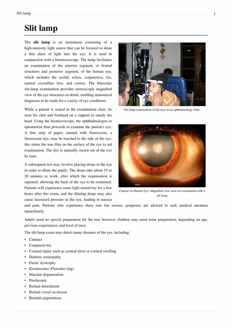
Slit lamp examination of the eyes in an ophthalmology clinic
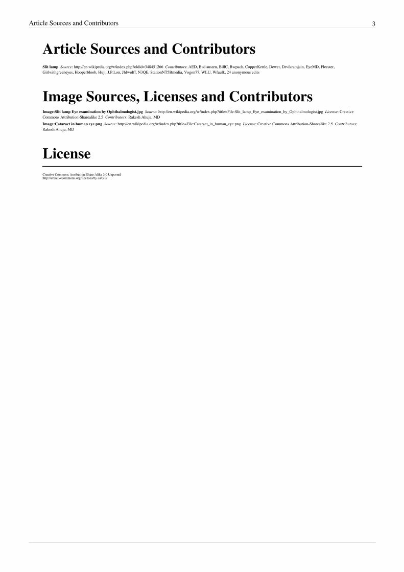
Cataract in Human Eye- Magnified view seen on examination with aslit lamp
The slit lamp is an instrument consisting of ahigh-intensity light source that can be focused to shinea thin sheet of light into the eye. It is used inconjunction with a biomicroscope. The lamp facilitatesan examination of the anterior segment, or frontalstructures and posterior segment, of the human eye,which includes the eyelid, sclera, conjunctiva, iris,natural crystalline lens, and cornea. The binocularslit-lamp examination provides stereoscopic magnifiedview of the eye structures in detail, enabling anatomicaldiagnoses to be made for a variety of eye conditions.
While a patient is seated in the examination chair, herests his chin and forehead on a support to steady thehead. Using the biomicroscope, the ophthalmologist oroptometrist then proceeds to examine the patient's eye.A fine strip of paper, stained with fluorescein, afluorescent dye, may be touched to the side of the eye;this stains the tear film on the surface of the eye to aidexamination. The dye is naturally rinsed out of the eyeby tears.
A subsequent test may involve placing drops in the eyein order to dilate the pupils. The drops take about 15 to20 minutes to work, after which the examination isrepeated, allowing the back of the eye to be examined.Patients will experience some light sensitivity for a fewhours after this exam, and the dilating drops may alsocause increased pressure in the eye, leading to nauseaand pain. Patients who experience these rare but serious symptoms are advised to seek medical attentionimmediately.
Adults need no special preparation for the test; however children may need some preparation, depending on age,previous experiences, and level of trust.The slit lamp exam may detect many diseases of the eye, including:• Cataract• Conjunctivitis• Corneal injury such as corneal ulcer or corneal swelling• Diabetic retinopathy• Fuchs' dystrophy• Keratoconus (Fleischer ring)• Macular degeneration• Presbyopia• Retinal detachment• Retinal vessel occlusion• Retinitis pigmentosa

Slit lamp 2
• Sjögren's syndrome• Uveitis• Wilson's disease (Kayser-Fleischer ring)
Article Sources and Contributors 3
Article Sources and ContributorsSlit lamp Source: http://en.wikipedia.org/w/index.php?oldid=348451266 Contributors: AED, Bad austen, BillC, Bwpach, CopperKettle, Dewet, Drvikramjain, EyeMD, Fleester,Girlwithgreeneyes, Hooperbloob, Huji, J.P.Lon, Jfdwolff, N3QE, StationNT5Bmedia, Vogon77, WLU, Wfaulk, 24 anonymous edits
Image Sources, Licenses and ContributorsImage:Slit lamp Eye examination by Ophthalmologist.jpg Source: http://en.wikipedia.org/w/index.php?title=File:Slit_lamp_Eye_examination_by_Ophthalmologist.jpg License: CreativeCommons Attribution-Sharealike 2.5 Contributors: Rakesh Ahuja, MDImage:Cataract in human eye.png Source: http://en.wikipedia.org/w/index.php?title=File:Cataract_in_human_eye.png License: Creative Commons Attribution-Sharealike 2.5 Contributors:Rakesh Ahuja, MD
LicenseCreative Commons Attribution-Share Alike 3.0 Unportedhttp:/ / creativecommons. org/ licenses/ by-sa/ 3. 0/