SIlver Nano Gram Negative
9
APPLIED AND ENVIRONMENTAL MICROBIOLOGY, Mar. 2007, p. 1712–1720 Vol. 73, No. 6 0099-2240/07/$08.00 0 doi:1 0.112 8/AEM.02 218-0 6 Copyright © 2007, American Society for Microbiology. All Rights Reserved. Does the Antibacterial Activity of Silver Nanoparticles Depend on the Shape of the Nanoparticle? A Study of the Gram-Negative Bacterium Escherichia coli Sukdeb Pal, Yu Kyung Tak, and Joon Myong Song* Research Institute of Pharmaceutical Sciences and College of Pharmacy, Seoul National University, Seoul 151-742, South Korea Received 21 September 2006/Accepted 9 January 2007 In this work we investigated the antibacterial properties of differently shaped silver nanoparticles against the gram-negative bacterium Escherichia coli, both in liquid systems and on agar plates. Energy-filtering trans- mission electron microscopy images revealed considerable changes in the cell membranes upon treatment, resulting in cell death. Truncated triangular silver nanoplates with a {111} lattice plane as the basal plane displa yed the stron gest biocidal action, compared with spheri cal and rod-s haped nanopart icles and with Ag (in the form of AgNO 3 ). It is proposed that nanoscale size and the presence of a {111} plane combine to promote this biocidal property. To our knowledge, this is the first comparative study on the bactericidal properties of silver nanoparticles of different shapes, and our results demonstrate that silver nanoparticles underg o a shape-dependent interaction with the gram- negat ive organism E. coli. Over the past few decades, inorganic nanoparticles, whose structures exhibit signific antly novel and improved physic al, chemical, and biological properties, phenomena, and function- ality due to their nanoscale size, have elicited much interest. Nano pha sic and nanost ruc tured mat erials are attracting a great deal of attention because of their potential for achieving specific processes and selectivity, especially in biological and pharmaceutical applications (5, 39). Disc overi es in the past decade have demonstr ated that the electromagnetic, optical, and catalytic properties of noble-metal nanocrystals are strongly influenced by shape and size (6, 26). This has motivated an upsurge in research on the synthesis routes that allow better control of shape and size (18, 34, 41), with projected applications in nanoelectronics and spectroscopy (14, 19, 36). Recent studies have demonstrated that specially formulated metal oxide nanoparticles have good antibacterial activity (33), and antimicrobial formulations comprising nanoparticles could be effective bactericidal materials (11, 12). Among inorganic antibacterial agents, silver has been em- ployed most extensively since ancient times to fight infections and control spoilage. The antibacterial and antiviral actions of silver, silver ion, and silver compounds have been thoroughly investigated (28, 29, 37). However, in minute concentrations, silver is nontoxic to human cells. The epidemiological history of silver has established its nontoxicity in normal use. Catalytic oxidat ion by metall ic silver and reaction with dissolv ed mono- valent silver ion probably contribute to its bactericidal effect (17). Microbes are unlikely to develop resistance against silver, as they do against conventional and narrow-target antibiotics, bec ause the metal attacks a bro ad range of target s in the organisms, which means that they would have to develop a host of mutations simultaneously to protect themselves. Thus, silver ions have been used as an antibacterial component in dental resin composites (15), in synthetic zeolites (22), and in coatings of medical devices (2). Recent literature reports encouraging results about the bac- tericidal activity of silver nanoparticles of either a simple or compo site nature (21, 31). Elechi guerr a and coworkers (9) found that silver nanoparticles undergo a size-dependent in- teraction with human immunodeficiency virus type 1, prefera- bly via binding to gp120 glycoprotein knobs. The size-depen- dent inter actio n of silver nanoparti cles with gram-negati ve bacteria has also been reported by the same group (25). How- ever, little is known about how the biological activity of silver nanoparticles changes as the shape of the particles changes. For these reason, we investigated the shape dependence of the antibacterial activity of silver nanoparticles against Esche- richia coli . Silver nanoparticles of different shapes were syn- thesized by solution phase routes, and their interactions with E. coli were studied. Energy-filtering transmission electron mi- croscopy (EFTEM) was used as a complementary technique to examine the treated cells. The size-dependent antimicrobial activity of silver nanoparticles has already been investigated (9, 25), while to our knowledge the effect of shape on the anti- bacterial activity of silver nanoparticles has not been reported previously. MATERIALS AND METHODS Materials. E. col i (ATCC 10536) was obta ined from the Amer ican Type Culture Collection (Manassas, VA). Difco nutrient broth (NB) (BD234000) and Difco nutrient agar (BD 213000) medium (Becton Dickinson and Co.) were used to grow and maintain the bacterial cultures per the supplier’s protocol. Chemi- cals such as silver nitrate, ascorbic acid, sodium citrate tribasic dihydrate, and cetyltrimethyl ammonium bromide (CTAB) were of the highest purity available (Sigma). These reagents were used as received without further purification. Preparation of silver nanoparticles. The silver nanoparticles used in this work were synthesized by two different methods. Both include seeding and growth. First, silver seeds were prepared by rapidly injecting 0.5 ml of 10 mM NaBH 4 into an aqueous solution (with continuous stirring) containing 0.5 ml of 0.01 M * Corre spond ing author. Maili ng address: Rese arch Insti tute of Phar mace utica l Sciences and Coll ege of Phar macy , Seoul Nati onal University, Seoul 151-742, South Korea. Phone: 82-2-880-7841. Fax: 82-2-871-2238. E-mail: [email protected] c.kr. Published ahead of print on 19 January 2007. 1712
Transcript of SIlver Nano Gram Negative
8/6/2019 SIlver Nano Gram Negative
http://slidepdf.com/reader/full/silver-nano-gram-negative 1/9
APPLIED AND ENVIRONMENTAL MICROBIOLOGY, Mar. 2007, p. 1712–1720 Vol. 73, No. 60099-2240/07/$08.000 doi:10.1128/AEM.02218-06Copyright © 2007, American Society for Microbiology. All Rights Reserved.
Does the Antibacterial Activity of Silver Nanoparticles Depend on theShape of the Nanoparticle? A Study of the Gram-Negative
Bacterium Escherichia coli
Sukdeb Pal, Yu Kyung Tak, and Joon Myong Song*
Research Institute of Pharmaceutical Sciences and College of Pharmacy, Seoul National University, Seoul 151-742, South Korea
Received 21 September 2006/Accepted 9 January 2007
In this work we investigated the antibacterial properties of differently shaped silver nanoparticles against thegram-negative bacterium Escherichia coli, both in liquid systems and on agar plates. Energy-filtering trans-mission electron microscopy images revealed considerable changes in the cell membranes upon treatment,resulting in cell death. Truncated triangular silver nanoplates with a {111} lattice plane as the basal planedisplayed the strongest biocidal action, compared with spherical and rod-shaped nanoparticles and with Ag
(in the form of AgNO3
). It is proposed that nanoscale size and the presence of a {111} plane combine topromote this biocidal property. To our knowledge, this is the first comparative study on the bactericidalproperties of silver nanoparticles of different shapes, and our results demonstrate that silver nanoparticles
undergo a shape-dependent interaction with the gram-negative organism E. coli.
Over the past few decades, inorganic nanoparticles, whosestructures exhibit significantly novel and improved physical,chemical, and biological properties, phenomena, and function-ality due to their nanoscale size, have elicited much interest.Nanophasic and nanostructured materials are attracting agreat deal of attention because of their potential for achievingspecific processes and selectivity, especially in biological andpharmaceutical applications (5, 39).
Discoveries in the past decade have demonstrated that theelectromagnetic, optical, and catalytic properties of noble-metalnanocrystals are strongly influenced by shape and size (6, 26).
This has motivated an upsurge in research on the synthesis routesthat allow better control of shape and size (18, 34, 41), withprojected applications in nanoelectronics and spectroscopy (14,19, 36).
Recent studies have demonstrated that specially formulatedmetal oxide nanoparticles have good antibacterial activity (33),and antimicrobial formulations comprising nanoparticles couldbe effective bactericidal materials (11, 12).
Among inorganic antibacterial agents, silver has been em-ployed most extensively since ancient times to fight infectionsand control spoilage. The antibacterial and antiviral actions of silver, silver ion, and silver compounds have been thoroughlyinvestigated (28, 29, 37). However, in minute concentrations,
silver is nontoxic to human cells. The epidemiological historyof silver has established its nontoxicity in normal use. Catalyticoxidation by metallic silver and reaction with dissolved mono- valent silver ion probably contribute to its bactericidal effect(17). Microbes are unlikely to develop resistance against silver,as they do against conventional and narrow-target antibiotics,because the metal attacks a broad range of targets in the
organisms, which means that they would have to develop a hostof mutations simultaneously to protect themselves. Thus, silverions have been used as an antibacterial component in dentalresin composites (15), in synthetic zeolites (22), and in coatingsof medical devices (2).
Recent literature reports encouraging results about the bac-tericidal activity of silver nanoparticles of either a simple orcomposite nature (21, 31). Elechiguerra and coworkers (9)found that silver nanoparticles undergo a size-dependent in-teraction with human immunodeficiency virus type 1, prefera-bly via binding to gp120 glycoprotein knobs. The size-depen-
dent interaction of silver nanoparticles with gram-negativebacteria has also been reported by the same group (25). How-ever, little is known about how the biological activity of silvernanoparticles changes as the shape of the particles changes.
For these reason, we investigated the shape dependence of the antibacterial activity of silver nanoparticles against Esche-
richia coli. Silver nanoparticles of different shapes were syn-thesized by solution phase routes, and their interactions with E. coli were studied. Energy-filtering transmission electron mi-croscopy (EFTEM) was used as a complementary technique toexamine the treated cells. The size-dependent antimicrobialactivity of silver nanoparticles has already been investigated (9,25), while to our knowledge the effect of shape on the anti-
bacterial activity of silver nanoparticles has not been reportedpreviously.
MATERIALS AND METHODS
Materials. E. coli (ATCC 10536) was obtained from the American TypeCulture Collection (Manassas, VA). Difco nutrient broth (NB) (BD234000) andDifco nutrient agar (BD 213000) medium (Becton Dickinson and Co.) were used
to grow and maintain the bacterial cultures per the supplier’s protocol. Chemi-cals such as silver nitrate, ascorbic acid, sodium citrate tribasic dihydrate, and
cetyltrimethyl ammonium bromide (CTAB) were of the highest purity available(Sigma). These reagents were used as received without further purification.
Preparation of silver nanoparticles. The silver nanoparticles used in this work
were synthesized by two different methods. Both include seeding and growth.First, silver seeds were prepared by rapidly injecting 0.5 ml of 10 mM NaBH4
into an aqueous solution (with continuous stirring) containing 0.5 ml of 0.01 M
* Corresponding author. Mailing address: Research Institute of Pharmaceutical Sciences and College of Pharmacy, Seoul NationalUniversity, Seoul 151-742, South Korea. Phone: 82-2-880-7841. Fax:82-2-871-2238. E-mail: [email protected].
Published ahead of print on 19 January 2007.
1712
8/6/2019 SIlver Nano Gram Negative
http://slidepdf.com/reader/full/silver-nano-gram-negative 2/9
AgNO3 and 20 ml of 0.001 M sodium citrate. The resultant solution was stirredfor 5 min and aged for 1.5 h.
In the first method, the spherical silver hydrosols were prepared by reducing
aqueous AgNO3 with sodium citrate at near-boiling temperature. In a typicalprocedure, an aqueous solution of AgNO3 (100 ml, 0.001 M) was brought toboiling, and then 3 ml of the silver seed solution and an aqueous solution of
sodium citrate were added so that the final concentration of sodium citrate in thereaction mixture became 0.001 M. The solution was heated until the color was
greenish yellow. The solution was cooled to room temperature. The silver nano-particles were purified by centrifugation. To remove excess silver ions, the silverpellet was washed three times with deionized water. A dried powder of the
nanosize silver was obtained by freeze-drying. To carry out interaction of the
silver nanoparticles with bacteria, the silver nanoparticle powder in the freeze-drying cuvette was resuspended in water; the suspension was homogenized witha Branson 2200 ultrasonicator. The resulting silver concentration was measuredby either inductively coupled plasma (ICP) emission spectroscopy (ES) (ICPS-
1000IV; Shimadzu, Kyoto, Japan) or ICP mass spectrometry (MS) (ELAN 6100;Perkin-Elmer SCIEX).
Elongated (rod-shaped) and truncated triangular silver nanoplates were syn-
thesized by a solution phase method for the large-scale preparation of truncatedtriangular nanoplates (8). To the particle growth solution containing 5 ml of 0.01
M AgNO3, 10 ml of 0.1 M ascorbic acid, 146 ml of 0.1 M CTAB, and 5 ml of silverseeds, 1 ml of 1 M NaOH was added to accelerate particle growth. The solutioncolor changed from light yellow to brown, red, and green within a few minutes.
The resultant solution was aged at 21°C for 12 h, 35°C for 5 min, and 21°C for24 h. The color of the aged solution changed from green to red.
The silver nanoplates were purified by centrifugation. The surfactants and thesmaller particles were separated by centrifugation at 2,100 g for 10 min. Theresultant precipitate was suspended in water and centrifuged again at 755 g for
10 min. Finally, the precipitate was suspended in water, and rods with largeraspect ratios were removed by centrifuging at 84 g for 10 min. The supernatantsolution contained the truncated triangular silver nanoplates. It was not possible
to quantify the precipitate by weight, as it was always associated with some water. The resulting silver level was quantified by ICP-ES or ICP-MS analysis
and reported in milligrams per liter or parts per million (detection limit of Ag, 0.1 ppb).
Organism preparation. E. coli ATCC 10536 was grown overnight in NB at
37°C. Washed cells were resuspended in NB, and optical density (OD) wasadjusted to 0.1, corresponding to 108 CFU/ml at 600 nm.
Bacterial growth or killing kinetics in presence of nanosilver. To examine thebacterial growth or killing kinetics in the presence of silver nanoparticles, E. coli
cells were grown in 100 ml of NB supplemented with different doses of nanosilver
(total silver content, 1, 6, 12, 12.5, 50, or 100 g), at 37°C with continuousagitation. The cylindrical sample containers were placed horizontally on anorbital shaker platform and agitated at 225 rpm. Growth or killing rates and
bacterial concentrations were determined by measuring OD at 600 nm. The OD
values were converted into concentration of E. coli cells (CFU per milliliter) (21,
31). OriginPro 7.5 software was used to analyze the experimental data and for
graphing (see Fig. 1 and 3 to 7).
Bacterial susceptibility to nanosilver. To examine the susceptibility of E. coli
to different silver nanoparticles, nutrient agar plates from a solution of agar were
prepared. A 100-l sample of bacterial suspension cultured in NB (with a
concentration of 105 or 107 CFU/ml of E. coli) was plated on a nutrient agar
plate. The plates were then supplemented with different amounts of nanosize
silver particles (1 to 100 g), and the plates were incubated further at 37°C. The
numbers of resultant colonies were counted after 24 h of incubation. Silver-free
plates incubated under the same conditions were used as controls. The counts
from three independent experiments corresponding to a particular sample wereaveraged.
EFTEM analysis was also carried out to examine sections of the treated
bacteria. Images of negatively stained particles were acquired as follows. Con-
centrated aqueous suspensions of E. coli cells were deposited on Formvar-coated
grids and allowed to sit for a few minutes. The grids were exposed to aqueous 1%
phosphotungstic acid for 30 s. The residual staining solution was then removed,
and the samples were immediately transferred to a microscope for inspection.
EFTEM images were acquired on a LIBRA 120 (Carl Zeiss) transmission elec-
tron microscope.
Characterization of silver nanoparticles. The synthesized nanoparticles were
characterized by UV-visible spectroscopy and EFTEM. EFTEM images were
observed with a LIBRA 120 (Carl Zeiss) transmission electron microscope. The
samples were prepared by placing a drop of homogeneous suspension on a
copper grid with a lacey carbon film and allowing it to dry in air. Mean particle
size was analyzed from the digitized images with Image Tool software. UV-
visible absorption spectra were recorded with an Optizen 2120 UV-visible spec-trophotometer (Mecasys, Daejeon, Korea) with a 1-cm quartz cell.
RESULTS
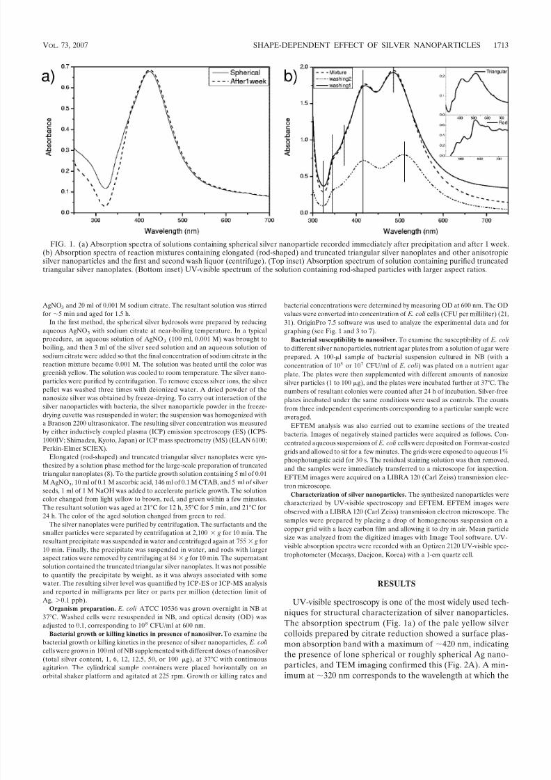
UV-visible spectroscopy is one of the most widely used tech-niques for structural characterization of silver nanoparticles.The absorption spectrum (Fig. 1a) of the pale yellow silvercolloids prepared by citrate reduction showed a surface plas-mon absorption band with a maximum of 420 nm, indicatingthe presence of lone spherical or roughly spherical Ag nano-particles, and TEM imaging confirmed this (Fig. 2A). A min-imum at 320 nm corresponds to the wavelength at which the
FIG. 1. (a) Absorption spectra of solutions containing spherical silver nanoparticle recorded immediately after precipitation and after 1 week.(b) Absorption spectra of reaction mixtures containing elongated (rod-shaped) and truncated triangular silver nanoplates and other anisotropicsilver nanoparticles and the first and second wash liquor (centrifuge). (Top inset) Absorption spectrum of solution containing purified truncated
triangular silver nanoplates. (Bottom inset) UV-visible spectrum of the solution containing rod-shaped particles with larger aspect ratios.
VOL. 73, 2007 SHAPE-DEPENDENT EFFECT OF SILVER NANOPARTICLES 1713
8/6/2019 SIlver Nano Gram Negative
http://slidepdf.com/reader/full/silver-nano-gram-negative 3/9
real and imaginary parts of the dielectric function of silveralmost vanish (32).
The optical absorption spectra of metal nanoparticles aredominated by surface plasmon resonances (SPR), which shiftto longer wavelengths with increasing particle size (4). The
position and shape of plasmon absorption of silver nanoclus-ters are strongly dependent on the particle size, dielectric me-dium, and surface-adsorbed species (20, 26). According toMie’s theory (24), only a single SPR band is expected in theabsorption spectra of spherical nanoparticles, whereas aniso-tropic particles could give rise to two or more SPR bandsdepending on the shape of the particles. The number of SPRpeaks increases as the symmetry of the nanoparticle decreases(32). Thus, spherical nanoparticles, disks, and triangular nano-plates of silver show one, two, and more peaks, respectively.
Even after several days, the aqueous dispersion of freeze-dried Ag nanoparticles, prepared by citrate reduction, dis-played UV-visible spectral characteristics of spherical Ag
nanoparticles (Fig. 1a), confirming the colloidal stability anduniformity of the silver hydrosol.On the other hand, interesting optical signatures were ob-
served for the sample synthesized by the soft template methodin the presence of CTAB micelles. The solution showed a redcolor; the corresponding absorption spectrum (Fig. 1b, “mix-ture” curve) displays three peaks at 348, 420, and 485 and ashoulder at 373 nm. The silver nanoparticles were separated bycentrifugation. The UV-visible spectra of all centrifuged sam-ples are also shown in Fig. 1b (curves for washing 1 and wash-ing 2). After centrifugation, the first and second wash liquorsshowed absorption maxima similar to that of the parent solu-tion. A broad hump could be identified at a longer wavelength(shifted to 509 nm for the second wash liquor), suggesting thepossible presence of a population of anisotropic Ag particles.
The UV-visible spectrum of the solution containing trun-cated triangular silver nanoparticles (Fig. 1b, top inset) showedtwo symmetrical absorption peaks at 418 and 514 nm. Also,a few humps can be seen at shorter (348 and 373 nm) as wellas longer (562 and 643 nm) wavelengths. The calculation of Schatz and Van Duyne (30) for the triangular prisms with thediscrete dipole approximation method suggests that the peaksat longer wavelengths are due to the in-plane dipole (514, 562,and 643 nm). The peak due to quadrupole plasmon resonance(460 nm) was expected to be very weak (8) and could not beseen. The shoulders at 373 and 348 nm are out-of-plane dipoleand quadrupole resonances, respectively. Formation of trian-
gular nanoplates is thus evident from the appearance of thesepeaks. Chen and Carroll (8) also showed that the blue shift of the in-plane dipole resonance to a lower wavelength (514 nm),in contrast to the calculated resonance peak at 770 nm for aperfect triangular nanoplate, indicates the truncated nature of
the triangular particles.The UV-visible spectra of the solution containing the parti-
cles with larger aspect ratios (Fig. 1b, lower inset) showedthree main absorption peaks at 480, 517, and 575 nm along with a few weak absorption peaks at the near-infrared (NIR)region. The absorption in the NIR region may be due to thein-plane dipole resonance mode associated with nanoplates with edge lengths greater than 100 nm (35) and the strongcoupling between the nanoparticles arising from aggregationof silver nanoparticles (16, 27). Absorption peaks for plates of a given thickness at longer wavelengths are also suggested tobe associated with the larger lateral size of the plates (7).
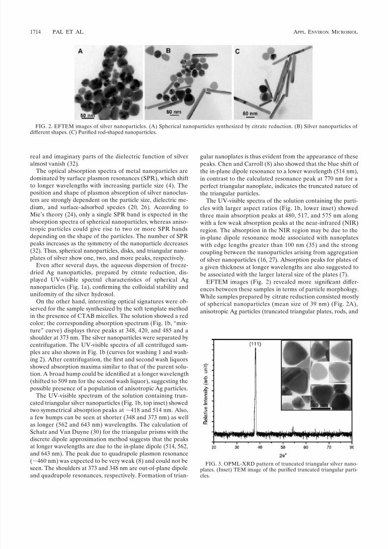
EFTEM images (Fig. 2) revealed more significant differ-
ences between these samples in terms of particle morphology.While samples prepared by citrate reduction consisted mostlyof spherical nanoparticles (mean size of 39 nm) (Fig. 2A),anisotropic Ag particles (truncated triangular plates, rods, and
FIG. 3. OPML-XRD pattern of truncated triangular silver nano-plates. (Inset) TEM image of the purified truncated triangular parti-cles.
FIG. 2. EFTEM images of silver nanoparticles. (A) Spherical nanoparticles synthesized by citrate reduction. (B) Silver nanoparticles of different shapes. (C) Purified rod-shaped nanoparticles.
1714 PAL ET AL. APPL. ENVIRON. MICROBIOL.

8/6/2019 SIlver Nano Gram Negative
http://slidepdf.com/reader/full/silver-nano-gram-negative 4/9
polyhedral plates) were more easily identified in other samples(Fig. 2B and C).
The presence of nanorods with an average edge lengthabove 100 nm was confirmed by EFTEM. Figure 2B showsrod-shaped nanoparticles with mean edge lengths of 133 nmand mean diameters of 16 nm. In Fig. 2C, rod-shaped nano-particles with larger aspect ratios, with a mean edge length of 192 nm and a mean diameter remaining at 16 nm, can be
seen.
Structural information on the truncated triangular nano-plates was obtained by oriented particulate monolayer X-raydiffraction (OPML-XRD). Figure 3 shows the XRD patternsof the nanoplates lying flat with their basal planes parallel tothe substrate. The remarkably intensive diffraction peak at a 2 value of 38.04 from the {111} lattice plane of face-centeredcubic silver unequivocally indicates that the particles are madeof pure silver and that their basal plane, i.e., the top crystal
plane, should be the {111} plane (8). It has been suggested (8)
FIG. 4. (A) Petri dishes initially supplemented with 107 CFU/ml of E. coli and incubated with different forms of silver nanoparticles at (a) 1,(b) 12.5, (c) 50, and (d) 100 g. (B) Number of E. coli colonies, expressed as log(1 number of colonies grown on plates under the conditionsused for panel A), as a function of the amount of silver nanoparticles in agar plates.
FIG. 5. Petri dishes initially supplemented with 105 CFU/ml of E. coli and incubated with silver at (a) 1, (b) 6, (c) 12.5, (d) 50, and (e) 100 gand corresponding graphs showing log(1 number of colonies grown on plates) as a function of the concentration of silver in agar plates. (A) Ag
(in the form of AgNO3); (B) spherical silver nanoparticles.
VOL. 73, 2007 SHAPE-DEPENDENT EFFECT OF SILVER NANOPARTICLES 1715
8/6/2019 SIlver Nano Gram Negative
http://slidepdf.com/reader/full/silver-nano-gram-negative 5/9
that this plane may possess the lowest surface tension. Figure3 (inset) shows the TEM image of purified truncated triangularparticles with an average edge length of 40 nm.
Figure 4A shows plates to which a 100-l bacterial suspen-sion (approximately 107 CFU/ml) was applied. The presence of nanoparticles at a certain level inhibited bacterial growth bymore than 90%. Interestingly, however, this level is signifi-cantly different for different nanoparticle shapes. For trun-cated triangular particles, almost complete inhibition of bac-terial growth was observed even at a total silver content of 1g. In the case of spherical nanoparticles, a total silver contentabove 12.5 g reduced the number of colonies significantly, while a total of 50 to 100 g of silver caused 100% inhibitionof bacterial growth. Rod-shaped nanoparticles and AgNO
3
showed inferior performance. Even in the presence of 100 gof rod-shaped particles, some colonies grew on the plate,though fewer than with an identical AgNO3 dose.
Figure 4B shows the corresponding results, expressed aslog(1 x) f( c), i.e., log(1 x) as a function of the totalamount of silver nanoparticles ( c), where x was the number of CFU grown on agar plates. The decrease in number of viablecells with increasing amounts of silver can be fitted with a
first-order exponential decay curve with a nonlinear regressioncoefficient ( R2) ranging from 0.96 to 1.
Figure 5 displays the number of bacterial colonies grown inthe presence of different amount of AgNO
3and spherical
nanoparticles when 100 l (approximately 105 CFU/ml) of sample was applied to each plate. The results clearly indicatethat at a given concentration of silver, inhibition of bacterialgrowth depends on the initial number of cells. An amount of 6g of spherical nanoparticles almost completely preventedbacterial growth (Fig. 5B). At silver (in the form of AgNO3)amounts of 12.5 g and above, 100% inhibition of bacterialgrowth was also observed (Fig. 5A). This is markedly differentfrom the results obtained when the initial number of bacterialcells was 107 CFU. In this case also, the results, expressed aslog (1 x) f( c), follow the first-order exponential decaycurve with a nonlinear regression coefficient ( R2) of 0.99(Fig. 5).
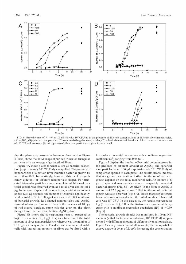
The bacterial growth kinetics was monitored in 100 ml NBmedium (initial bacterial concentration, 107 CFU/ml) supple-mented with different amounts of different silver nanoparticles.Figure 6 clearly shows that at all amounts, the nanoparticlescaused a growth delay of E. coli; increasing the concentration
FIG. 6. Growth curve of E. coli in 100 ml NB with 10
7
CFU/ml in the presence of different concentrations of different silver nanoparticles.(A) AgNO3; (B) spherical nanoparticles; (C) truncated triangular nanoparticles; (D) spherical nanoparticles with an initial bacterial concentrationof 105 CFU/ml. Amounts (in micrograms) of silver nanoparticles are given in each panel.
1716 PAL ET AL. APPL. ENVIRON. MICROBIOL.

8/6/2019 SIlver Nano Gram Negative
http://slidepdf.com/reader/full/silver-nano-gram-negative 6/9
of nanoparticles increased this growth delay. This is in line withfindings reported previously (31). A trend similar to that seenin the plate study can be noticed in the broth study. At a givenconcentration, truncated triangular nanoparticles are found tobe more effective in delaying the growth of E. coli than spher-ical particles, which again demonstrated better antibacterialactivity than AgNO
3. A dose of 10 g of truncated triangular
particles completely inhibited growth even after 24 h (Fig. 6C), whereas 100 g of silver (in the form of AgNO3) and spherical
nanoparticles caused only a prolonged growth delay, 8 to10 h (Fig. 6A and B). As expected, bacterial growth was found to be dependent on
the initial number of cells present in the broth. Figure 6Ddepicts the result of bacterial growth monitored in 100 ml of NB (initial cell numbers, 105 CFU/ml) supplemented with dif-ferent doses of spherical silver nanoparticles. A comparison with Fig. 6B shows that spherical nanoparticles exhibited sig-nificantly superior antibacterial property at a given nanopar-ticle dose when the initial E. coli concentration was reduced. Asimilar trend was observed for other nanoparticles as well(data not shown).
Figure 7a depicts the killing activity for E. coli (in 100 ml of NB supplemented with 107 CFU/ml of cells) in the presence of the mother liquor (the reaction mixture of triangular, rod-shaped, and polyhedral silver nanoparticles, before centrifuga-tion), containing different amounts of total silver (Ag and Agnanoparticles) as well as a substantial amount of CTAB. Thekilling activity was found to be sustained, as no cell growth wasseen even after 26 h of incubation. Figure 7b shows the com-parative killing activities for E. coli when the bacterial suspen-sion was treated with the same mother liquor containing 12 gof total silver immediately after preparation (see experimentalsection) and after 1 and 2 weeks of aging at room temperature.The killing activity of the silver hydrosol was found to diminish with increased aging time. The Fig. 7b inset shows the dynam-ics of E. coli growth as observed in the presence of 12 g of
different silver nanoparticles (10 g of truncated triangularnanoparticles) and ionic silver (AgNO
3) in 100 ml of NB sup-
plemented with 107 CFU/ml. Interestingly, instead of a growthcurve (Fig. 7b, inset), as seen in the presence of purified silvernanoparticles, a decay curve was obtained when the E. coli
suspension was treated with the unpurified mother solution of truncated triangular and rod-shaped nanoparticles, finallyleading to complete killing of the bacterial cells.
The surface structures of both the untreated (Fig. 8A) and
the treated bacterial cells (Fig. 8B) were examined usingEFTEM. The treated bacterial cells were significantly changed,and major damage was observed in the outer membrane (Fig.8C). Nanoparticles that accumulated in the membrane as wellas some penetrating the cells can also be discerned in theEFTEM micrograph.
DISCUSSION
The size of metallic nanoparticles ensures that a significantlylarge surface area of the particles is in contact with the bacte-rial effluent. Considering a hypothetical case with sphericalparticles of uniform size, a reduction in the particle size from10 m to 10 nm will increase the contact surface area by 109.Such a large contact surface is expected to enhance the extentof bacterial elimination. However, smallness in itself is not thegoal. Synthesis and characterization of nanoscaled materials interms of novel physicochemical properties is of great interest inthe formulation of bactericidal materials.
Although growth on agar plates is a more ready means of distinguishing antimicrobial properties of silver nanoparticlesof different shapes, in this study liquid-growth experimentsshowed basically similar results. But a previous study (31)pointed out a distinct difference between these two methods.In our study, complete inhibition of bacterial growth was ob-served on agar plates supplemented with nanoparticles (Fig. 4and 5). It is noteworthy that inhibition depends on the con-
FIG. 7. (a) Killing activity for E. coli (in 100 ml NB supplemented with 107 CFU/ml) in the presence of a mixture of triangular, rod-shaped,and polyhedral silver nanoparticles (before purification), containing different amounts of total silver (Ag and Ag nanoparticles) and CTAB. Totalsilver amounts are shown in each panel. (b) Decay curve of E. coli growth in 100 ml NB with 107 CFU/ml treated with the mother solution of truncated triangular and rod-shaped nanoparticles (total amount of silver, 12 g). (Inset) Comparative graph of the dynamics of E. coli growth in
the presence of 12 g of spherical silver nanoparticles, 10 g of triangular nanoparticles, and 12 g of ionic silver (AgNO3) in 100 ml NBsupplemented with 107 CFU/ml. Axes and units for the inset are same as those for the main graph and have been omitted for simplicity.
VOL. 73, 2007 SHAPE-DEPENDENT EFFECT OF SILVER NANOPARTICLES 1717

8/6/2019 SIlver Nano Gram Negative
http://slidepdf.com/reader/full/silver-nano-gram-negative 7/9
centration of the silver nanoparticles as well as on the initialbacterial number. On the other hand, silver nanoparticles inliquid medium, even at high concentrations, caused mainly agrowth delay of E. coli (Fig. 6). Growth of bacterial cellsresumed rapidly with a decrease in the concentration of nano-particles. Sondi and Salopek-Sondi (31) showed that interac-tion of these particles with intracellular substances from thelysed cells caused their coagulation and the particles werethrown out of the liquid system.
The decay curves shown in Fig. 7a can be explained similarly.In the mother liquor of truncated triangular and rod-shapednanoparticles, silver particles with different shapes werepresent in the micelles of the cationic quaternary ammoniumsurfactant CTAB. The surfaces of E. coli ATCC 10536 cellshave a negative charge. Thus, the addition of a nanoparticlesolution with a certain amount of cationic surfactant to thebacterial suspension probably led to coagulation of the nano-particles via a charge neutralization mechanism. The low col-loidal stability of the nanoparticles resulted in a decrease of effective silver concentration in the broth medium. However,most likely due to a synergistic effect of the cationic surfactantand silver nanoparticles, a decay curve was obtained, finally
leading to complete cell death. Coagulation via a charge neu-tralization mechanism is most effective at an optimum coagu-lant dose under a particular set of experimental conditions(temperature, pH, and mixing speed). Most probably in ourstudy, under the given experimental conditions, the nanopar-ticle levels of 6 to 12 g correspond to the optimum surfactantconcentration for effective coagulation, and a slower killingcurve was obtained. At a level lower than the optimum nano-particle level (and hence the optimum surfactant level) (e.g., 1g of silver), complete charge neutralization does not happen, whereas at a higher level (e.g., 50 or 100 g), due to chargereversal the nanoparticles regain their colloidal stability, lead-ing to poor or ineffective coagulation. Consequently, in thosecases the killing is either much faster or immediate. A controlexperiment with the same amount of surfactant dose (but with-out silver nanoparticles) produced similar types of graphs (datanot shown), though with a much lower killing rate, and con-firmed the previous observations.
Reports on the mechanism of inhibitory action of silver ionson microorganisms show that upon Ag treatment, DNA losesits replication ability (10) and expression of ribosomal subunitproteins as well as some other cellular proteins and enzymes
FIG. 8. EFTEM images of E. coli cells. (A) Untreated E. coli. Flagella can be seen. (B) E. coli grown on agar plates supplemented with Ag
(AgNO3). Arrows indicate partially damaged membranes. These cells are viable. (C) E. coli treated with triangular silver nanoplates. Silver
nanoparticles appear as dark irregular pits on the cell surface. (D) E. coli treated with spherical silver nanoparticles. (E) Enlarged image of partof the bacterial cell membrane treated with triangular silver nanoparticles. The cell membrane is damaged in multiple locations.
1718 PAL ET AL. APPL. ENVIRON. MICROBIOL.
8/6/2019 SIlver Nano Gram Negative
http://slidepdf.com/reader/full/silver-nano-gram-negative 8/9
essential to ATP production becomes inactivated (40). It hasalso been hypothesized that Ag primarily affects the functionof membrane-bound enzymes, such as those in the respiratorychain (3, 23).
However, the mechanism of bactericidal actions of silvernanoparticles is still not well understood. In a previous report(31) on the bactericidal activity of silver nanoparticles, it wasshown that the interaction between silver nanoparticles andconstituents of the bacterial membrane caused structuralchanges in and damage to membranes, finally leading to celldeath.
We speculate that the action of silver nanoparticles isbroadly similar to that of silver ion. It may be anticipated thata bacterial cell in contact with silver nanoparticles takes insilver ions, which inhibit a respiratory enzyme(s), facilitatingthe generation of reactive oxygen species and consequentlydamaging the cell. The uptake of silver can be recognized byirregular pits (Fig. 8C). Silver (soft acid) has a greater tendencyto react with sulfur- or phosphorus-containing soft bases, suchas R-S-R, R-SH, RS, or PR3. Thus, sulfur-containing proteins
in the membrane or inside the cells and phosphorus-containingelements like DNA are likely to be the preferential sites forsilver nanoparticle binding (3, 23). Figure 8C is an enlargedimage of a part of a severely damaged cell membrane treated with truncated triangular silver nanoplates. It has been sug-gested (31) that disruption of membrane morphology may causea significant increase in permeability, leading to uncontrolledtransport through the plasma membrane and, finally, cell death.
Although the findings regarding interaction with E. coli weresimilar among nanoparticles with markedly different shapes,the inhibition results were not the same. The differences in theobserved trends in E. coli inhibition can be explained in termsof the percent active facets present in nanoparticles of different
shapes. The OPML-XRD pattern (Fig. 3) shows that the topbasal plane of a truncated triangular nanoplate is a {111} surface.Spherical silver nanoparticles (generally with a cubooctahedral ormultiple-twinned decahedral or quasi-spherical morphology) pre-dominantly have {100} facets along with small percentage of {111} facets, while in case of the rod-like silver nanoparticles(e.g., pentagonal rods), side surfaces are bound by {100} and theends by {111} facets (38). It has been demonstrated that thereactivity of silver is favored by high-atom-density facets such as{111} (1, 13). Morones et al. (25) proved the faceting of theparticles as well as the direct interaction of the {111} facets withthe bacterial surface. Thus, a high reactivity of the truncatedtriangular nanoplates, as found in this study, in comparison toother particles that contain fewer than {111} facets, like sphericalor rod-shaped particles, is expected.
In conclusion, we have found that silver nanoparticles un-dergo shape-dependent interaction with the gram-negativebacterium E. coli. The interactions of silver nanoparticles withbiosystems are just beginning to be understood, and theseparticles are increasingly being used as microbicidal agents. Itmay be speculated that silver nanoparticles with the same sur-face areas but with different shapes may also have differenteffective surface areas in terms of active facets. Though atpresent we are unable to give an estimation of how the surfaceareas of different nanoparticles influence their killing activityor to relate the bacterial killing capacity of silver nanoparticles with their effective surface areas, our results provide a basis for
the measurement of shape-dependent bacterial activity of sil- ver nanoparticles. However, it is imperative that the flexibilityof nanoparticle preparation methods and their influence onother bacterial strains be explored.
ACKNOWLEDGMENT
This research was sponsored by the Korean Ministry of Science andTechnology under contract M10640020002-06N4002-00211 (“A Studyon Advanced Molecular Recognition Technology” program) withSeoul National University.
REFERENCES
1. Ajayan, P. M., and L. D. Marks. 1988. Quasimelting and phases of smallparticles. Phys. Rev. Lett. 60:585–587.
2. Bosetti, M., A. Masse, E. Tobin, and M. Cannas. 2002. Silver coated materialsfor external fixation devices: in vitro biocompatibility and genotoxicity. Bio-materials 23:887–892.
3. Bragg, P. D., and D. J. Rainnie. 1974. The effect of silver ions on therespiratory chains of Escherichia coli. Can. J. Microbiol. 20:883–889.
4. Brause, R., H. Moeltgen, and K. Kleinermanns. 2002. Characterization of laser-ablated and chemically reduced silver colloids in aqueous solution byUV/VIS spectroscopy and STM/SEM microscopy. Appl. Phys. B 75:711–716.
5. Brigger, I., C. Dubernet, and P. Couvreur. 2002. Nanoparticles in cancer
therapy and diagnosis. Adv. Drug Delivery Rev. 54:631–651.6. Burda, C., X. Chen, R. Narayanan, and M. A. El-Sayed. 2005. Chemistry andproperties of nanocrystals of different shapes. Chem. Rev. 105:1025–1102.
7. Callegari, A., D. Tonti, and M. Chergui. 2003. Photochemically grown silvernanoparticles with wavelength-controlled size and shape. Nano Lett. 3:1565–1568.
8. Chen, S., and D. L. Carroll. 2002. Synthesis and characterization of trun-cated triangular silver nanoplates. Nano Lett. 2:1003–1007.
9. Elechiguerra, J. L., J. L. Burt, J. R. Morones, A. Camacho-Bragado, X.Gao, H. H. Lara, and M. J. Yacaman. 29 June 2005. Interaction of silver nano-particles with HIV-1. J. Nanobiotechnol. 3:6. http://www.jnanobiotechnology.com/content/3/1/6.
10. Feng, Q. L., J. Wu, G. Q. Chen, F. Z. Cui, T. M. Kim, and J. O. Kim. 2000. A mechanistic study of the antibacterial effect of silver ions on Escherichia coli and Staphylococcus aureus. J. Biomed. Mater. Res. 52:662–668.
11. Fresta, M., G. Puglisi, G. Giammona, G. Cavallaro, N. Micali, and P. M.Furneri. 1995. Pefloxacine mesilate-loaded and ofloxacin-loaded polyethyl-cyanoacrylate nanoparticles—characterization of the colloidal drug carrier
formulation. J. Pharm. Sci.84:
895–902.12. Hamouda, T., M. Hayes, Z. Cao, R. Tonda, K. Johnson, W. Craig, J. Brisker,and J. Baker. 1999. A novel surfactant nanoemulsion with broad-spectrumsporicidal activity against Bacillus species. J. Infect. Dis. 180:1939–1949.
13. Hatchett, D. W., and S. Henry. 1996. Electrochemistry of sulfur adlayers onthe low-index faces of silver. J. Phys. Chem. 100:9854–9859.
14. Hermanson, K. D., S. O. Lumsdon, J. P. Williams, E. W. Kaler, and O. D. Velev. 2001. Dielectrophoretic assembly of electrically functional microwiresfrom nanoparticle suspensions. Science 294:1082–1086.
15. Herrera, M., P. Carrion, P. Baca, J. Liebana, and A. Castillo. 2001. In vitroantibacterial activity of glass-ionomer cements. Microbios 104:141–148.
16. Hranisavljevic, J., N. M. Dimitrijevic, G. A. Wurtz, and G. P. Wiederrecht.2002. Photoinduced charge separation reactions of J-aggregates coated onsilver nanoparticles. J. Am. Chem. Soc. 124:4536–4537.
17. James, G. V. 1971. Water treatment, 4th ed., p. 38. CRC Press, Cleveland, OH.18. Jana, N. R., L. Gearheart, and C. J. Murphy. 2001. Wet chemical synthesis
of high aspect ratio cylindrical gold nanorods. J. Phys. Chem. B 105:4065–4067.
19. Knoll, B., and F. Keilmann. 1999. Near-field probing of vibrational absorp-
tion for chemical microscopy. Nature 399:134–137.20. Kreibig, U., and M. Vollmer. 1995. Optical properties of metal clusters.
Springer, Berlin, Germany.21. Li, P., J. Li, C. Wu, Q. Wu, and J. Li. 2005. Synergistic antibacterial effects
of -lactam antibiotic combined with silver nanoparticles. Nanotechnology16:1912–1917.
22. Matsumura, Y., K. Yoshikata, S. Kunisaki, and T. Tsuchido. 2003. Mode of bactericidal action of silver zeolite and its comparison with that of silvernitrate. Appl. Environ. Microbiol. 69:4278–4281.
23. McDonnell, G., and A. D. Russell. 1999. Antiseptics and disinfectants: ac-tivity, action, and resistance. Clin. Microbiol. Rev. 12:147–179.
24. Mie, G. 1908. Contributions to the optics of turbid media, especially colloidalmetal solutions. Ann. Phys. 25:377–445.
25. Morones, J. R., J. L. Elechiguerra, A. Camacho, K. Holt, J. B. Kouri, J. T.Ramirez, and M. J. Yacaman. 2005. The bactericidal effect of silver nano-particles. Nanotechnology 16:2346–2353.
26. Mulvaney, P. 1996. Surface plasmon spectroscopy of nanosized metal parti-cles. Langmuir 12:788–800.
VOL. 73, 2007 SHAPE-DEPENDENT EFFECT OF SILVER NANOPARTICLES 1719

8/6/2019 SIlver Nano Gram Negative
http://slidepdf.com/reader/full/silver-nano-gram-negative 9/9
27. Novak, J. P., and D. L. Feldheim. 2000. Assembly of phenylacetylene-bridgedsilver and gold nanoparticle arrays. J. Am. Chem. Soc. 122:3979–3980.
28. Oka, M., T. Tomioka, K. Tomita, A. Nishino, and S. Ueda. 1994. Inactivation of enveloped viruses by a silver-thiosulfate complex. Metal-Based Drugs 1:511.
29. Oloffs, A., C. Crosse-Siestrup, S. Bisson, M. Rinck, R. Rudolvh, and U.Gross. 1994. Biocompatibility of silver-coated polyurethane catheters andsilver-coated Dacron® material Biomaterials 15:753–758.
30. Schatz, G. C., and R. P. Van Duyne. 2002. Electromagnetic mechanism of surface-enhanced spectroscopy, p. 759–774. In J. M. Chalmers and P. R.
Griffiths (ed.), Handbook of vibrational spectroscopy. Wiley, New York, NY.31. Sondi, I., and B. Salopek-Sondi. 2004. Silver nanoparticles as antimicrobial
agent: a case study on E. coli as a model for Gram-negative bacteria. J.Colloid Interface Sci. 275:177–182.
32. Sosa, I. O., C. Noguez, and R. G. Barrera. 2003. Optical properties of metalnanoparticles with arbitrary shapes. J. Phys. Chem. B 107:6269–6275.
33. Stoimenov, P. K., R. L. Klinger, G. L. Marchin, and K. J. Klabunde. 2002.Metal oxide nanoparticles as bactericidal agents. Langmuir 18:6679–6686.
34. Sun, Y. G., B. Mayers, T. Herricks, and Y. N. Xia. 2003. Polyol synthesis of uniform silver nanowires: a plausible growth mechanism and the supportingevidence. Nano Lett. 3:955–960.
35. Sun, Y. G., B. Mayers, and Y. N. Xia. 2003. Transformation of silver nano-
spheres into nanobelts and triangular nanoplates through a thermal process.Nano Lett. 3:675–679.
36. Tessier, P. M., O. D. Velev, A. T. Kalambur, J. F. Rabolt, A. M. Lenhoff, andE. W. Kaler. 2000. Assembly of gold nanostructured films templated bycolloidal crystals and use in surface-enhanced Raman spectroscopy. J. Am.Chem. Soc. 122:9554–9555.
37. Tokumaru, T., Y. Shimizu, and C. L. Fox. 1984. Antiviral activities of silversulfadiazine and ocular infection. Res. Commun. Chem. Pathol. Pharmacol.8:151–158.
38. Wiley, B., Y. Sun, B. Mayers, and Y. Xia. 2005. Shape-controlled synthesis of metal nanostructures: the case of silver. Chem. Eur. J. 11:454–463.39. Wu, X., H. Liu, J. Liu, K. N. Haley, J. A. Treadway, J. P. Larson, E. Ge, F.
Peale, and M. P. Bruchez. 2003. Immunofluorescent labeling of cancermarker Her2 and other cellular targets with semiconductor quantum dots.Nat. Biotechnol. 21:41–46.
40. Yamanaka, M., K. Hara, and J. Kudo. 2005. Bactericidal actions of a silverion solution on Escherichia coli, studied by energy-filtering transmissionelectron microscopy and proteomic analysis. Appl. Environ. Microbiol. 71:7589–7593.
41. Zhou, Y., S. H. Yu, X. P. Cui, C. Y. Wang, and Z. Y. Chen. 1999. Formationof silver nanowires by a novel solid-liquid phase arc discharge method.Chem. Mat. 11:545–546.
1720 PAL ET AL. APPL. ENVIRON. MICROBIOL.