Performance Evaluation of the Verigene Gram-Positive and Gram-Negative Blood Culture Test for
1
GRAM POSITIVE & GRAM NEGATIVE
BACTERIA
Lecturer name: Dr. Khalifa Binkhamis & Dr. Fawzia Alotaibi
Department of Pathology, Microbiology Unit
(Foundation Block, Microbiology)

Objectives: By the end of this lecture, the student should
able to:
• Recall the general basic characteristics of
bacteria
• Differentiate between gram positive and
gram negative bacteria.
• Recall the different groups, genera and
species of gram positive bacteria (cocci and
bacilli (rods))
2

• Recall the different groups, genera and
species of gram negative bacteria (cocci
and bacilli (rods))
• Recall the common infections and diseases
caused by these organisms
• Recall the common identification
characteristics of these groups and
organisms
• Recall the different non gram sustainable
bacteria3

4
Bacterial cells
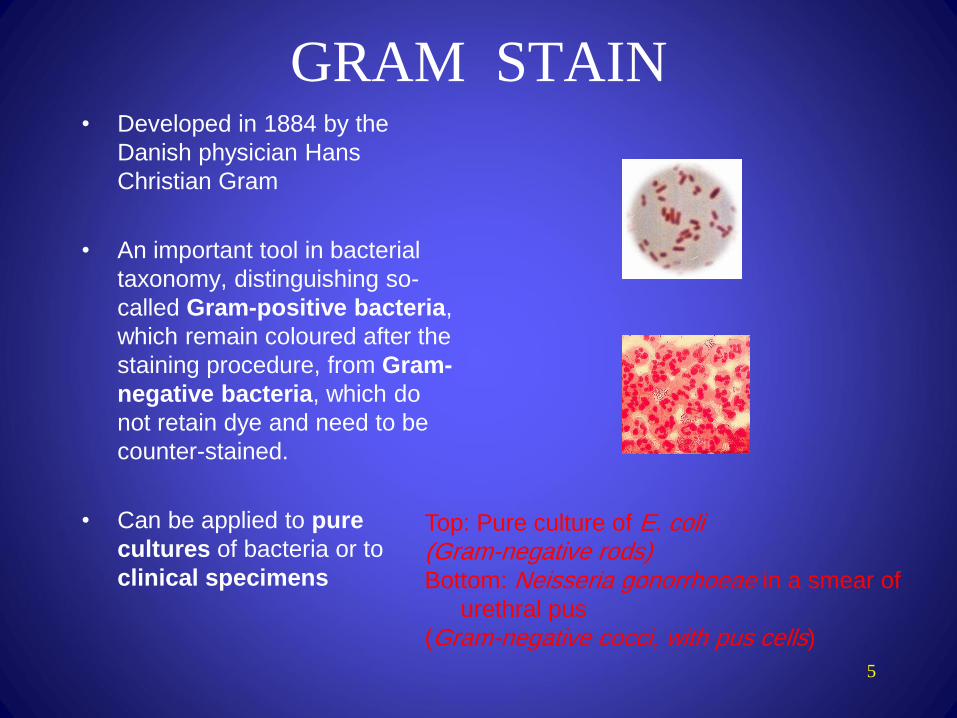
GRAM STAIN
5
• Developed in 1884 by the
Danish physician Hans
Christian Gram
• An important tool in bacterial
taxonomy, distinguishing so-
called Gram-positive bacteria,
which remain coloured after the
staining procedure, from Gram-
negative bacteria, which do
not retain dye and need to be
counter-stained.
• Can be applied to pure
cultures of bacteria or to
clinical specimens
Top: Pure culture of E. coli (Gram-negative rods)Bottom: Neisseria gonorrhoeae in a smear of
urethral pus
(Gram-negative cocci, with pus cells)
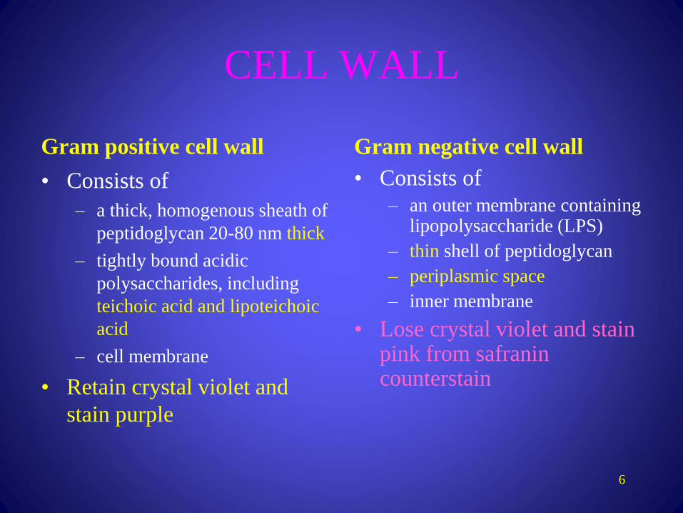
CELL WALL
6
Gram positive cell wall
• Consists of
– a thick, homogenous sheath of
peptidoglycan 20-80 nm thick
– tightly bound acidic
polysaccharides, including
teichoic acid and lipoteichoic
acid
– cell membrane
• Retain crystal violet and
stain purple
Gram negative cell wall
• Consists of
– an outer membrane containing lipopolysaccharide (LPS)
– thin shell of peptidoglycan
– periplasmic space
– inner membrane
• Lose crystal violet and stain pink from safranin counterstain
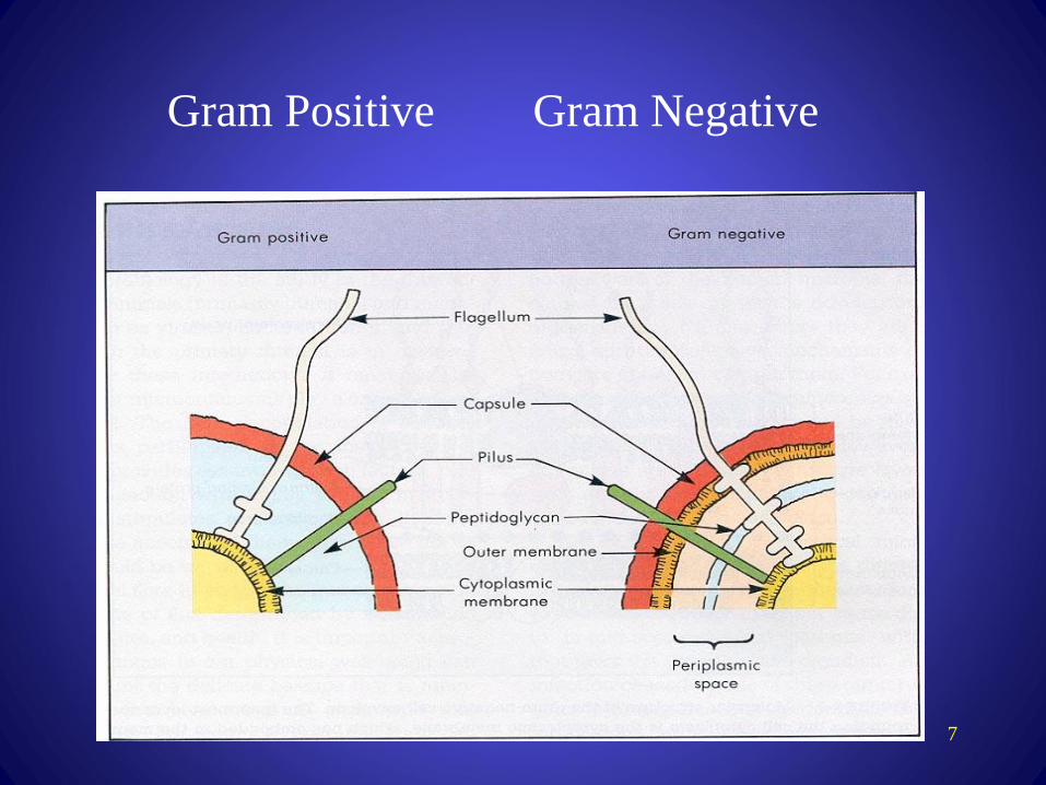
7
Gram Positive Gram Negative
8
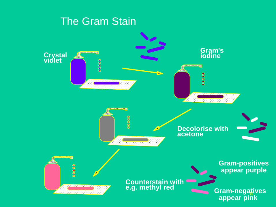
Crystal violet
Gram's iodine
Decolorise with acetone
Counterstain withe.g. methyl red
Gram-positives appear purple
Gram-negatives appear pink
The Gram Stain

9

10
Gram-positive rods
Gram-negative rods
Gram-positive cocci
Gram-negative cocci
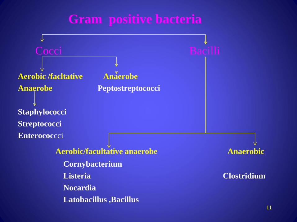
Gram positive bacteria
Cocci Bacilli
Aerobic /facltative Anaerobe
Anaerobe Peptostreptococci
Staphylococci
Streptococci
Enterococcci
Aerobic/facultative anaerobe Anaerobic
Cornybacterium
Listeria Clostridium
Nocardia
Latobacillus ,Bacillus 11

12
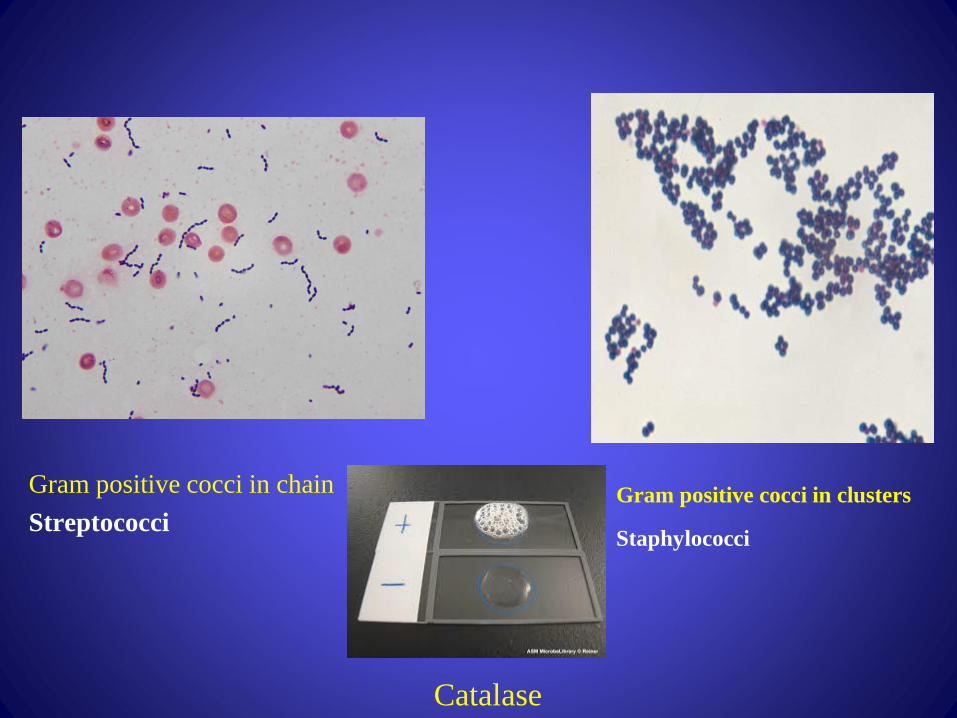
Gram positive cocci in chain
StreptococciGram positive cocci in clusters
Staphylococci
Catalase
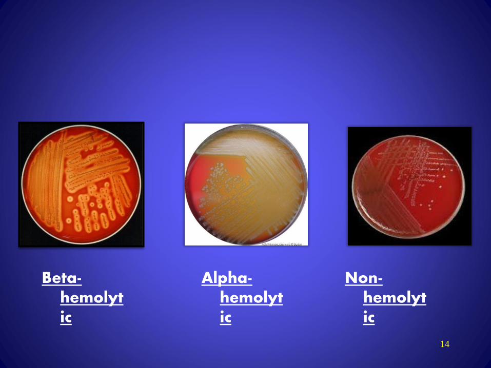
14
Beta-hemolytic
Alpha-hemolytic
Non-hemolytic
15
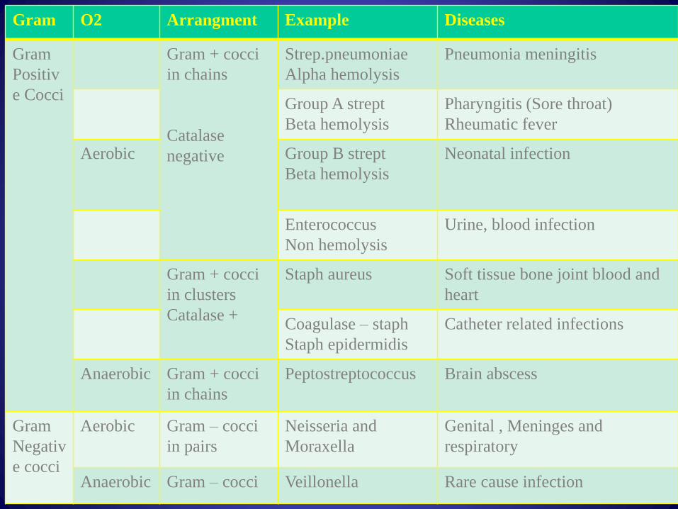
Gram O2 Arrangment Example Diseases
Gram
Positiv
e Cocci
Gram + cocci
in chains
Catalase
negative
Strep.pneumoniae
Alpha hemolysis
Pneumonia meningitis
Group A strept
Beta hemolysis
Pharyngitis (Sore throat)
Rheumatic fever
Aerobic Group B strept
Beta hemolysis
Neonatal infection
Enterococcus
Non hemolysis
Urine, blood infection
Gram + cocci
in clusters
Catalase +
Staph aureus Soft tissue bone joint blood and
heart
Coagulase – staph
Staph epidermidis
Catheter related infections
Anaerobic Gram + cocci
in chains
Peptostreptococcus Brain abscess
Gram
Negativ
e cocci
Aerobic Gram – cocci
in pairs
Neisseria and
Moraxella
Genital , Meninges and
respiratory
Anaerobic Gram – cocci Veillonella Rare cause infection

16
17
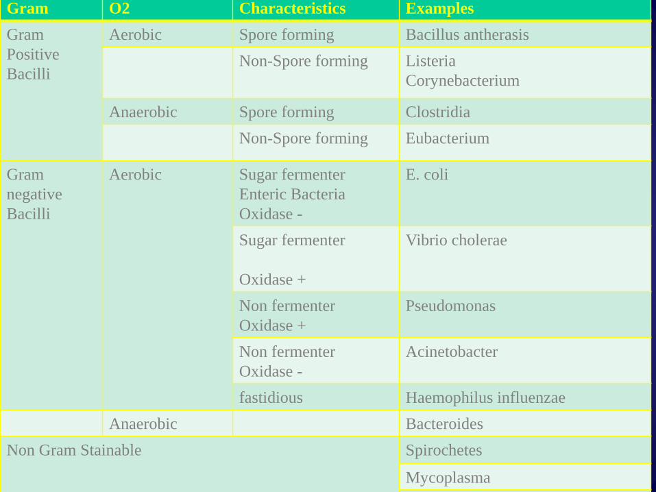
Gram O2 Characteristics Examples
Gram
Positive
Bacilli
Aerobic Spore forming Bacillus antherasis
Non-Spore forming Listeria
Corynebacterium
Anaerobic Spore forming Clostridia
Non-Spore forming Eubacterium
Gram
negative
Bacilli
Aerobic Sugar fermenter
Enteric Bacteria
Oxidase -
E. coli
Sugar fermenter
Oxidase +
Vibrio cholerae
Non fermenter
Oxidase +
Pseudomonas
Non fermenter
Oxidase -
Acinetobacter
fastidious Haemophilus influenzae
Anaerobic Bacteroides
Non Gram Stainable Spirochetes
Mycoplasma
Chlamydia
18

19
• Staphylococci
– Catalase-positive
– Gram-positive cocci in clusters
• Staphylococcus aureus
– coagulase-positive, most important pathogen
• Staph. epidermidis
– and other coagulase negative staphylococci e.g. S
saprophiticus
Gram-positive Cocci

Streptococci• Catalase-negative
• Gram-positive cocci in chains or pairs
• Divided by type of hemolysis.
• Alpha hemolytic:
– S. viridans- oral flora - infective endocarditis
– S. pneumoniae-important cause of community acquired pneumonia
• Beta hemolytic:
– S. pyogenes, group A streptococcus
• Important cause of pharyngitis and cellulitis
20
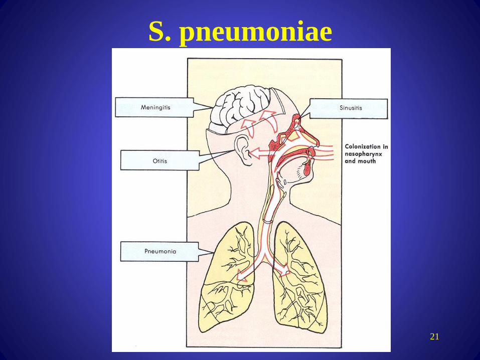
S. pneumoniae
21
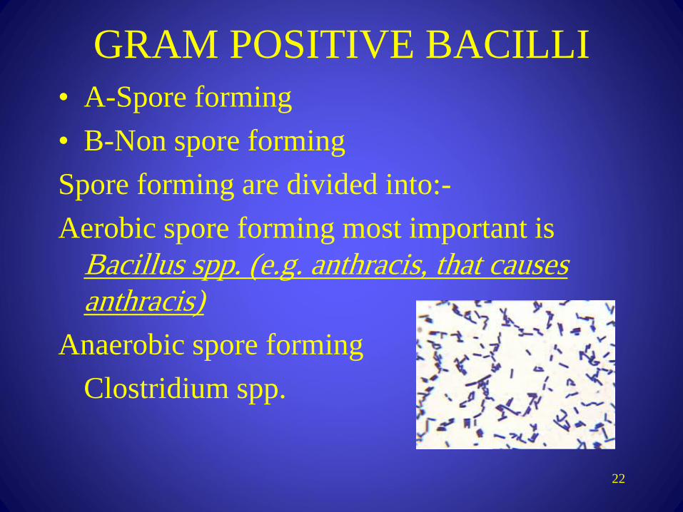
GRAM POSITIVE BACILLI
• A-Spore forming
• B-Non spore forming
Spore forming are divided into:-
Aerobic spore forming most important is
Bacillus spp. (e.g. anthracis, that causes
anthracis)
Anaerobic spore forming
Clostridium spp.
22

GRAM POSITIVE BACILLIAnaerobic gram positive bacilli
• C. tetani - Tetanus C. perfringens
Gas gangrene
• C. botulinum - botulism
– Descending weakness-->paralysis
– diplopia, dysphagia-->respiratory failure
23

GRAM POSITIVE BACILLI
Aerobic gram positive bacilli
• Corynebacterium diphtheriae
– Fever, pharyngitis, cervical LAD
– thick, gray, adherent membrane
– sequelae-->airway obstruction, myocarditis
24

25
Gram-Negative Cocci
• Neisseria gonorrhoeae
– The Gonococcus
• Neisseria meningitidis
– The Meningococcus
• Both Gram-negative intracellular diplococci
• Moraxella catarrhalis

Gram-Negative Rods
• Enteric Bacteria they
ferment sugars most
important are;
– E. coli
– Salmonella
– Shigella
– Yersinia and Klebsiella pneumoniae
– Proteus
Gram-Negative Rods
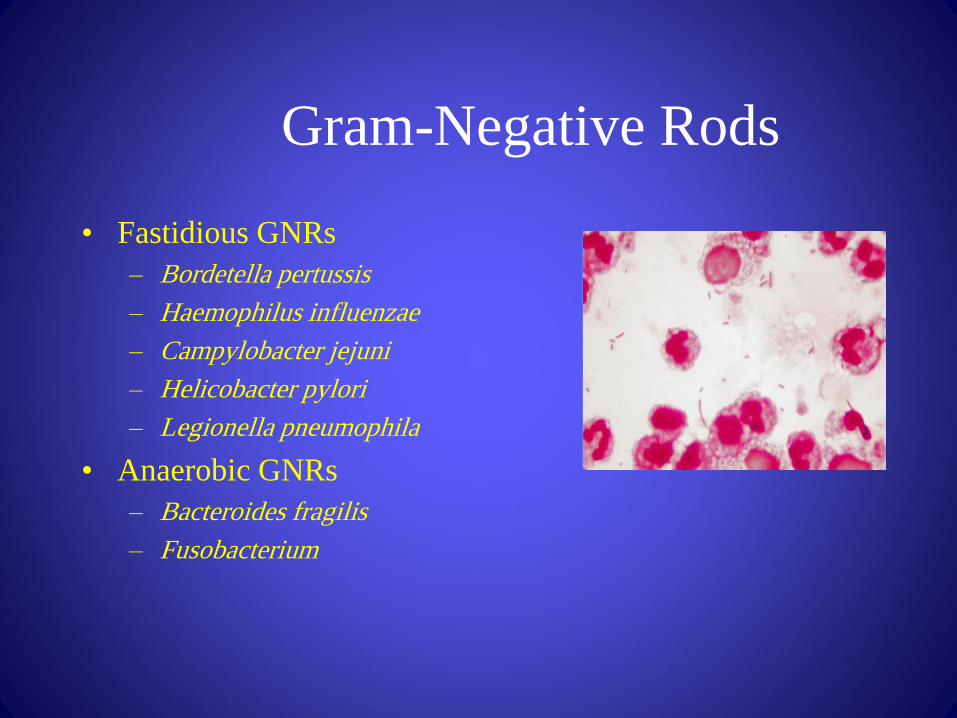
• Fastidious GNRs
– Bordetella pertussis
– Haemophilus influenzae
– Campylobacter jejuni
– Helicobacter pylori
– Legionella pneumophila
• Anaerobic GNRs
– Bacteroides fragilis
– Fusobacterium

Non fermentative gram negative
rods i.e. they do not ferment
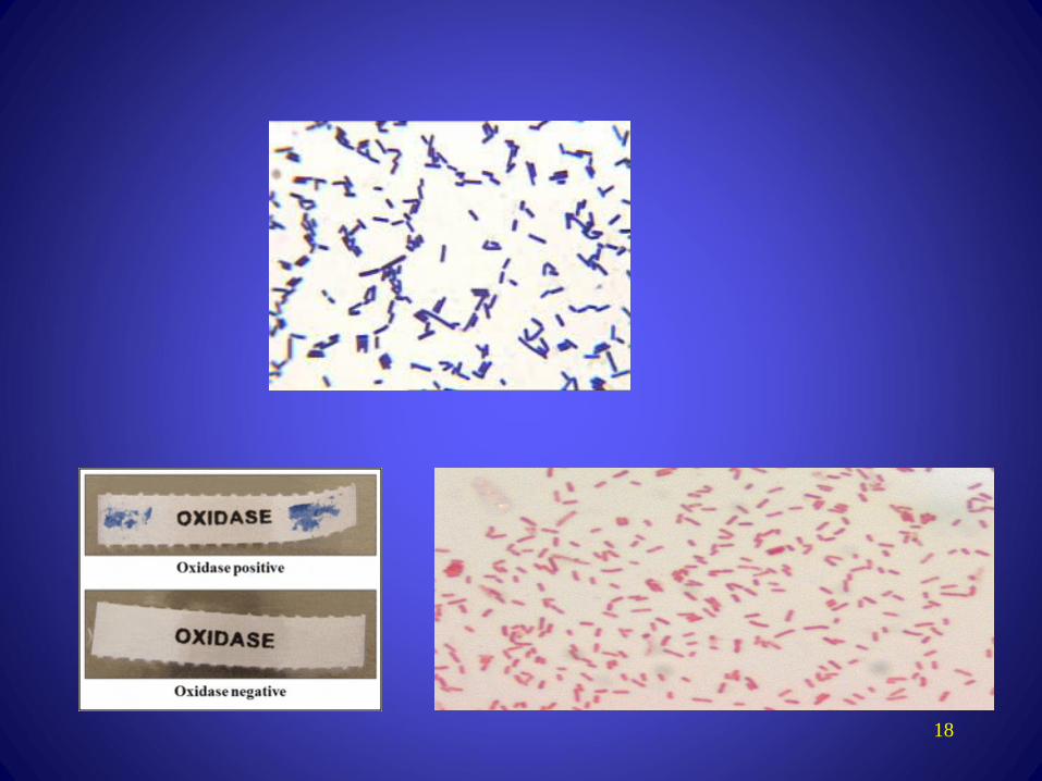
sugars e.g.❖ Oxidase positive: Pseudomonas, causes
infection in immunocompromised patients
❖ Oxidise negative non fermentative e.g.
Acinobacter spp.
28

Oxidise positive comma shaped
and also fermentative most
important is Vibrio cholerae that
causes cholera which is a disease
characterized by severe diarrhea
and dehydration
29

Non-Gram-stainable bacteria
• Unusual gram-positives
• Some spirochaetes (e.g Treponema
pallidum (cause of syphilis))
• Bacteria with no cell wall
• Obligate intra-cellular bacteria

Unusual Gram-positives
• Mycobacteria
– Contain mycolic acid in cell wall

Non-Gram-stainable bacteria
No cell wall
• Mycoplasmas
– Smallest free-living organisms
– No cell wall
– M. pneumonia, M. genitalium
Obligate intra-cellular
• Chlamydia
– C. pneumoniae, C.
trachomatis
• Rickettsia
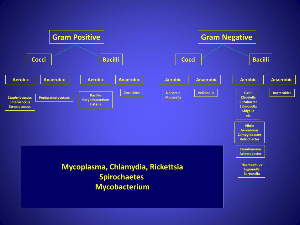
Gram Positive Gram Negative
Cocci Bacilli
Aerobic Anaerobic Aerobic Anaerobic
Cocci Bacilli
Aerobic Anaerobic Aerobic Anaerobic
Peptostreptococcus
E.coliKlebsiella
CitrobacterSalmonella
Shigellaetc
VeillonellaNeisseriaMoraxella
ClostridiumBacillus
CorynebacteriumListeria
StaphylococcusEnterococcusStreptococcus
VibrioAeromonas
CampylobacterHelicobacter
PseudomonasAcinetobacter
HaemophilusLegionellaBartonella
Mycoplasma, Chlamydia, RickettsiaSpirochaetes
Mycobacterium
Bacteroides