Section 11: Extracellular Macromolecules Fibrous proteins: keratin, collagen and elastin 02/21/06.
22
Section 11: Extracellular Macromolecules • Fibrous proteins: keratin, collagen and elastin 02/21/06
-
Upload
jaylyn-reaver -
Category
Documents
-
view
218 -
download
0
Transcript of Section 11: Extracellular Macromolecules Fibrous proteins: keratin, collagen and elastin 02/21/06.

Section 11: Extracellular Macromolecules
Section 11: Extracellular Macromolecules
• Fibrous proteins: keratin, collagen and elastin
• Fibrous proteins: keratin, collagen and elastin
02/21/06

Selected Extracellular and Cytoskeletal Proteins
• Connective Tissue Fibrous Proteins– collagen– elastin– keratin – fibronectin
• Other Fibrous Proteins– fibrin– myosin (partially)
• Cytoskeleton Proteins– actin– keratin – intermediate filaments– microtubules
1

Cell Adhesion
• Receptors and Integrin are transmembrane proteins
Fibronectin
FibronectinReceptor
Epithelial Cell
Fibroblast
Proteoglycans
Laminin:Entactin
Laminin Receptor
Integrin
ExtracellularMatrix:
Collagen IV
2

Cell adhesion proteinsCell adhesion proteins
Laminin FibronectinLaminin Fibronectin
© 2000 by Geoffrey M. Cooper
Integrin
3
A chain
Collagenbinding
B2 chainB1 chain
Cellbinding Cell
binding
Enactinbinding
Proteoglycan binding
Collagen bindingCell binding
Proteoglycan binding
Matrix binding
Actin
Integrin
Extracellular matrix
Plasma membrane

Association Between Cell and
Extracellular Matrix
• Some of the fibrous proteins are transmembane and connect (and communicate) to the cytoskeleton (actin, keratin, microtubule, tailin, vinculin).
Fig. 11-22, Lehninger.4

Elastin• Elastin (64-66kD) is rich in prolines and non-polar side
chains, and one third of its amino acids are glycine.• As a result, its has uncommon secondary structure
(more random structure than found in other proteins). It does not have a stable tertiary structure.
• Elastin is very resilient. It can be stretched to lengths many times greater than in its relaxed state. It can also be compressed.
• Elastin is common in many connective tissues, along with collagen, especially if the tissue undergoes physical stress. It surrounds arteries, is in the lung and in ligaments.
5

Elastin Structure and Function• Elastin interconverts between a number of conformations, both
disordered (upper two on left) and -spiral (bottom left).• After cross-linking, when elastin is stretched (or compressed) it
is less stable and it returns to the disordered conformations.
6 (Fig. 4-28, Rawn) (Fig. 4-30, Rawn)

Elastin Cross-linking
• Some lysine residues in elastin are deaminated and oxidized to the aldehyde level.
• They combine with each other and with other lysines to form lysinonorleucine and desmosine cross-links
NH
CH
O
CH2 CH2 CH2 CH2NH CH2
CH2CH2
NH
CH
O
CH2
lysinonorleucine
+CHNH
CH
CH2
CH2
CH2 CH
CH2
CH2
CH2
CH2
CH
CH2CH2CH
NH
NH
NH
OO
O
CH2
CHNH
O
CH2
desmosine
7

Keratin• Keratin is rich in cysteines.• Its secondary structure is mostly -helical.• The helices form coiled coils (on right).• The coiled coils pack into higher order
elongated structures.• Keratin properties depend strongly on the
degree of disulfide cross-linking.– With low levels of cross-linking, it is flexible (hair,
skin).– It can be made very hard with additional cross-linking
(claws, horns).
• Extracellular via whithering of keratin-filled cells.• Intracellular: cytoskeletal intermediate filaments.
Fig. 3.34
8
2 nm

Keratin Supramolecular Structure• Two coiled coils bind together to
form a protofibril (below).• Protofibrils assemble into
various microfibrils (on the right).
Fig. 4-5Fig. 4-6Rawn
9

Keratin Cross-linking
• The structure of keratin is strengthened by disulfide cross-links from one helix to another.
CH CH2 S
NH
O
CHCH2
NH
S
O
CH CH2 SH
NH
O
disulfidecross-link
CHCH2
NH
SH
O
two cysteines
10

Collagen Types Collagen Types Fibrils – long triple helicesI. Skin, tendon, bone, dentin II. Cartilage and vitreous humor III. Skin, muscles, blood vessels (frequently found with type I)V. Fetal tissues, placenta, interstitial tissues XI. Cartilage
Fibril associated – interrupted triple helicesIX. Cartilage, vitreous, humor XII. Embryonic skin and tendons XIV. Fetal skin and tendons
Fibril associated -- beadedVI. Most interstitial tissues
SheetsIV. All basal laminae VIII. Endothelial cells, X. Cartilage growth plate
Fibrils – long triple helicesI. Skin, tendon, bone, dentin II. Cartilage and vitreous humor III. Skin, muscles, blood vessels (frequently found with type I)V. Fetal tissues, placenta, interstitial tissues XI. Cartilage
Fibril associated – interrupted triple helicesIX. Cartilage, vitreous, humor XII. Embryonic skin and tendons XIV. Fetal skin and tendons
Fibril associated -- beadedVI. Most interstitial tissues
SheetsIV. All basal laminae VIII. Endothelial cells, X. Cartilage growth plate
11

Collagen• Collagen has glycine in every third position, is rich in
proline, and contains hydroxyproline and hydroxylysine residues.
• Collagen does not have secondary structure, but three highly extended strands interact to form a triple helix.
• Collagen triple helices form a rod-like fibril or sheet aggregate that is somewhat flexible, not extensible, and can be very strong.
• Cross-linking increases its strength.• It is common to connective tissue and is present in
bone, dentin and cementum.• Collagen is the most abundant protein in the
biosphere.
12

Collagen Triple Helix• Prolines, especially hydroxylated prolines,
keep the individual chains extended, and increase Tm (keep it above body temperature).
• The small size of the glycine sidechain in every third position allows the three strands to come close together.
• There are interstrand hydrogen bonds.• It is not very extensible because it is
already extended (3.1 per residue vs 1.5 for -helix).
• Glycosylation of hydroxylysines appear to modulate fibril or sheet formation by the triple helices.
Fig. 11-5Stryer 3rd
13
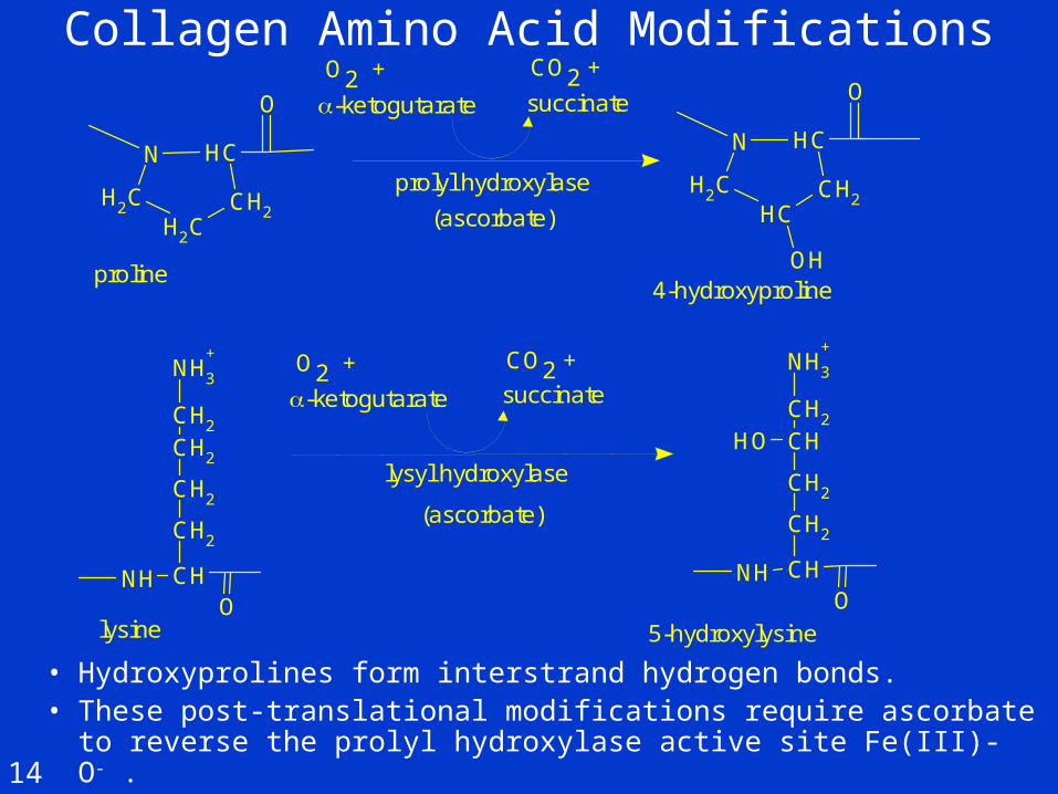
Collagen Amino Acid Modifications
• Hydroxyprolines form interstrand hydrogen bonds.• These post-translational modifications require ascorbate to
reverse the prolyl hydroxylase active site Fe(III)-O- .
N CH
CH2CH2
CH2
O
N CH
CH2CH2
CH
O
OH
NH3
+
CH2
CH2
CH2
CH2
CHNHO
NH3
+
CH2
CH
CH2
CH2
CHNHO
OH
-ketogutarate succinate
-ketogutarate succinate
prolyl hydroxylase
lysyl hydroxylase
(ascorbate)
(ascorbate)
O2 + CO2 +
O2 + CO2 +
proline4-hydroxyproline
lysine 5-hydroxylysine
14

Glycosylation of Hydroxylysine
• The hydroxylysines are modified by sequential glycosylations, giving lysyl-gal-(12)-glc
• Activated sugar complexes are usually UDP-sugars.• Higher levels of glycosylation favor formation of
sheet structures by the collagen triple helices.
galactosyl transferase
glucosyltransferase
UDP-galactose UDP-glucoseNH3
+
CH2
CH
CH2
CH2
CHNHO
OH
O
CH2
O
OHOH
OH
OH
H
H
H H
H
O
CH2O
O
OH
OH
OH
H
HH
H
H
CH
CH2
CH2
CH2
CH
NH3
+
NH
O
15
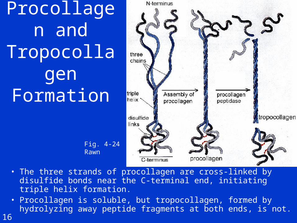
Procollagen and
Tropocollagen Formation
• The three strands of procollagen are cross-linked by disulfide bonds near the C-terminal end, initiating triple helix formation.
• Procollagen is soluble, but tropocollagen, formed by hydrolyzing away peptide fragments at both ends, is not.
Fig. 4-24Rawn
16

Aggregation and Cross-linking• Tropocollagen
spontaneously aggregates into elongated staggered arrays, shown in two dimensions at right.
• Hydroxylysine glycosylation determines fibril or sheet formation.
• Cross-linking strengthens the structure (lower).
• In bone, dentin and cementum, biomineralization begins in the gaps (hole zones) between the individual tropocollagens (type I).
Fig. 4-24, Rawn
17

Collagen Cross-linking
• Lysines and hydroxylysines are used in cross-linking.• Other than oxidation to the aldehyde level, the reactions
appear to be non-enzymatic.
Aldol Cross-link
Aldehyde Derivatives
Lysines
lysyl oxidase
NH
CH
O
CH2 CH2 CH2 CH2NH3
+
NH
CH
O
CH2 CH2 CH2
H
O
NH
CH
O
CH2 CH2 CH2
H
C CH2
NH
CH
O
CH2
O O
NH3
+CH2 CH2
CH2
NH
CH
O
CH2
CH2CH2
NH
CH
O
CH2
O
Hhydroxypyridimiumcross-link(from 3 lysines)
H
H
CHN
CH
CH2
CH
CH2
CHNH
O
O
CH2
CH2
CH
NH
O
CH2
CH NH
O
CH2
O
+
18

Fibroblast to Mature Collagen Fiber
• Procollagen in vesicles is transported to the cell membrane in vesicles, and then secreted via exocytosis.
• Proteolysis, assembly, and lysine oxidation leading to cross-linking occurs outside the cell.
1. Polypeptide synthesis
2. Hydroxylation and glycosylation
3. Triple helix formation
4. Secretion into extracellular matrix
Procollagen bundles
Tropocollagen bundles
Collagen fiber
Maturecollagen fiber
5. Hydrolysis of peptide bonds
6. Assembly near the cell surface
7. Cross-link formation
fibroblast cell
19

Collagen Degradation
• Collagenase cuts the 1000 aa triple helix into 250 and 750 aa fragments which melt and are proteolyzed.
• Some animal tissues (for example tadpole tails) have collagenases that are used to degrade collagen during growth and remodeling.
• The collagenase of Clostridium histolyticum destroys host connective tissue, helping to make it a highly invasive bacterium.
• In periodontal disease, host collagenases help break down periodontal ligament (collagens type I and III).
20

Next topic:
Section 12: Mineralized tissues.
Calcium and phosphate metabolism


![Self‐Assembled Proteins and Peptides as Scaffolds for …yoksis.bilkent.edu.tr/pdf/files/12074.pdf[ 4,5 ] Fibrous proteins such as silks and elastin dominate the area of protein](https://static.fdocuments.us/doc/165x107/610eb8637c961f43826a8886/selfaassembled-proteins-and-peptides-as-scaffolds-for-45-fibrous-proteins.jpg)