
Sandra M Cordo (PhD) Laboratorio de Virología · Los microdominios rafts son plataformas...
43
Sandra M Cordo (PhD) Laboratorio de Virología
Transcript of Sandra M Cordo (PhD) Laboratorio de Virología · Los microdominios rafts son plataformas...

Sandra M Cordo (PhD)
Laboratorio de Virología

virus + lípidos


Estructuras celulares lipídicas
membrana plasmática
gotas lipídicas
Virus
entrada
replicación
salida

Estructuras celulares lipídicas
membrana plasmática (microdominios rafts)
gotas lipídicas (lipid droplets)
Virus
entrada
replicación
salida

Glycerophospholipids Sphingolipids & Cholesterol
Principales grupos de lípidos en membranas
celulares
Estructuras celulares lipídicas:Microdominios de membrana

Glycerophospholipids Sphingolipids & Cholesterol
Estructuras celulares lipídicas:Microdominios rafts

Los (glico)esfingolipídos se asocian preferencialmente con colesterol para formar “lipid rafts”
liquid ordered liquid disordered
liquid disordered
From Simons and Ikonen, 1997Glycerophospholipids Sphingolipids & Cholesterol

“raft” domains morphogenesis

“raft” protein targeting mechanisms
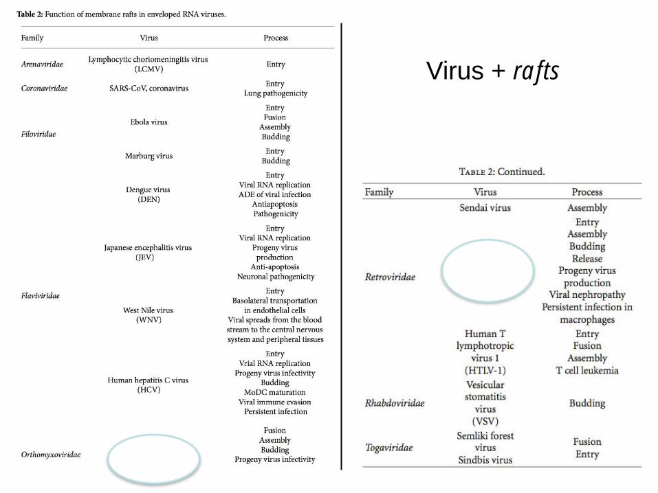
Virus + rafts
All content in this shaded area was uploaded by Abdul A Waheed on Oct 17, 2015
Figures
The Role of Lipids in Retrovirus Replication
Article in Viruses 2(5):1146-1180 · May 2010DOI: 10.3390/v2051146 · Source: PubMed
Abstract
Retroviruses undergo several critical steps to complete a replication cycle. These include the complex processesof virus entry, assembly, and budding that often take place at the plasma membrane of the host cell. Both virusentry and release involve membrane fusion/Xssion reactions between the viral envelopes and host cellmembranes. Accumulating evidence indicates important roles for lipids and lipid microdomains in virus…
33.51 · NCI-Frederick1st Abdul A Waheed
42.91 · National Institutes of Health2nd Eric O Freed
See all ›267 Reads
See all ›60 Citations Download Recommend
Home 1 More1 7 Figures
Article · May 2010 · Viruses
The Role of Lipids in Retrovirus Replication
Recommend publication
Figure
Caption
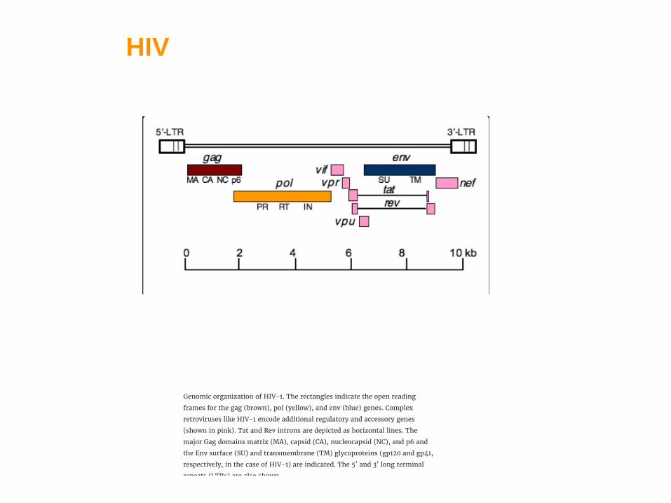
Genomic organization of HIV-1. The rectangles indicate the open reading
frames for the gag (brown), pol (yellow), and env (blue) genes. Complex
retroviruses like HIV-1 encode additional regulatory and accessory genes
(shown in pink). Tat and Rev introns are depicted as horizontal lines. The
major Gag domains matrix (MA), capsid (CA), nucleocapsid (NC), and p6 and
the Env surface (SU) and transmembrane (TM) glycoproteins (gp120 and gp41,
respectively, in the case of HIV-1) are indicated. The 5′ and 3′ long terminal
repeats (LTRs) are also shown.
Recommend
Comment
All content in this shaded area was uploaded by Abdul A Waheed on Oct 17, 2015
Figures
The Role of Lipids in Retrovirus Replication
Article in Viruses 2(5):1146-1180 · May 2010DOI: 10.3390/v2051146 · Source: PubMed
Abstract
Retroviruses undergo several critical steps to complete a replication cycle. These include the complex processesof virus entry, assembly, and budding that often take place at the plasma membrane of the host cell. Both virusentry and release involve membrane fusion/Xssion reactions between the viral envelopes and host cellmembranes. Accumulating evidence indicates important roles for lipids and lipid microdomains in virus…
33.51 · NCI-Frederick1st Abdul A Waheed
42.91 · National Institutes of Health2nd Eric O Freed
See all ›267 Reads
See all ›60 Citations Download Recommend
Home 1 More1 7 Figures
Article · May 2010 · Viruses
The Role of Lipids in Retrovirus Replication
Recommend publication
Figure
Caption
Genomic organization of HIV-1. The rectangles indicate the open reading
frames for the gag (brown), pol (yellow), and env (blue) genes. Complex
retroviruses like HIV-1 encode additional regulatory and accessory genes
(shown in pink). Tat and Rev introns are depicted as horizontal lines. The
major Gag domains matrix (MA), capsid (CA), nucleocapsid (NC), and p6 and
the Env surface (SU) and transmembrane (TM) glycoproteins (gp120 and gp41,
respectively, in the case of HIV-1) are indicated. The 5′ and 3′ long terminal
repeats (LTRs) are also shown.
Recommend
Comment
HIV

HIV

Human Immunodeficiency Virus Type 1 Envelope Glycoproteins That Lack Cytoplasmic Domain Cysteines: Impact on Association with Membrane Lipid
Rafts and Incorporation onto Budding Virus Particles Jayanta Bhattacharya, Paul J. Peters, and Paul R. Clapham*
HIV

The cytoplasmic domain of gp41.
Jayanta Bhattacharya et al. J. Virol. 2004;78:5500-5506
Palmitoylation

Expression and Membrane Association of gp41 envelope mutants

Jayanta Bhattacharya et al. J. Virol. 2004;78:5500-5506
Association of gp41 envelope mutants. with lipid rafts.
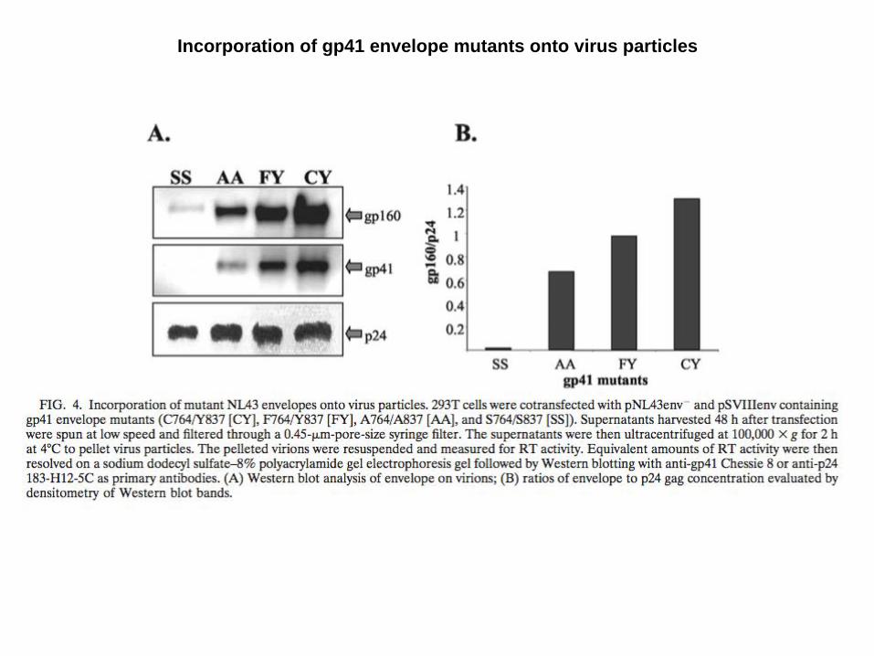
Incorporation of gp41 envelope mutants onto virus particles

FLU
Palmitoylation
Advances in VirologyVolume 2011, doi:10.1155/2011/370606 Association of Influenza Virus Proteins with Membrane RaftsMichael Veit and Bastian Thaa

<< Prev FIG. 1. Next >>PMC full text: Microbiol Mol Biol Rev. 2003 Jun; 67(2): 226–237.doi: 10.1128/MMBR.67.2.226-237.2003Copyright/License ► Request permission to reuse
FIG. 1.
Model of influenza virus assembly and budding within membrane rafts. The eight RNPs (one for eachgenomic RNA) are associated with M1 in the nucleus and are exported to the cytoplasm. After synthesis inthe ER, the transmembrane HA, NA, and possibly M2 are joined together in the TGN, because of NA-HAinteractions and because of their affinity to lipid rafts, before reaching the plasma membrane. There, theM1-RNP1 to M1-RNP8 complexes bind to the HA- and NA-enriched raft membranes because M1 has someaffinity to membranes as well as to NA and HA cytoplasmic tails. Then the virus buds away from themembrane rafts. Membrane rafts are represented as shaded grey regions within the lipid bilayer.
Images in this article
Click on the image to see a larger version.
FLU

Lateral Distribution of the Transmembrane Domain of Influenza Virus
Hemagglutinin Revealed by Time-resolved Fluorescence ImagingTHE JOURNAL OF BIOLOGICAL CHEMISTRY VOL.284,NO.23,pp.15708–15716,,2009
FLU

Transporte a membrana
Interacción-proximidad

Modulation of cell surface transport and lipid raft localization by the cytoplasmic tail of the influenza virus hemagglutinin
Cellular MicrobiologyVolume 18, Issue 1, pages 125-136, 31 AUG 2015 DOI: 10.1111/cmi.12491

Modulation of cell surface transport and lipid raft localization by the cytoplasmic tail of the influenza virus hemagglutinin
Cellular MicrobiologyVolume 18, Issue 1, pages 125-136, 31 AUG 2015 DOI: 10.1111/cmi.12491

Conclusiones LRs
La membrana plasmática es una superficie de grandes dimensiones que le ofrece al virus múltiples posibilidades de interacción.
Es una estructura heterogénea desde el punto de vista estructural como funcional. Su alta especialización esta compartimentalizada e interconectada.
Los microdominios rafts son plataformas biológicas de gran utilidad en el proceso de ensamblado y brotación. Las proteínas virales que los utilizan tienen determinantes moleculares específicos para utilizarlas.
El conocimiento detallado de los mecanismos implicados permitirán explorar nuevos blancos de intervención anti-viral
Ej: drogas anti-colesterol : SP01A entry inhibitor (Samaritan Pharmaceuticals-2009)
MCBD (microbicida anti HIV)

Novel contraceptive microbicides
107
(low pH) mechanisms (Zeitlin et al., 2001). Spermatozoa haveweak intracellular buffering capacity, and even small variations inextracellular pH are rapidly reflected in intracellular changes,which cause fast sperm immobilization (<1 min at pH 4), and, ifpersistent, membrane damage and cell death (Hamamah et al.,1996; Olmsted et al., 2000). Low pH also inactivates HIV andother STD pathogens (Graves et al., 1980; Martin et al., 1985;Croughan and Behbehani, 1988; O’Connor et al., 1995; Zeitlinet al., 2001), providing the mechanistic basis for the contraceptiveand antimicrobial properties of BufferGel™.
Another common structure that is susceptible to functionalalteration are the so-called lipid rafts. They are detergent-resistantmembrane microdomains enriched in cholesterol, sphingolipidsand glycosylphosphatidylinositol (GPI)-anchored proteins(Simons and Ikonen, 1997). Lipid rafts have been described inboth sperm and HIV.
In sperm, lipid rafts associated with caveolin-1 localize toregions of the plasma membrane involved in acrosome reactionand flagellar motility (Travis et al., 2001; Trevino et al., 2001).These distinct areas of the plasma membrane have been linked tocapacitation-related signal transduction and to consequent activa-tion of mechanisms leading to the acquisition of fertilization capa-city (Travis and Kopf, 2002). Membrane cholesterol efflux is acritical event associated with sperm capacitation, increasing mem-brane fluidity and protein–protein interaction and triggering signaltransduction (Davis, 1974; Cross, 1998; Visconti et al., 1999;Shadan et al., 2004; Buffone et al., 2005).
Lipid rafts are also critical in the HIV life cycle. They particip-ate in viral entry, replication and assembly (Campbell et al.,2001). Lipid rafts are cell membrane microdomains through whichHIV buds out (Nguyen and Hildreth, 2000). The high concentra-tion of cholesterol and sphingolipids in lipid rafts would explaintheir high levels in the HIV envelope. Moreover, the inhibition ofcholesterol synthesis provokes a decrease in HIV particle forma-tion in infected cells (Raulin, 2002). Efficient virus entry wouldalso require intact lipid rafts (Viard et al., 2002). It has beenhypothesized that lipid rafts serve as a site of recruitment forgp120/gp41-CD4/coreceptor complexes in a limited area of the
cell surface. Host cell signal transduction may also be activated asa result of HIV infection through lipid rafts, leading to enhancedviral replication via cell- and virus-derived regulatory factors(Campbell et al., 2001).
Removing cholesterol from the sperm membrane and HIVenvelope or host cell plasma membrane has shown to dramaticallyalter their functional capacities. Beta-cyclodextrin, a cyclic hep-tasaccharide that acts as a powerful cholesterol acceptor, has beenused to effect both changes. Incubated with sperm, methylβ-cyclodextrin (MBCD) induces capacitation and tyrosine phos-phorylation and enhances zona binding, acrosomal reaction andfertilizing ability (Choi and Toyoda, 1998; Osheroff et al., 1999;Parinaud et al., 2000). However, profound depletion of cholesteroldisrupts lipid raft functionality and increases membrane fragility,having an overall negative impact on sperm function that could beused for contraceptive purposes. Premature tyrosine phosphoryla-tion and acrosome reaction induced by cholesterol efflux wouldcontribute to the contraceptive effects.
Incubated with HIV virions, host cells and infected cells,β-cyclodextrins have also been shown to reduce viral infectivity(Nguyen and Hildreth, 2000; Liao et al., 2001; Graham et al.,2003). Experiments with other cholesterol acceptors as well ascholesterol analogues have confirmed these results (Maziere et al.,1994; Campbell et al., 2004).
Together, these data show that the disruption of lipid raft func-tionality or membrane cholesterol balance, such as that effected byβ-cyclodextrins, could serve as a viable strategy to achieve theinhibition of sperm fertilization and HIV infection (Figure 3). Inspite of these data and perhaps because of their failure to preventsimian immunodeficiency virus transmission in a monkey model(Ambrose et al., 2004), β-cyclodextrins remain within the realm ofbasic science and preclinical studies and have not progressed toclinical testing.
Sperm and microbial membranes are susceptible to oxidativedamage. Reactive oxygen species (ROS), such as hydrogen perox-ide (H2O2), superoxide anion (O2•−) and hydroxyl radical (OH•),can be spermicidal as well as virucidal (Bell et al., 1992; Chaseand Klebanoff, 1992; Chaki and Misro, 2002). Although small
Figure 3. Effects of methyl β-cyclodextrin (MBCD) on human immunodeficiency virus (HIV) replication and sperm acrosome reaction. HIV-1 (IIIB) was incubatedwith target cells and multiple concentrations of MBCD for 2 h. Then compound and virus were removed, and infectivity was detected by measuring virus-induced celldeath after a 6 day culture. Compound cytotoxicity and virus cytopathicity were assessed with a colorimetric XTT assay (reduction of 2, 3-bis-[2-methoxy-4-nitro-5-sulfophenyl]-2H-tetrazolium-5-carboxanilide sodium salt). Sperm were incubated with calcium ionophore (A23187, positive control) for 30 min, or medium (negativecontrol) for 90 min, or MBCD (10 mM) for 10–90 min. Acrosome reaction was detected by fluorescein isothiocyanate-PSA (pisum sativum agglutinin) (FITC-PSA) (Crosset al., 1986). MBCD displayed anti-HIV and acrosome reaction-inducing activities, which constitute the basis for its potential contraceptive and microbicidal properties.
HIV Cell Entry
0
20
40
60
80
100
120
0.9
2.8 9 32 100
320
1000
3200
1000
0
MBCD (ug/mL)
%co
ntro
l
Cytotoxicity
Antiviral Activity
Control A23187 Methyl-β-CD
Sperm Acrosome Reaction
0
10
20
30
40
90 30 10 30 60 90
Per
cent
at Rutgers University Libraries/Technical Services on A
ugust 21, 2015http://hum
upd.oxfordjournals.org/D
ownloaded from
Exploiting common targets in human fertilization and HIV infection: development of novel contraceptive microbicides
G. F. Doncel Human Reproduction Update, Vol.12, No.2 pp. 103–117, 2006
CONRAD, Department of Obstetrics and Gynecology, The Jones Institute for Reproductive Medicine, Eastern Virginia Medical School, Norfolk, VA, USA
10mM

Estructuras celulares lipídicas: gotas lipídicas

gotas lipídicas = lipid droplets

Virus + lipid droplets

Another virus with marked differences with cellular membranes is HCV, whose particles show a unique lipid composition in comparison with all other viruses analyzed to date. In addition, the lipid content of the HCV envelope is also different from that of the cells in which it was produced (cholesteryl esters comprise almost half of the total HCV lipids), resembling the composition of VLDL and LDL [160]. This finding is compatible with the association of HCV assembly with the VLDL pathway that leads to the formation of lipo-viro-particles [4,147].
HCVSteatosis hepática causada por HCV

HCV
Trends in Molecular Medicine 2015 21, 34-42DOI: (10.1016/j.molmed.2014.11.003)
DGAT1 interacúa con NS5A y Core facilitando su localización en LDs.

NS5A localization at LDs is dependent on DGAT1 activity.
Gregory Camus et al. J. Biol. Chem. 2013;288:9915-9923

DGAT1 is required for interaction of NS5A and core and forms a tripartite complex with these viral proteins.
Gregory Camus et al. J. Biol. Chem. 2013;288:9915-9923

DENV
Dengue Virus Capsid Protein Usurps Lipid Droplets for Viral Particle Formation
Samsa MM, Mondotte JA, Iglesias NG, Assunc ̧a ̃o-Miranda I, Barbosa-Lima G, et al. (2009) Dengue Virus Capsid Protein Usurps Lipid Droplets for Viral Particle Formation. PLoS Pathog 5(10): e1000632.

DENV
C localiza en LDs
LDs aumentan con la infección (48 hs p.i.) y con la expresión de C


LDs as target for DENV inhibition
Fatty acid synthase (FAS) inhibitor:
C75

Cell Host Microbe. 2010 November 18; 8(5): 422–432. Dengue virus induced autophagy regulates lipid metabolism Nicholas S. Heaton and Glenn Randall* Department of Microbiology, The University of Chicago, Chicago, IL 60637, United States
DENV

Lipid droplet area decreases as the number of GFP-LC3 puncta increase in DENV-infected cells

Conclusiones LDs
Las gotas lipídicas o LDs son organelas de reservorio energetico y estructural y plataformas importantes para la regulación de procesos biológicos, interconectándo compartimentos celulares.
Los virus hacen uso de los LDs localizando proteínas y procesos específicos (replicación, ensamblado)
El conocimiento detallado de los mecanismos involucrados en la relación virus-LDs permitirán explorar nuevos blancos de intervención anti-viral
Ej: drogas anti-obesidad: C75 (FAS)
TOFA (ACC1: acetyl-CoA carboxylase 1)
Journal of General Virology (2013), 94, 1310–1317
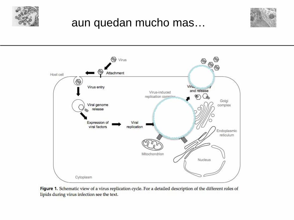
aun quedan mucho mas…


gracias x su atención!


