Salivary glands
55
Upload Upload By : Ahmed Ali Abbas By : Ahmed Ali Abbas Babylon University College of Dentistry Babylon University College of Dentistry download download this file from Website on this file from Website on Google Google TheOptimalSmile.wix.com TheOptimalSmile.wix.com Then choose Lectures Then choose Lectures Then Second Stage Then Second Stage Then choose the lecture you need Then choose the lecture you need
Transcript of Salivary glands

UploadUpload By : Ahmed Ali Abbas By : Ahmed Ali Abbas
Babylon University College of Dentistry Babylon University College of Dentistry
downloaddownload this file from Website on this file from Website on Google Google
TheOptimalSmile.wix.comTheOptimalSmile.wix.com Then choose Lectures Then choose Lectures Then Second StageThen Second Stage Then choose the lecture you needThen choose the lecture you need

THE SALIVARY GLANDSBy :Ahmed Ali Abbas

Saliva
complex fluid found lubricating the mucosa and teeth of the oral cavity
secreted by the salivary glands Major Minor

Types of Saliva
salivary glands, their cells and ducts are greatly responsible for the modification and kind of saliva being secreted
It is of three types: Serous Saliva Viscous Saliva Mixed Saliva


Serous Saliva
Content: Amylase protein polysaccharides
Cell: Serous Cells “watery saliva” Glands that secrete this type:
Parotid Gland Von Ebner’s glands

Viscous Saliva
Content: Mucins (glycoproteins) Carbohydrates
Cell: Mucous Cells Thick and viscous Glands that secrete this type:
Sublingual Gland Minor Salivary Glands (except Von Ebner’s
glands)

Mixed Saliva
simply the combination of the aforementioned types of saliva
Able to do a multitude of significant functions Secreted by:
Submandibular Gland Sublingual Gland
Cells: Serous Cells Mucous Cells

Functions of Saliva
Main function: maintaining the well-being of the mouth
Other important functions: Protection Buffering Action Digestion Facilitation of Taste Defensive Action against Microbes Ionic Exchange between Tooth Surface

Functions of Saliva

MAJOR SALIVARY GLANDS

Major Salivary Glands
Functional Unit Parenchyma Stroma
In some books: salivon Three pairs:
Parotid glands Submandibular glands Sublingual glands

Parotid Glands
In front of the ear behind the ramus of mandible
Purely serous Ptyalin and IgA Stensen’s duct-
opens opposite the upper second molar

Parotid Glands
Nerve supply: CN V Sympathetic control:
superior cervical ganglion
Parasympathetic control: CN IX, oticganglion

Submandibular Glands
Inner aspect of the mandible below the floor of the oral cavity
Mixed, 90% serous 10% mucous Secretes lysozymes Wharton’s duct: opens at each side of the
sublingual folds
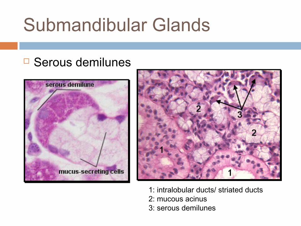
Submandibular Glands
Serous demilunes
1: intralobular ducts/ striated ducts2: mucous acinus3: serous demilunes

Submandibular Glands
Nerve supply: CN V Sympathetic
control: superior cervical ganglion
Parasympathetic control: CN VII, chorda tympani, submandibular ganglion

Sublingual Glands
Floor of the mouth, below the tongue
Mixed, more mucous Ducts of Rivinus Bartholin’s duct:
opens into Wharton’sduct
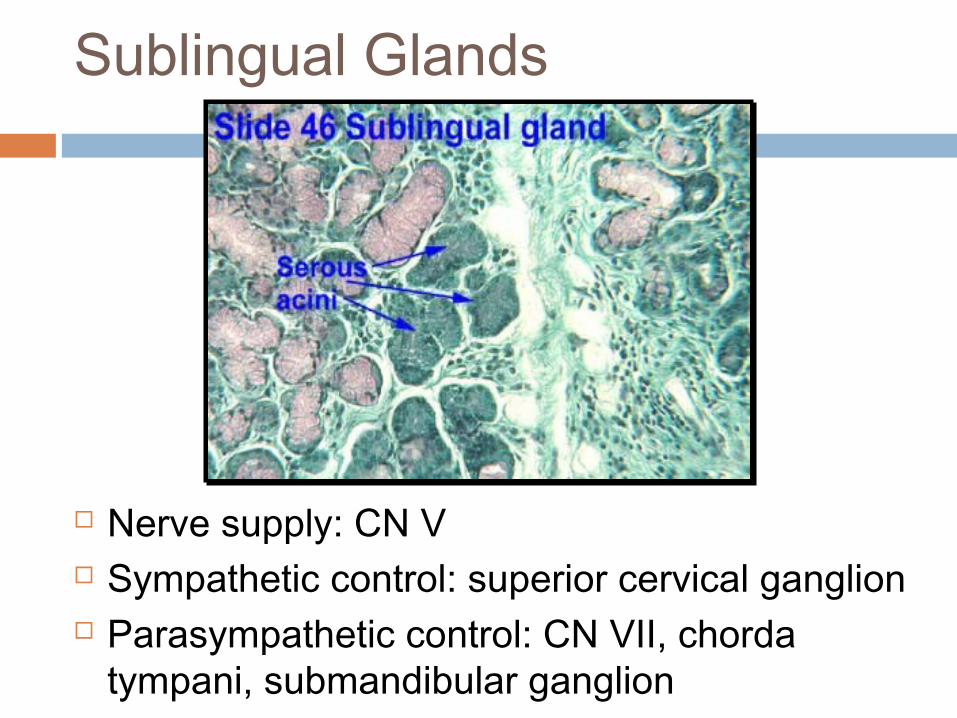
Sublingual Glands
Nerve supply: CN V Sympathetic control: superior cervical ganglion Parasympathetic control: CN VII, chorda
tympani, submandibular ganglion

MINOR SALIVARY GLANDS

MINOR SALIVARY GLANDS
Over 600 present in the oral cavity Types
Mucous producing minor salivary glands Serous fluid producing minor salivary glands

Mucous Producing Minor Salivary Glands Submucosa of the oral mucosa 1-2 mm in diameter Not encapsulated Number of acini connected in a tiny lobule

Serous Fluid Producing Minor Salivary Glands Aka. Von Ebner’s Glands Lipid hydrolysis and perception of taste Located around the foliate and circumvallate
papillae

Labial Glands
Inner surface of lips Mixed saliva Cells have distinct
mucoalbuminouscharacter
Terminal portions often form typicaldemilunes

Minor Buccal Glands
Continuation of the labial glands in the cheek Lie within the vicinity of the opening of the
parotid duct Drain into the third molar region Therefore, it is known as molar glands

Glossopalatine Glands
Pure mucous glands Continuation of the lesser sublingual glands
(posteriorly) Ascend in the mucosa of the glossopalatine
fold
Palatine Glands
Pure mucous glands Occupy the roof of the oral cavity Divided into the glands of the:
Hard palate Soft palate and uvula

Glands of the Tongue
Anterior lingual gland Anterior part- chiefly mucous Posterior part- branching tubules lined with
mucous cells and capped with demilunes of serous cells
Posterior lingual glands Base of the tongue- purely mucous Glands of the vallate papillae (Von Ebner’s
glands)- purely serous, opens into the trough (depression) of the vallate papillae

Glands of the Tongue

Von Ebner’s Glands

STRUCTURES OF SALIVARY GLANDS

STRUCTURES OF SALIVARY GLANDS Parenchyma of glands consists of:
Secretory Portions Branching Duct Sytem
Lobules Septae Capsule


SECRETORY PORTIONS
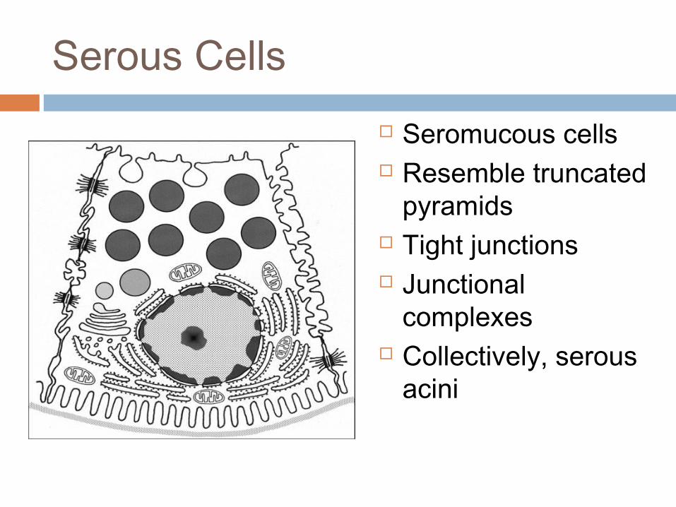
Serous Cells Seromucous cells Resemble truncated
pyramids Tight junctions Junctional
complexes Collectively, serous
acini


Apical Cytoplasm of Serous Cells containing secretory granules
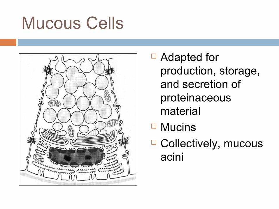
Mucous Cells
Adapted for production, storage, and secretion of proteinaceous material
Mucins Collectively, mucous
acini

Mucous Cells in tubulard secretory end pieces.

Mucous cells showing the serous demilunes.

Myoepithelial Cells
Flattened stellate cells
Desmosomes

Myoepithelial Cells

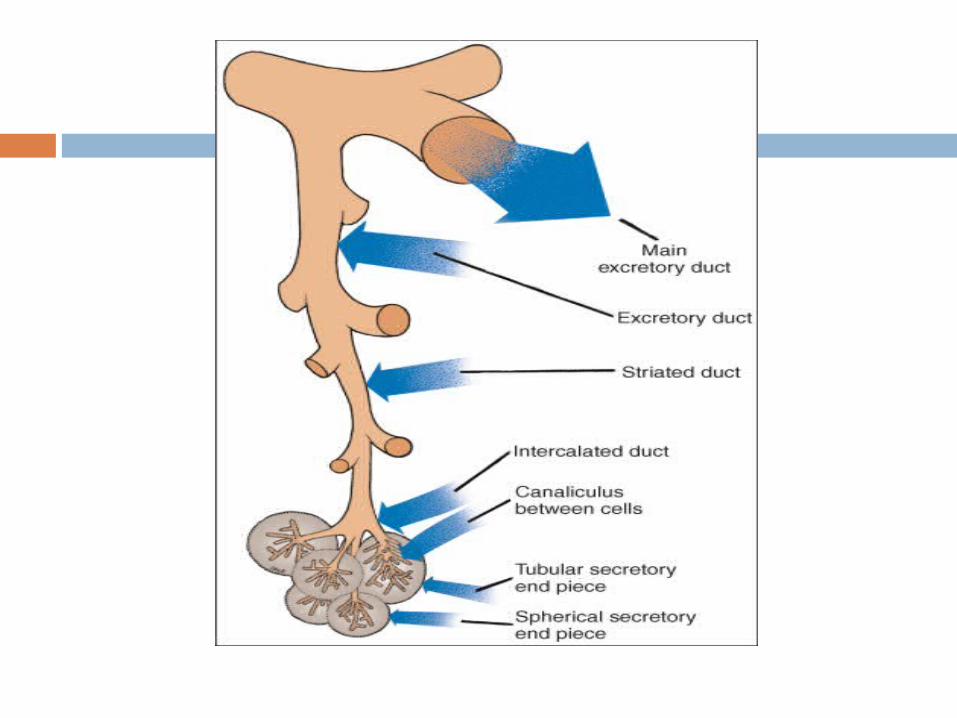
DUCTAL SYSTEM

Intercalated Ducts
Classified as intralobular duct
Smallest branch of the system of ducts
Prominent in Salivary Glands
Frequent in Parotid Gland

Striated Ducts
Formed by union of Intercalated Ducts

Terminal Excretory Ducts
Special eosinophilic cells

Terminal Excretory Ducts
Interlobular excretory duct

Development of Salivary Glands Derived from the oral mucosa Arise in weeks 5-6 of embryonic life

Development of Salivary GlandsPRIMORDIA TIME OF
DEVELOPMENT
EMBRYONIC ORIGIN
REGION
Parotid gland primordia (anlage)
5th to 6th week Ectoderm Labiogingival sulcus
Submandibular gland primordia
6th week Endoderm Hyoid arch
Sublingual gland primordia
7th to 8th week Endoderm Linguogingival sulcus
Intraoral minor salivary glands
3rd month

Development of Salivary Glands

AGE CHANGES

Age Changes
the aging salivary glands are known to undergo structural changes
The lobule structure becomes less ordered The acini vary more in size and eventually atrophy Interlobular ducts become more prominent and the
percentage of fibroadipose tissue increases
Age Changes
Changes in the salivary glands (submandibular,parotid (less) and minor salivary glands) Shrinkage of cells Dilation of ducts Oncocytic transformation Increased adiposity Fibrosis Focal microcalcifications with obstruction Chronic inflammation

THE END THANK YOU FOR LISTENING…