Rom J Morphol Embryol R J M E ORIGINAL PAPER Romanian ... · Rom J Morphol Embryol 2013, 54(2 ......
10
Rom J Morphol Embryol 2013, 54(2):385–394 ISSN (print) 1220–0522 ISSN (on-line) 2066–8279 ORIGINAL PAPER Histopathological change of the endocrine pancreas in male albino rat treated with the atypical antipsychotic clozapine E. A. ABDELRAHIM Department of Histology, Qena Faculty of Medicine, South Valley University, Egypt Abstract Clozapine is an atypical antipsychotic. It is the best option in treatment of resistant schizophrenia. It causes many metabolic side effects, including diabetes mellitus. This study aims to detect the histopathological change of the rat pancreas under chronic clozapine treatment. Twenty adult male albino rats were divided into two groups; control; received daily 2 mL tap water by intragastric tube for 21 days, and treated; received instead 25 mg/kg clozapine in 2 mL tap water. Animals were sacrificed and specimens from the pancreatic tails were processed for histopathological and morphometric studies. In treated animals, the islets were large and irregular. Most interlobular and interlobar ducts were dilated and some showed a related area of endocrine-like cells, which was sometimes immunoreactive to the anti- insulin antibody. Masson’s trichrome revealed increased interlobular and interacinar collagen fibers. PAS stain revealed an increase in the thickness of the islet capillary basement membrane. Immunohistochemical staining with anti-insulin antibody showed strong staining of the islets of treated animals. Morphometric study revealed a highly significant increase in the number of both islets and interlobular ducts/lobule and in the diameter of both islets and interlobular ducts. It also revealed a highly significant increase in the diameter of the periductal fibrotic area and in the thickness of the islets capillary basement membrane in treated animals. In conclusion, chronic clozapine treatment caused changes in the pancreas similar to those occur in diabetes in the form of islets hyperplasia overlapped by atrophic changes as fibrosis and thickening of islets capillaries. Keywords: clozapine, pancreas, islets of Langerhans, diabetes mellitus. Introduction Clozapine is an atypical antipsychotic drug with unique pharmacological and therapeutic properties [1]. It is a tricyclic dibenzodiazepine derivative [2] that acts as an antagonist at adrenergic, cholinergic, dopaminergic, histaminergic and serotonergic receptors [3]. Clozapine is the best treatment option in several clinical circumstances, including treatment of resistant schizophrenia [4]. Unlike the typical antipsychotic drug, haloperidol, clozapine does not cause extrapyramidal side effects [1]. However, clozapine treatment is associated with many serious life shortening side effects including weight gain, dyslipidemia, metabolic syndrome and cardiovascular diseases, glucose intolerance and type 2 diabetes mellitus [5, 6]. Possible mechanisms of the clozapine-associated metabolic side effects especially those concerning impaired glucose homeostasis may be indirect through induction of peripheral insulin resistance. However, direct impairment of pancreatic beta-cell function by histaminic or α 2 -adrenergic receptor antagonism, direct suppression of insulin secretion by selective impairment of cholinergic-stimulated insulin release [7], or by direct toxic effects may be included [8]. On the other hand, atypical antipsychotics might not be an independent risk factor for the development of diabetes mellitus, but hasten its onset in patients bearing other risk factors [8]. Up until now, it is not clear if clozapine treatment directly affects glucose metabolism or simply increases known risk factors for diabetes such as weight gain [9]. Most studies concerning the adverse effects of the commonly used atypical antipsychotic clozapine were concentrated on the physiological mechanisms or biochemical assay. This study aimed at detection of the histopathological changes of the endocrine tissues of the pancreas of male albino rat under chronic clozapine treatment using histological, histochemical, immuno- histochemical and morphometric methods. Materials and Methods Animals and drug Twenty adult male albino rats weighing about 180– 200 g were used in this study. They were purchased from the animal house of the Faculty of Medicine, Assiut University, Egypt. The animals were treated according to the international laws of the animal’s rights. They were kept under normal day and night cycles at room temperature, fed rat chow ad libitum and allowed free access of water. They were divided equally into two groups; control group received daily for 21 days 2 mL tap water by intragastric tube, and treated; received instead 25 mg/kg body weight clozapine dissolved in 2 mL tap water [10, 11]. Clozapine is present in the market in the form of 25 mg tablets named Leponex from Novartis Company, Egypt. The 25 mg tablet was dissolved in 10 mL tap water and the calculated dose for the 200 g rat was 5 mg which equals 2 mL daily of the resultant solution. R J M E Romanian Journal of Morphology & Embryology http://www.rjme.ro/
Transcript of Rom J Morphol Embryol R J M E ORIGINAL PAPER Romanian ... · Rom J Morphol Embryol 2013, 54(2 ......
Rom J Morphol Embryol 2013, 54(2):385–394
ISSN (print) 1220–0522 ISSN (on-line) 2066–8279
OORRIIGGIINNAALL PPAAPPEERR
Histopathological change of the endocrine pancreas in male albino rat treated with
the atypical antipsychotic clozapine
E. A. ABDELRAHIM
Department of Histology, Qena Faculty of Medicine, South Valley University, Egypt
Abstract Clozapine is an atypical antipsychotic. It is the best option in treatment of resistant schizophrenia. It causes many metabolic side effects, including diabetes mellitus. This study aims to detect the histopathological change of the rat pancreas under chronic clozapine treatment. Twenty adult male albino rats were divided into two groups; control; received daily 2 mL tap water by intragastric tube for 21 days, and treated; received instead 25 mg/kg clozapine in 2 mL tap water. Animals were sacrificed and specimens from the pancreatic tails were processed for histopathological and morphometric studies. In treated animals, the islets were large and irregular. Most interlobular and interlobar ducts were dilated and some showed a related area of endocrine-like cells, which was sometimes immunoreactive to the anti-insulin antibody. Masson’s trichrome revealed increased interlobular and interacinar collagen fibers. PAS stain revealed an increase in the thickness of the islet capillary basement membrane. Immunohistochemical staining with anti-insulin antibody showed strong staining of the islets of treated animals. Morphometric study revealed a highly significant increase in the number of both islets and interlobular ducts/lobule and in the diameter of both islets and interlobular ducts. It also revealed a highly significant increase in the diameter of the periductal fibrotic area and in the thickness of the islets capillary basement membrane in treated animals. In conclusion, chronic clozapine treatment caused changes in the pancreas similar to those occur in diabetes in the form of islets hyperplasia overlapped by atrophic changes as fibrosis and thickening of islets capillaries.
Keywords: clozapine, pancreas, islets of Langerhans, diabetes mellitus.
Introduction
Clozapine is an atypical antipsychotic drug with unique pharmacological and therapeutic properties [1]. It is a tricyclic dibenzodiazepine derivative [2] that acts as an antagonist at adrenergic, cholinergic, dopaminergic, histaminergic and serotonergic receptors [3].
Clozapine is the best treatment option in several clinical circumstances, including treatment of resistant schizophrenia [4]. Unlike the typical antipsychotic drug, haloperidol, clozapine does not cause extrapyramidal side effects [1]. However, clozapine treatment is associated with many serious life shortening side effects including weight gain, dyslipidemia, metabolic syndrome and cardiovascular diseases, glucose intolerance and type 2 diabetes mellitus [5, 6].
Possible mechanisms of the clozapine-associated metabolic side effects especially those concerning impaired glucose homeostasis may be indirect through induction of peripheral insulin resistance. However, direct impairment of pancreatic beta-cell function by histaminic or α2-adrenergic receptor antagonism, direct suppression of insulin secretion by selective impairment of cholinergic-stimulated insulin release [7], or by direct toxic effects may be included [8]. On the other hand, atypical antipsychotics might not be an independent risk factor for the development of diabetes mellitus, but hasten its onset in patients bearing other risk factors [8].
Up until now, it is not clear if clozapine treatment directly affects glucose metabolism or simply increases
known risk factors for diabetes such as weight gain [9]. Most studies concerning the adverse effects of the
commonly used atypical antipsychotic clozapine were concentrated on the physiological mechanisms or biochemical assay. This study aimed at detection of the histopathological changes of the endocrine tissues of the pancreas of male albino rat under chronic clozapine treatment using histological, histochemical, immuno-histochemical and morphometric methods.
Materials and Methods
Animals and drug
Twenty adult male albino rats weighing about 180–200 g were used in this study. They were purchased from the animal house of the Faculty of Medicine, Assiut University, Egypt. The animals were treated according to the international laws of the animal’s rights. They were kept under normal day and night cycles at room temperature, fed rat chow ad libitum and allowed free access of water. They were divided equally into two groups; control group received daily for 21 days 2 mL tap water by intragastric tube, and treated; received instead 25 mg/kg body weight clozapine dissolved in 2 mL tap water [10, 11]. Clozapine is present in the market in the form of 25 mg tablets named Leponex from Novartis Company, Egypt. The 25 mg tablet was dissolved in 10 mL tap water and the calculated dose for the 200 g rat was 5 mg which equals 2 mL daily of the resultant solution.
R J M ERomanian Journal of
Morphology & Embryologyhttp://www.rjme.ro/
E. A. Abdelrahim
386
Tissue preparation and staining techniques
Animals were anaesthetized with ether and perfused by intracardiac infusion of saline until the flowing blood was cleared. The perfusion was completed with 10% neutral phosphate buffered formaldehyde. Specimens from the pancreatic tails of all animals used in this study, both the control and the treated were obtained, fixed in the same perfusate used, dehydrated, cleared and embedded in paraffin. Paraffin blocks were sectioned at 5–7 µm and stained with Harris Hematoxylin and Eosin for histopathological examination. Masson’s trichrome stain was done for demonstration of collagen fibers. Periodic Acid Schiff (PAS) method was used to demonstrate the basement membrane of the islet capillaries. Specimen preparation and staining techniques were done according to Drury RAB and Wallington EA [12]. The observed histopathological changes of the pancreatic tissues of the treated animals were detected through a comparative examination of the corresponding tissues of the normal control animals.
Some sections from all specimens (both control and treated) were picked upon coated slides for the immuno-histochemical study which was done using the anti-insulin antibody; insulin Ab-6 (INS04 + INS05) Mouse Monoclonal Antibody (Thermo Fisher Scientific, Fremont, CA, USA) which is a cocktail especially designed for sensitive detection of insulin. Sections were pretreated with hydrogen peroxide block for 15 minutes to reduce non-specific background staining due to endogenous peroxidase, washed by phosphate buffer, followed by incubation in the ultra V block for 5 minutes at room temperature to block the non-specific background staining. The primary antibody (anti-insulin antibody; insulin Ab-6) was diluted at 1:200 in antibody diluent. Sections were incubated in the primary antibody for 30 minutes at room temperature. The positive control was the normal pancreatic tissue of the normal control animals themselves. The reaction was visualized using; UltraVision ONE Detection System, HRP Polymer & DAB Plus Chromogen (Thermo Fisher Scientific, Fremont, CA, USA). The procedure was done according to the manufacture instructions. After completion of the reaction, counter-staining was done using Hematoxylin. The changes in the immunohistochemical reaction of the pancreatic tissues of the treated animals were detected through a comparative examination of the corresponding tissues of the normal control animals. Some sections upon the coated slides were stained first by PAS stain followed by immunostaining with the anti-insulin antibody by the same procedure described in the immunostaining alone. This method is used to demonstrate the islet capillary basement membrane in immunostained sections and it was done according to Gulubova MV et al. [13].
Morphometric study
Many morphometric evaluations were made in this study including the following:
1. Manual counting of the number of islets, as well as the number of interlobular ducts/pancreatic lobule. The number was counted in at least five lobules for each animal used in this study both control and treated, and the average value for each animal was estimated to be
used in the statistical study. 2. Measuring the inner diameter of the interlobular
duct. 3. Measuring the diameter of the islets of Langerhans. 4. Measuring the diameter of the periductal area of
fibrosis surrounding the interlobular ducts in sections stained with Masson’s trichrome.
5. Measuring the thickness of the islets capillary basement membrane in both PAS and combined PAS/ anti-insulin antibody stained sections.
For the previous parameters from (2) to (5) at least five measures for each animal both control and treated were taken, and the average value for each animal was estimated to be used in the statistical study.
6. Measuring the diameter of the area occupied by endocrine-like cells related to the interlobular ducts in treated animals in sections stained with both Hematoxylin and Eosin, and with the immunostaining using the anti-insulin antibody. The diameter of all of these areas was measured whether they were immunoreactive or not.
All measures from (2) to (6) were done using Leica EZ4D stereomicroscope (Leica DM 1000) with internal digital camera using both ×10 and ×20 objective lenses, except in measuring the capillary basement membrane thickness where the ×40 objective lens was used. The microscope is equipped with Leica EC3 digital camera, which is programmed with LAS EZ (Leica Application Suite Educational Zoom) software image analyzer.
Statistical analysis
Statistical analysis of data was done using Microsoft Excel 2007 through the descriptive statistics (summary statistics) and analysis of variance (ANOVA) single factor test. P-value <0.05 was considered statistically significant and p-value <0.001 was considered statistically highly significant.
Results
Histopathological examination
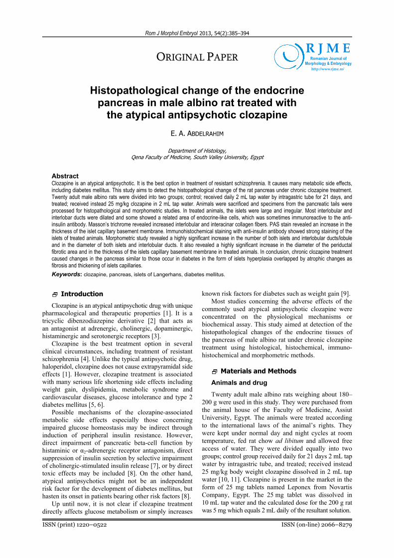
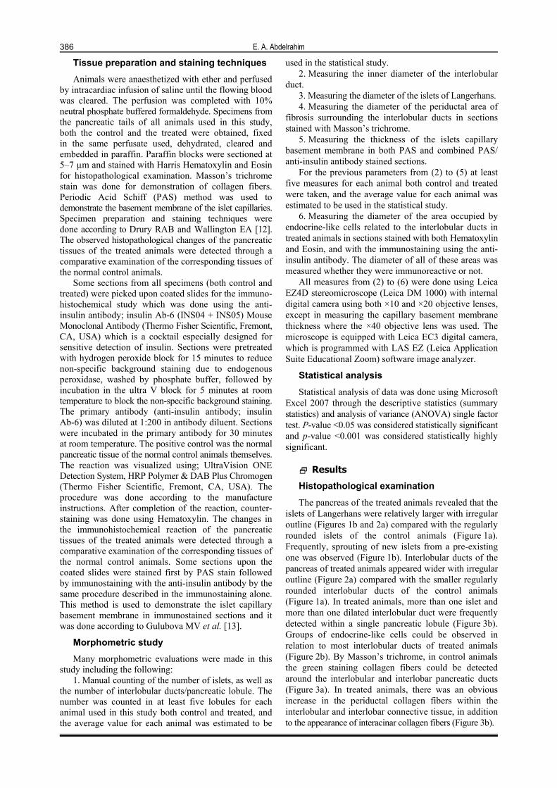
The pancreas of the treated animals revealed that the islets of Langerhans were relatively larger with irregular outline (Figures 1b and 2a) compared with the regularly rounded islets of the control animals (Figure 1a). Frequently, sprouting of new islets from a pre-existing one was observed (Figure 1b). Interlobular ducts of the pancreas of treated animals appeared wider with irregular outline (Figure 2a) compared with the smaller regularly rounded interlobular ducts of the control animals (Figure 1a). In treated animals, more than one islet and more than one dilated interlobular duct were frequently detected within a single pancreatic lobule (Figure 3b). Groups of endocrine-like cells could be observed in relation to most interlobular ducts of treated animals (Figure 2b). By Masson’s trichrome, in control animals the green staining collagen fibers could be detected around the interlobular and interlobar pancreatic ducts (Figure 3a). In treated animals, there was an obvious increase in the periductal collagen fibers within the interlobular and interlobar connective tissue, in addition to the appearance of interacinar collagen fibers (Figure 3b).
Histopathological change of the endocrine pancreas in male albino rat treated with the atypical antipsychotic clozapine
387
Figure 1 – (a) A photomicrograph of the pancreas of a control animal showing; a rounded islet of Langerhans (L) with an adjacent regularly rounded interlobular duct (d) within a pancreatic lobule. (b) A photomicrograph of the pancreas of a treated animal showing; a large irregularly shaped islet of Langerhans. It looks that there is a sprouting of a new islet (↑) from a preexisting one. HE stain, ×100.
Figure 2 – (a) A photomicrograph of the pancreas of a treated animal showing; a large irregularly shaped islet of Langerhans (L) with an adjacent dilated irregularly rounded interlobular duct (d). HE stain, ×100. (b) A magnified part of the previous photomicrograph showing a clearly obvious endocrine-like group (↑) of cells related to a dilated interlobular duct (d). HE stain, ×200.
Figure 3 – (a) A photomicrograph of the pancreas of a control animal showing the green stained periductal collagen fibers within the interlobular (↑) and interlobar connective tissue (arrowhead). Islet of Langerhans (L). (b) A photo-micrograph of the pancreas of a treated animal showing the relative increase in periductal collagen fibers within the interlobular connective tissue (↑) and the appearance of interacinar collagen fibers (arrowhead). Notice: The presence of two islets (L) and two dilated interlobular ducts within a pancreatic lobule. Masson’s trichrome stain, ×100.
PAS staining showed highly positive thick basement membranes of the islets capillaries of treated animals (Figure 4b), compared with the mild positive relatively thin basement membranes of the islets capillaries of control animals (Figure 4a). Immunohistochemical staining with the anti-insulin antibody; Ab-6 revealed that the positive staining of the islets of control animals was
concentrated mainly in their centre (Figure 5a). In treated animals, there was an obvious increase in the intensity of the positive reaction which seemed to occupy the whole islet mass (Figure 5b). Combined PAS/anti-insulin antibody revealed an obvious thickening of the basement membrane of the islets capillaries of treated animals (Figure 6a) compared with the control (Figure 6b).

E. A. Abdelrahim
388
Moreover, the immunostaining of the islets themselves was deep brown in treated animals (Figure 6a) compared with the orange brown color observed in the control islets (Figure 6b). In the same staining method, small groups of endocrine-like cells related to the wall of dilated inter-
lobular and interlobar ducts observed in treated animals and showed an obvious immunoreactivity with the anti-insulin antibody (Figure 7, a–c), while few others were negative (Figure 7a).
Figure 4 – (a) A photomicrograph of the pancreas of a control animal showing mildly positive basement membrane of the capillary network (↑) within an islet of Langerhans. (b) A photomicrograph of the pancreas of a treated animal showing; a relative increase in the positivity of the basement membrane of most blood capillaries (↑) within a large elongated islet of Langerhans. PAS stain, ×200.
Figure 5 – (a) A photomicrograph of the pancreas of a control animal showing the brown staining of the cytoplasm of beta-cells of an islet. Notice that the stained cells are mainly concentrated at the center of the islet. (b) A photo-micrograph of the pancreas of a treated animal showing a deep brown staining of the cytoplasm of nearly all the cells of an islet. Immunostaining with anti-insulin Ab-6, ×200.
Figure 6 – (a) A photomicrograph of the pancreas of a control animal showing thebasement membrane of a blood capillary (arrow) within an islet. Notice the orange brown color of the immunostained beta-cells. (b) A photomicrograph of the pancreas of a treated animal showing the thickened basement membrane of a blood capillary (arrow) within an islet. Notice the dark brown color of the immuno-stained beta-cells. Combined PAS/immuno-staining with anti-insulin Ab-6, ×400.

Histopathological change of the endocrine pancreas in male albino rat treated with the atypical antipsychotic clozapine
389
Figure 7 – Three photomicrographs of the pancreas of the treated animals. (a) Two groups of endocrine-like cells related to a dilated interlobular duct (d). Notice that one of these groups is not immunoreactive (E) while the other shows some immunoreactive cells (arrow). (b) An immunoreactive group of endocrine-like cells (arrow) between two elongated inter-lobar ducts (d). (c) An immunoreactive group of endocrine-like cells (arrow) arises between two elongated interlobar ducts (d) and tends to migrate between the acini of the exocrine pancreas. Combined PAS/immunostaining with anti-insulin Ab-6, ×400.
Morphometric results
Statistical analysis of the morphometric data revealed that:
▪ The number of islets in control animals was ranged from 0 to 2 islets/lobule with the mean (average) value 0.9±0.57 (SD). In comparison, the number of islets in treated animals was ranged from 2 to 5 islets/lobule with the mean (average) value 3.5±0.85 (SD) (Figure 8, a and b). P-value was 2.26 E-07 (0.000000226), which was considered statistically highly significant.
▪ The number of interlobular ducts in control animals was ranged from 1 to 2 interlobular ducts/lobule with the mean (average) value 1.3±0.48 (SD). In comparison, the number of interlobular ducts in treated animals was ranged from 2 to 4 interlobular ducts/lobule with the mean (average) value 3.1±0.57 (SD) (Figure 9, a and b). P-value was 4.72 E-07 (0.000000472), which was considered statistically highly significant.
▪ The diameter of islets in control animals was ranged from 88.61 to 163.16 µm with the mean (average) value 123.93±24.36 µm (SD). In comparison, the diameter of islets in treated animals was ranged from 186.88 to 365.65 µm with the mean (average) value 260.504± 60.56 µm (SD) (Figure 10, a and b). P-value was 7.22 E-06 (0.00000722), which was considered statistically highly significant.
▪ The diameter of interlobular ducts in control animals was ranged from 33.23 to 63.65 µm with the mean
(average) value 44.676±9.43 µm (SD). In comparison, the diameter of interlobular ducts in treated animals was ranged from 94.64 to 231.8 µm with the mean (average) value 146.793±45.77 µm (SD) (Figure 11, a and b). P-value was 1.85 E-06 (0.00000185), which was considered statistically highly significant.
▪ The diameter of the periductal area of fibrosis in control animals was ranged from 9.14 to 32.99 µm with the mean (average) value 18.63±7.75 µm (SD). In comparison, the diameter of the periductal area of fibrosis in treated animals was ranged from 15.8 to 63.06 µm with the mean (average) value 39.466±15.87 µm (SD) (Figure 12, a and b). P-value was 0.001533, which was considered statistically highly significant.
▪ The thickness of the islet capillary basement membrane in PAS and in combined PAS/anti-insulin antibody stained sections in control animals was ranged from 0.97 to 1.31 µm with the mean (average) value 1.142±0.12 µm (SD). In comparison, the thickness of the islet capillary basement membrane in treated animals was ranged from 1.32 to 3.32 µm with the mean (average) value 1.888±0.58 µm (SD) (Figure 13, a and b). P-value was 0.000849, which was considered statistically highly significant.
▪ The diameter of the area occupied by endocrine-like cells related to the interlobular and interlobar ducts in treated animals was ranged from 17.89 up to 99.65 µm with the mean value 50.49±29.058 µm.

E. A. Abdelrahim
390
(a) (b)
Figure 8 – (a) The number of islets/lobule in both control and treated animals. (b) The mean value of the number of islets/lobule in both control and treated animals.
(a) (b)
Figure 9 – (a) The number of interlobular ducts/lobule in both control and treated animals. (b) The mean value of the number of interlobular ducts/lobule in both control and treated animals.
(a) (b)
Figure 10 – (a) The diameter of islets in μm in both control and treated animals. (b) The mean value of the diameter of islets in µm in both control and treated animals.
(a) (b)
Figure 11 – (a) The diameter of interlobular ducts in µm in both control and treated animals. (b) The mean value of the diameter of interlobular ducts in µm in both control and treated animals.

Histopathological change of the endocrine pancreas in male albino rat treated with the atypical antipsychotic clozapine
391
(a) (b)
Figure 12 – (a) The diameter of the periductal area of fibrosis in µm in sections stained by Masson’s trichrome from both control and treated animals. (b) The mean value of the diameter of the periductal area of fibrosis in µm in sections stained by Masson’s trichrome from both control and treated animals.
(a) (b)
Figure 13 – (a) The thickness of the capillary basement membrane in µm in both control and treated animals. (b) Mean value of the thickness of the capillary basement membrane in µm in both control and treated animals.
Discussion
In this study, the histopathological examination of the pancreatic islets of clozapine-treated animals revealed signs of hyperplasia in the form of large sized irregularly-shaped islets, sprouting of new islets from a pre-existing one and more than one islet within a single pancreatic lobule. Morphometric study also revealed a highly significant increase in the number of both islets and interlobular ducts/pancreatic lobule and in the diameter of both islets and interlobular ducts in treated animals. Despite the fact that the pancreatic islets of Langerhans are multi-hormonal endocrine micro-organs that contain four principle types of hormone secreting cells, the predominant cell type is the insulin-secreting beta-cells [14, 15]. It seems that islets hyperplasia usually results from beta-cell hyperplasia. There are two types of beta-cell hyperplasia, primary and secondary. Primary beta-cell hyperplasia is associated with hyperinsulinemia and hypoglycemia, while secondary hyperplasia is usually associated with insulin resistance and hyperglycemia [16].
Previous studies reported that one possible patho-mechanism for hyperglycemia induced by the new generation antipsychotics including clozapine is the induction of insulin resistance [17]. Others added that beside their major role in increasing insulin resistance, these drugs could markedly and selectively impair cholinergic-stimulated insulin secretion by blocking muscarinic M2 receptors, which could be one of the contributing factors to their higher risk for producing hyperglycemia and diabetes leading to inadequate compensation by the beta-cells [18]. It was also reported
that the clozapine-associated hyperglycemia was reversible after withdrawal of the drug suggesting that a functional defect rather than a beta-cell destruction should be implicated [18]. From the previous studies, it is suggested that the clozapine-associated beta-cell hyperplasia observed in this study is related to the secondary type, which is associated with insulin resistance and hyper-glycemia [16].
Secondary beta-cell hyperplasia was observed in the early stages of type 2 diabetes, where beta-cells adapted to insulin resistance by increasing cell mass and function by several mechanisms including hypertrophy and proliferation of pre-existing beta-cells, formation of new cells from progenitor cells, and increased insulin production and secretion. Failure of beta-cells to adequately expand in settings of increase insulin demand results in hyperglycemia and diabetes [19]. Hyperplastic changes of the pancreatic islets were also experimentally observed in sand rat with obesity-diabetes syndrome. It was observed that the number of islets was higher than normal (polynesia), with the islets themselves enlarged (macronesia) and double islets in the secretory ducts of the exocrine pancreas were frequent [20].
Hyperplastic and proliferative changes occur under the effect of suitable stressors or stimuli. They result from the formation of new beta-cells that can take place by replication of already differentiated beta-cells (beta-cell plasticity) or neogenesis from putative islet stem cells within the islets or the pancreatic ductal epithelium [21] or even pancreatic acinar cells [22].
Beta-cell plasticity is a unique property of these cells to adapt their number and volume (beta-cell mass), and

E. A. Abdelrahim
392
their function to the increased secretory demand linked to insulin resistance. This is well documented in physiological (pregnancy) as well in pathophysiological conditions (obesity, acromegaly) [23]. Moreover, it was observed that in mice, in addition to increased self-duplication of beta-cells on increase metabolic demand, there is also a conversion of glucagon secreting alpha-cells into insulin secreting beta-cells. Such inter-endocrine spontaneous adult cell conversion could give a great hope in diabetes therapies [24].
Another frequent observation in this study was the presence of small groups of endocrine-like cells related to the dilated interlobular and interlobar ducts of treated animals. Some of them were immunoreactive to the anti-insulin antibody. This finding is in accordance with many authors who suggested that there are pancreatic progenitor cells in the exocrine pancreatic ducts that have the potential to grow and differentiate into endocrine cells [25]. Such extra-islet precursor cells may be important for beta-cell regeneration in beta-cell-depleted segments [26].
Pancreatic-duct cells may serve as a source of regeneration in adult rats after partial pancreatectomy by undergoing a reproducible dedifferentiation followed by redifferentiation into beta-cells [27]. This histopathological picture was previously described as nesidioblastosis, which is defined as the transformation of the exocrine ductal epithelium into endocrine tissue, which can be hormonally active or inactive [28]. Nesidioblastosis may occur in the pathogenesis of diabetes [29].
Moreover, clozapine may cause upregulation of some genes that result in increased levels of insulin, triglycerides and leptin in the plasma of clozapine-treated patients [1]. It was observed that chronic treatment with clozapine caused upregulation of several genes including the glucose-dependent insulinotropic polypeptide (GIP) gene by over 200% in the rat striatum [1]. Upregulation of the same gene observed also in the intestine of clozapine-treated patients. GIP is an insulinotropic agent with stimulatory effects on insulin synthesis and release from the pancreas. However, changes in brain GIP levels were found to be unrelated to the metabolic adverse effects of clozapine. Conversely, the adverse metabolic effects associated with clozapine treatment may be related to its ability to increase intestinal gene expression for GIP [1].
Unfortunately, many factors can contribute to failure of beta-cells to undergo unlimited hyperplasia. Prolonged exposure of islet cells to high plasma levels of glucose and free fatty acids in cases of insulin resistance and type 2 diabetes results in what is named as gluco-lipotoxicity. The mechanisms by which hyperglycemia and hyperlipidemia exert their deleterious effects on the beta-cells include the generation of Reactive Oxygen Species (ROS) and Reactive Nitrogen Species (RNS) and Advanced Glycosylation End (AGE) products [23].
In this study, Masson’s trichrome stain revealed a highly significant increase in the periductal collagen fibers within the interlobar and interlobular connective tissue, and the appearance of interacinar collagen fibers. Interlobular and interacinar fibrosis and compensatory enlargement of some islets were observed in spontaneous diabetes of young cattle [30]. Also, in long-term diabetic
dogs, atrophy, hyperplastic nodules, and interacinar and interlobular fibrosis were observed suggesting that insulinopenia may cause injurious effects on exocrine pancreas producing interacinar fibrosis [31]. Intralobular and interacinar fibrosis was also observed in pancreatic autopsy of diabetic patients [32]. A similar observation was detected in pancreas of old rats and was described as interstitial fibrosis which is composed of cystic ductal hyperplasia associated with increase in collagen fibers [33].
It was suggested that pancreatic stellate cells might participate in the development of pancreatic fibrosis. These cells are similar to hepatic stellate cells. They are located in the interlobular and interacinar regions [34]. High concentrations of glucose and insulin as in case of type 2 diabetes contribute to pancreatic stellate cells activation and proliferation leading to the fibrotic changes. Also, invasions of the pancreatic islets by these cells result in fibrotic islet destruction which leads to the limited capacity of beta-cell proliferation and the accelerated apoptosis in diabetic patients [35].
Endocrine cells of islets of Langerhans are embedded in a network of specialized capillaries, which regulates islets blood flow [36]. Many cellular and developmental processes, including pancreatic beta-cell growth and differentiation, stem and progenitor cell proliferation and cancer cell metastasis are vascular-based processes. Blood capillaries in these processes supply cells by what is known as vascular niche. Vascular niche provides basement membranes to cells that are unable to form their own. It was suggested that cells require a vascular niche when they retain a high degree of plasticity [37].
PAS stain was used in this study to delineate the basement membranes of the islets capillaries. An obvious and highly significant thickening of the capillary basement membranes was noticed in the present study in treated animals. Vascular basement membrane thickening is a fundamental structural alteration of small blood vessels in cases of diabetes [38]. It was considered the major and earliest morphological characteristic of diabetic microangiopathy in many organs such as the retina, kidneys, muscle and skin of both diabetic humans and diabetic animals [39]. It was previously observed over two decades of research that hyperglycemia was the primary causal factor mediating basement membrane thickening. The major contributing factor to basement membrane thickening was the excess synthesis of its components [38]. A previous immunohistochemical study of galactosemic rats indicated that there was an increase in labeling densities of collagen type IV and laminin in thickened basement membranes of retinal capillaries along with the expression of interstitial collagens like collagen type III and an abnormal collagen that weakly cross-reacts with antibody to collagen type I [40]. Regarding the endocrine pancreas, basement membrane components especially in humans may provide an optimal microenvironment for beta-cell proliferation and differentiation where laminins are the functionally most important components of the basement membrane [41]. Moreover, laminins were identified as endothelial signals that promote insulin gene expression and proliferation of beta-cells [42]. However, thickening of the basement membrane could affect the exchange of

Histopathological change of the endocrine pancreas in male albino rat treated with the atypical antipsychotic clozapine
393
materials between the capillary lumen and the surrounding tissues [43].
In this study, immunohistochemical staining with anti-insulin antibody revealed increased amount and intensity of positive beta-cells within the islets of treated animals. They appeared deep brown in color instead of the orange brown color of the positive cells of the control. Enlarged islets of spontaneous diabetes in young cattle showed prominent immunohistochemical staining for insulin consisted of beta-cells with cytosolic edema [30]. A similar finding was observed in islets of diabetic patients and was attributed to the staining not only of the stimulated beta-cells but to the staining of the surrounding extracellular matrix as well. The staining of this matrix was attributed to the deposition of extra-cellular amyloid composed mainly of insulin and pro-insulin [44]. It was also observed that the number of beta-cells with abnormal expression of proinsulin in the whole cytoplasm was significantly higher in obese type 2 diabetic patients than in normoglycemic control subjects [45].
Collectively, the histopathological changes observed in the endocrine pancreas of the clozapine-treated rats in the present study were very similar to those occur in type 2 diabetes mellitus. For this reason, it is important for patients under clozapine treatment to be adequately monitored for the occurrence of glucose metabolism abnormalities at least at a three months interval during therapy as recommended by Schwenkreis P and Assion HJ [8]. Also, a careful selection of patients is a good preventive measure before starting clozapine treatment, avoiding those with high-risk factors for diabetes such as obese patients or those with family history of diabetes [46, 47].
Conclusions
Histopathological findings in this study using different staining methods, in addition to the morphometric study suggested that chronic clozapine treatment causing changes in pancreatic tissues very similar to those occur in diabetic islets. It seemed that the response of the pancreas to clozapine treatment occurred by the same manner as that in type 2 diabetes. At first, the islets respond to the impaired insulin secretion and the peripheral insulin resistance, caused by clozapine, by the compensatory hyperplastic changes of the islets themselves and by the production of new beta-cells from extra-endocrine sites such as the exocrine duct epithelium. On continuation of the treatment, overlapping atrophic changes started to appear due to the prolonged exposure of the pancreatic tissues to high levels of glucose and fatty acids, because of insulin resistance, leading to gluco-lipotoxicity. The atrophic changes included peri-ductal and interacinar fibrosis, and thickening of the capillary basement membrane. They may finally lead to failure of islet compensation and may lastly ended by islet atrophy and destruction.
References [1] Sondhi S, Castellano JM, Chong VZ, Rogoza RM,
Skoblenick KJ, Dyck BA, Gabriele J, Thomas N, Ki K, Pristupa ZB, Singh AN, MacCrimmon D, Voruganti P, Foster J, Mishra RK, cDNA array reveals increased expression of glucose-dependent insulinotropic polypeptide
following chronic clozapine treatment: role in atypical antipsychotic drug-induced adverse metabolic effects, Pharmacogenomics J, 2006, 6(2):131–140.
[2] Iqbal MM, Rahman A, Husain Z, Mahmud SZ, Ryan WG, Feldman JM, Clozapine: a clinical review of adverse effects and management, Ann Clin Psychiatry, 2003, 15(1):33–48.
[3] Buchanan RW, Clozapine: efficacy and safety, Schizophr Bull, 1995, 21(4):579–591.
[4] Raja M, Clozapine safety, 35 years later, Curr Drug Saf, 2011, 6(3):164–184.
[5] Sapunar ZJ, Muñoz NS, Vásquez AT, Risk of type 2 diabetes mellitus among users of atypical antipsychotic drugs or conventional treatment: systematic review and meta-analysis, Rev Med Chil, 2009, 137(11):1417–1426.
[6] Gautam S, Meena PS, Drug-emergent metabolic syndrome in patients with schizophrenia receiving atypical (second-generation) antipsychotics, Indian J Psychiatry, 2011, 53(2): 128–133.
[7] Johnson DE, Yamazaki H, Ward KM, Schmidt AW, Lebel WS, Treadway JL, Gibbs EM, Zawalich WS, Rollema H, Inhibitory effects of antipsychotics on carbachol-enhanced insulin secretion from perifused rat islets: role of muscarinic antagonism in antipsychotic-induced diabetes and hyper-glycemia, Diabetes, 2005, 54(5):1552–1558.
[8] Schwenkreis P, Assion HJ, Atypical antipsychotics and diabetes mellitus, World J Biol Psychiatry, 2004, 5(2):73–82.
[9] Henderson DC, Cagliero E, Copeland PM, Borba CP, Evins E, Hayden D, Weber MT, Anderson EJ, Allison DB, Daley TB, Schoenfeld D, Goff DC, Glucose metabolism in patients with schizophrenia treated with atypical antipsychotic agents: a frequently sampled intravenous glucose tolerance test and minimal model analysis, Arch Gen Psychiatry, 2005, 62(1):19–28.
[10] Chiodo LA, Bunney BS, Typical and atypical neuroleptics: differential effects of chronic administration on the activity of A9 and A10 midbrain dopaminergic neurons, J Neurosci, 1983, 3(8):1607–1619.
[11] Zahorodna A, Bobula B, Grzegorzewska M, Tokarski K, Hess G, The influence of repeated administration of clozapine and haloperidol on the effects of the activation of 5-HT(1A), 5-HT(2) and 5-HT(4) receptors in rat frontal cortex, J Physiol Pharmacol, 2004, 55(2):371–379.
[12] Drury RAB, Wallington EA, Carleton’s histology technique, 5th edition, Oxford University Press, Oxford–New York–Toronto, 1980, 241–242.
[13] Gulubova MV, Yovchev Y, Vlaykova T, Hadjipetkov P, Prangova DK, Popharitov A, Application of light microscopical and ultrastructural immunohistochemistry in the study of goblet cell carcinoid in the appendix, World J Surg Oncol, 2008, 6:15.
[14] Fawcett DW, Bloom and Fawcett: a textbook of histology, 12th edition, Chapman and Hall, New York–London, 1994, 694.
[15] Junqueira LC, Carneiro J, Basic histology, text & atlas, 10th edition, McGraw–Hill Co., Inc., 2005, 420.
[16] Rother KI, Carney JA, Couce M, Charlesworth J, Butler PC, Islet amyloid polypeptide in pancreatic tissue of children with persistent hyperinsulinemic hypoglycemia caused by primary islet hyperplasia and nesidioblastosis, J Clin Endocrinol Metab, 1995, 80(6):1956–1959.
[17] Engl J, Tschoner A, Laimer M, Rettenbacher M, Wolfgang Fleischhacker W, Patsch JR, Ebenbichler C, Antipsychotic drug-induced changes in metabolism, Wien Klin Wochenschr, 2006, 118(7–8):196–206.
[18] Scheen AJ, De Hert MA, Abnormal glucose metabolism in patients treated with antipsychotics, Diabetes Metab, 2007, 33(3):169–175.
[19] Chang-Chen KJ, Mullur R, Bernal-Mizrachi E, Beta-cell failure as a complication of diabetes, Rev Endocr Metab Disord, 2008, 9(4):329–343.
[20] Hahn von Dorsche H, Schäfer H, Titlbach M, Histophysiology of the obesity-diabetes syndrome in sand rats, Adv Anat Embryol Cell Biol, 1994, 130:1–95.
[21] Duvillié B, Currie C, Chrones T, Bucchini D, Jami J, Joshi RL, Hill DJ, Increased islet cell proliferation, decreased apoptosis, and greater vascularization leading to beta-cell hyperplasia in mutant mice lacking insulin, Endocrinology, 2002, 143(4): 1530–1537.

E. A. Abdelrahim
394
[22] Baeyens L, Bonné S, Bos T, Rooman I, Peleman C, Lahoutte T, German M, Heimberg H, Bouwens L, Notch signaling as gatekeeper of rat acinar-to-beta-cell conversion in vitro, Gastroenterology, 2009, 136(5):1750–1760.
[23] Cerasi E, Ktorza A, Anatomical and functional plasticity of pancreatic beta-cells and type 2 diabetes, Med Sci (Paris), 2007, 23(10):885–894.
[24] Thorel F, Népote V, Avril I, Kohno K, Desgraz R, Chera S, Herrera PL, Conversion of adult pancreatic alpha-cells to beta-cells after extreme beta-cell loss, Nature, 2010, 464(7292):1149–1154.
[25] Park MK, Han C, Lee KH, Hong SH, Kim HS, Lee YJ, Jeong IK, Noh JH, Yang TY, Lee MS, Kim KW, Lee MK, Effects of activin A on pancreatic ductal cells in streptozotocin-induced diabetic rats, Transplantation, 2007, 83(7):925–930.
[26] Waguri M, Yamamoto K, Miyagawa JI, Tochino Y, Yamamori K, Kajimoto Y, Nakajima H, Watada H, Yoshiuchi I, Itoh N, Imagawa A, Namba M, Kuwajima M, Yamasaki Y, Hanafusa T, Matsuzawa Y, Demonstration of two different processes of beta-cell regeneration in a new diabetic mouse model induced by selective perfusion of alloxan, Diabetes, 1997, 46(8):1281–1290.
[27] Li WC, Rukstalis JM, Nishimura W, Tchipashvili V, Habener JF, Sharma A, Bonner-Weir S, Activation of pancreatic-duct-derived progenitor cells during pancreas regeneration in adult rats, J Cell Sci, 2010, 123(Pt 16):2792–2802.
[28] Martínez Valls JF, Ascaso JF, Ferrández A, Hernández A, González Bayo E, Carmena R, Hyperplasia of the pancreatic islets or nesidioblastosis in adults? Apropos 2 cases, Med Clin (Barc), 1990, 95(9):341–343.
[29] Kon YC, Loh KC, Chew SP, Wong D, Yap WM, Lee YS, Low CH, Hypoglycaemia from islet cell hyperplasia and nesidioblastosis in a patient with type 2 diabetes mellitus – a case report, Ann Acad Med Singapore, 2000, 29(5):682–687.
[30] Taniyama H, Shirakawa T, Furuoka H, Osame S, Kitamura N, Miyazawa K, Spontaneous diabetes mellitus in young cattle: histologic, immunohistochemical, and electron microscopic studies of the islets of Langerhans, Vet Pathol, 1993, 30(1): 46–54.
[31] Yasuda H, Kim CI, Kakudo K, Morino H, Kitamura H, Harano Y, Shigeta Y, Light and electron microscopic changes of the exocrine pancreas in diabetic dogs induced by streptozotocin, Acta Pathol Jpn, 1982, 32(5):783–792.
[32] Putzke HP, Friedrich G, Pancreatopathy in diabetes mellitus, Zentralbl Allg Pathol, 1986, 131(1):37–41.
[33] Riccillo FL, Bracamonte MI, Cónsole GM, Gómez Dumm CL, Histomorphological and quantitative immunohistochemical changes in the rat pancreas during aging, Biocell, 2004, 28(2):127–134.
[34] Bachem MG, Schneider E, Gross H, Weidenbach H, Schmid RM, Menke A, Siech M, Beger H, Grünert A, Adler G, Identification, culture, and characterization of pancreatic stellate cells in rats and humans, Gastroenterology, 1998, 115(2):421–432.
[35] Kim JW, Ko SH, Cho JH, Sun C, Hong OK, Lee SH, Kim JH, Lee KW, Kwon HS, Lee JM, Song KH, Son HY, Yoon KH, Loss of beta-cells with fibrotic islet destruction in type 2 diabetes mellitus, Front Biosci, 2008, 13:6022–6033.
[36] Ballian N, Brunicardi FC, Islet vasculature as a regulator of endocrine pancreas function, World J Surg, 2007, 31(4):705–714.
[37] Nikolova G, Strilic B, Lammert E, The vascular niche and its basement membrane, Trends Cell Biol, 2007, 17(1):19–25.
[38] Roy S, Ha J, Trudeau K, Beglova E, Vascular basement membrane thickening in diabetic retinopathy, Curr Eye Res, 2010, 35(12):1045–1056.
[39] Stitt AW, Anderson HR, Gardiner TA, Archer DB, Diabetic retinopathy: quantitative variation in capillary basement membrane thickening in arterial or venous environments, Br J Ophthalmol, 1994, 78(2):133–137.
[40] Das A, Frank RN, Zhang NL, Samadani E, Increases in collagen type IV and laminin in galactose-induced retinal capillary basement membrane thickening – prevention by an aldose reductase inhibitor, Exp Eye Res, 1990, 50(3): 269–280.
[41] Otonkoski T, Banerjee M, Korsgren O, Thornell LE, Virtanen I, Unique basement membrane structure of human pancreatic islets: implications for beta-cell growth and differentiation, Diabetes Obes Metab, 2008, 10(Suppl 4):119–127.
[42] Nikolova G, Jabs N, Konstantinova I, Domogatskaya A, Tryggvason K, Sorokin L, Fässler R, Gu G, Gerber HP, Ferrara N, Melton DA, Lammert E, The vascular basement membrane: a niche for insulin gene expression and Beta cell proliferation, Dev Cell, 2006, 10(3):397–405.
[43] Nagata M, Katz ML, Robison WG Jr, Age-related thickening of retinal capillary basement membranes, Invest Ophthalmol Vis Sci, 1986, 27(3):437–440.
[44] Schneider HM, Störkel FS, Will W, The influence of insulin on local amyloidosis of the islets of Langerhans and insulinoma, Pathol Res Pract, 1980, 170(1–3):180–191.
[45] Sempoux C, Guiot Y, Dubois D, Moulin P, Rahier J, Human type 2 diabetes: morphological evidence for abnormal beta-cell function, Diabetes, 2001, 50(Suppl 1):S172–S177.
[46] Nasrallah HA, Factors in antipsychotic drug selection: tolerability considerations, CNS Spectr, 2003, 8(11 Suppl 2): 23–25.
[47] Haupt DW, Differential metabolic effects of antipsychotic treatments, Eur Neuropsychopharmacol, 2006, 16(Suppl 3): S149–S155.
Corresponding author Eman A. Abdelrahim, Lecturer, MD, PhD, Department of Histology, Qena Faculty of Medicine, South Valley University, 83523 Egypt; Phone +201114321558, Fax 20965337571, e-mail: [email protected] Received: August 30th, 2012
Accepted: May 8th, 2013


![Rom J Morphol Embryol 2011, 52(1):69–74 R J M E … · Rom J Morphol Embryol 2011, 52(1) ... blished by the World Health Organization (WHO) Classification [1], ... rehydrated in](https://static.fdocuments.us/doc/165x107/5b6443407f8b9a687e8d1c3f/rom-j-morphol-embryol-2011-5216974-r-j-m-e-rom-j-morphol-embryol-2011.jpg)