Rom J Morphol Embryol R J M E CASE REPORT ... - Journal of Morphology · Visceral and tissular...
7
Rom J Morphol Embryol 2016, 57(3):1099–1105 ISSN (print) 1220–0522 ISSN (online) 2066–8279 CASE REPORT Visceral and tissular reactivity in acute heart failure due to supraventricular tachyarrhythmia in a young patient MARIA CRISTINA BEZNĂ 1) , DOINA CÂRSTEA 1) , MARINELA BEZNĂ 2) , OCTAVIAN ISTRĂTOAIE 1) , IONELA CRISTINA DELIU 2) , PETRU RĂZVAN MELINTE 3) 1) Department of Cardiology, University of Medicine and Pharmacy of Craiova, Romania 2) Department of Internal Medicine, University of Medicine and Pharmacy of Craiova, Romania 3) Department of Human Anatomy, University of Medicine and Pharmacy of Craiova, Romania Abstract Observation of major pathological alterations in a young person involves etiological and clinical justifications, in order to properly assess, treat and control these conditions. The aim of this paper is to present severe, acute pathological lesions, installed in a young person, secondary to hypodiastolic heart failure, due to persistent supraventricular tachyarrhythmia, triggered by a post-traumatic external stimulus, with complete remission post-electrical conversion. Pathological and clinical modification are revealed, in a young person, shortly after a minor thoracic trauma, in the absence of traumatic injury but with high-frequency palpitations onset and progressive installation of vascular, visceral and interstitial stasis modifications, as well as of vascular and tissular hypoperfusion with reactive vasoconstriction. These clinical and paraclinical aspects were: stasis hepatomegaly with hepatojugular reflux, pulmonary congestion with stasis rales, peripheral edema, transudative polyserositis – pericarditis, hydrothorax, ascites, dilatation of inferior vena cava and suprahepatic veins, decrease of arterial blood pressure, tissue and cutaneous vasoconstriction. Anatomical and clinical aspects, with major alterations (V th degree hepatomegaly, polyserositis, peripheral edema, tachyarrhythmic heart contractions, hypotension, pallor accentuated by vasoconstriction) acutely installed in a previously healthy young person, require a rapid lesions diagnosis and emergency treatment due to vital risk, control of acute heart failure manifestations remission and proper monitoring. Differential diagnosis was focused on determining possible aspects like: acute heart failure (of various etiology), internal post-traumatic lesions or hemorrhages, tuberculosis polyserositis, collagenosis, nephrotic syndrome, protein deficiencies, neoplasia with hepatic determinations, hematological diseases (lymphomas, leukemias), considered in young patients. Severe visceral, vascular and tissular pathological alterations were reactively induced in a young person, by stasis and hypoperfusion due to hypodiastolic heart failure caused by persistent supraventricular tachyarrhythmia triggered post-traumatic, on a proarrhythmic structural heart. Keywords: tissue reactivity, tachyarrhythmia, acute heart failure, visceral stasis. Introduction Important pathological modifications may be installed due to the outbreak of a heart failure with acute or subacute evolution, in the presence of severe cardiac dysrhythmia, involving hemodynamic impairment [1]. Congestion and visceral, vascular, tissular stasis, aggra- vated by the persistence of cardiac arrhythmic dysfunction worsen anatomical and clinical modifications [2]. Post-traumatic complications, although apparently minor ones, with absence of musculoskeletal and musculo- cutaneous or visceral injuries, may be caused, in rare cases, by morpho-functional disorders, such as cardiac pathology, involving visceral and systemic morphological repercussions through cardiac dysfunction. Arrhythmia correction and resume to normal para- meters of myocardial function, unaltered previously, except by dysrhythmia (absence of myocarditis or carditis by other causes), progressively reduced perturbations at visceral and tissular levels [3], merging to normal status and disappearance of heart failure modifications at organic level, in a period of time similar to the installation one. The occurrence of supraventricular arrhythmia on an anatomically healthy heart (but prone to developing arrhythmias) may lead to a slowly progressive installation of anatomo-pathological modifications, initially subclinical, and afterwards with clinical impact, presenting heart failure severe manifestations; precocious dysrhythmia control may correct systemic and visceral impact caused by cardiac pump dysfunction [4]. Although the major causes of arrhythmia are multiple [5] (in adults and elderly predominantly ischemic heart disease, hypertension, lung diseases; in adults and young people, especially carditis: infectious, valvular [6], inflammatory, rheumatic, colla- genotic, congenital, toxic, genetic), they can sometimes be triggered by particular post-traumatic conditions (even by apparently minor ones). Aim The aim of this paper is the analysis of anatomo- pathological implications in visceral, vascular systemic and tissular reactivity, in induction and remission of these conditions, caused by acute/subacute heart failure, due to persistent supraventricular arrhythmia, tardive corrected, in a young person without cardiac confirmed preexisting heart pathology. Case presentation Anatomical and clinical conditions are evaluated in a young patient, aged 20 years, admitted in the emergency department for: marked asthenia, palpitation, dyspnea, R J M E Romanian Journal of Morphology & Embryology http://www.rjme.ro/
Transcript of Rom J Morphol Embryol R J M E CASE REPORT ... - Journal of Morphology · Visceral and tissular...
Rom J Morphol Embryol 2016, 57(3):1099–1105
ISSN (print) 1220–0522 ISSN (online) 2066–8279
CCAASSEE RREEPPOORRTT
Visceral and tissular reactivity in acute heart failure due to supraventricular tachyarrhythmia in a young patient
MARIA CRISTINA BEZNĂ1), DOINA CÂRSTEA1), MARINELA BEZNĂ2), OCTAVIAN ISTRĂTOAIE1), IONELA CRISTINA DELIU2), PETRU RĂZVAN MELINTE3)
1)Department of Cardiology, University of Medicine and Pharmacy of Craiova, Romania 2)Department of Internal Medicine, University of Medicine and Pharmacy of Craiova, Romania 3)Department of Human Anatomy, University of Medicine and Pharmacy of Craiova, Romania
Abstract Observation of major pathological alterations in a young person involves etiological and clinical justifications, in order to properly assess, treat and control these conditions. The aim of this paper is to present severe, acute pathological lesions, installed in a young person, secondary to hypodiastolic heart failure, due to persistent supraventricular tachyarrhythmia, triggered by a post-traumatic external stimulus, with complete remission post-electrical conversion. Pathological and clinical modification are revealed, in a young person, shortly after a minor thoracic trauma, in the absence of traumatic injury but with high-frequency palpitations onset and progressive installation of vascular, visceral and interstitial stasis modifications, as well as of vascular and tissular hypoperfusion with reactive vasoconstriction. These clinical and paraclinical aspects were: stasis hepatomegaly with hepatojugular reflux, pulmonary congestion with stasis rales, peripheral edema, transudative polyserositis – pericarditis, hydrothorax, ascites, dilatation of inferior vena cava and suprahepatic veins, decrease of arterial blood pressure, tissue and cutaneous vasoconstriction. Anatomical and clinical aspects, with major alterations (Vth degree hepatomegaly, polyserositis, peripheral edema, tachyarrhythmic heart contractions, hypotension, pallor accentuated by vasoconstriction) acutely installed in a previously healthy young person, require a rapid lesions diagnosis and emergency treatment due to vital risk, control of acute heart failure manifestations remission and proper monitoring. Differential diagnosis was focused on determining possible aspects like: acute heart failure (of various etiology), internal post-traumatic lesions or hemorrhages, tuberculosis polyserositis, collagenosis, nephrotic syndrome, protein deficiencies, neoplasia with hepatic determinations, hematological diseases (lymphomas, leukemias), considered in young patients. Severe visceral, vascular and tissular pathological alterations were reactively induced in a young person, by stasis and hypoperfusion due to hypodiastolic heart failure caused by persistent supraventricular tachyarrhythmia triggered post-traumatic, on a proarrhythmic structural heart.
Keywords: tissue reactivity, tachyarrhythmia, acute heart failure, visceral stasis.
Introduction
Important pathological modifications may be installed due to the outbreak of a heart failure with acute or subacute evolution, in the presence of severe cardiac dysrhythmia, involving hemodynamic impairment [1]. Congestion and visceral, vascular, tissular stasis, aggra-vated by the persistence of cardiac arrhythmic dysfunction worsen anatomical and clinical modifications [2].
Post-traumatic complications, although apparently minor ones, with absence of musculoskeletal and musculo-cutaneous or visceral injuries, may be caused, in rare cases, by morpho-functional disorders, such as cardiac pathology, involving visceral and systemic morphological repercussions through cardiac dysfunction.
Arrhythmia correction and resume to normal para-meters of myocardial function, unaltered previously, except by dysrhythmia (absence of myocarditis or carditis by other causes), progressively reduced perturbations at visceral and tissular levels [3], merging to normal status and disappearance of heart failure modifications at organic level, in a period of time similar to the installation one.
The occurrence of supraventricular arrhythmia on an anatomically healthy heart (but prone to developing arrhythmias) may lead to a slowly progressive installation of anatomo-pathological modifications, initially subclinical,
and afterwards with clinical impact, presenting heart failure severe manifestations; precocious dysrhythmia control may correct systemic and visceral impact caused by cardiac pump dysfunction [4]. Although the major causes of arrhythmia are multiple [5] (in adults and elderly predominantly ischemic heart disease, hypertension, lung diseases; in adults and young people, especially carditis: infectious, valvular [6], inflammatory, rheumatic, colla-genotic, congenital, toxic, genetic), they can sometimes be triggered by particular post-traumatic conditions (even by apparently minor ones).
Aim
The aim of this paper is the analysis of anatomo-pathological implications in visceral, vascular systemic and tissular reactivity, in induction and remission of these conditions, caused by acute/subacute heart failure, due to persistent supraventricular arrhythmia, tardive corrected, in a young person without cardiac confirmed preexisting heart pathology.
Case presentation
Anatomical and clinical conditions are evaluated in a young patient, aged 20 years, admitted in the emergency department for: marked asthenia, palpitation, dyspnea,
R J M ERomanian Journal of
Morphology & Embryologyhttp://www.rjme.ro/

Maria Cristina Beznă et al.
1100
abdominal volume distension, pseudotumoral hepato-megaly, inferior limbs edema.
The patient had no significant medical history, des-cribing an apparently minor thorax-abdominal trauma, two weeks priory, without significant post-traumatic injuries, without any implications at surgical and radio-logical control conducted immediately post-trauma.
After a few days, the patient described palpitations and fine tremors of the head and body, noticed by family and considered as result of emotional shock. In the follo-wing week, besides palpitations, the patient experienced progressive installation of marked asthenia, dyspnea at increasingly smaller efforts, inferior limbs edema, abdo-minal volume distension with abdominal pain, persistent palpitations and diffuse chest pain.
At physical examination, the following conditions were assessed: alteration of general status, intense cutaneous pallor, accentuated by vasoconstriction, underrepresented adipose tissue, high frequency tachycardia (heart rate 234–256 beats/minute), deafened heart sounds, gallop, weak-perceptible peripheral pulse with high frequency (over 200 beats/minute), low blood pressure (BP 75/50 mmHg), asthenic thorax with percussion dullness and bilaterally (predominantly basal left) decreased vesicular murmur, bilaterally subcrepitant rales, with acute pulmonary edema appearance, mobile abdomen with respiratory movements, with movable dullness due to ascites, liver top edge into intercostal space VI, on the medial-clavicular straight line and bottom edge at 5 cm below costal margin, rounded, sensitive, with hepatojugular reflux, impalpable spleen, lower limbs edema, slightly psychical confusion.
Carried out urgently and also in evolution of this con-dition, paraclinical explorations revealed important data.
The initial electrocardiogram (ECG) aspect revealed wide QRS complexes supraventricular tachycardia, due to aberrant conduction, with a rate of 235 beats/minute (Figure 1); widening of QRS complexes, during tachy-arrhythmia, was caused by a functional block. After electrical conversion (electric shock), the frequency decreased, recording at first sinus tachycardia with a frequency of 103 beats/minute (Figure 2), followed by cardiac rhythm normalization; initially, there were also observed T wave subepicardic ischemic changes and left atrial dilation (increased P wave duration), with norma-lization of electric activity (including QRS complexes with normalized morphology) within the following days. Emergency cardiac ultrasound (post-electric conversion) described tachycardia, atrial stasis dilatation, presence of pericardial fluid in moderate quantity (Figure 3), without any ventricular, valvular, verrucous, endocardial, con-genital defects or post-traumatic chordae ruptures, with an ejection fraction (EF) of 50%.
Thorax-abdominal ultrasound revealed bilateral pleural fluid, in important quantity, predominantly within left pleural cavity, pericardial fluid, ascites in moderate quantity (Figure 4); hepatic volume was significantly increased (anterior-posterior left lobe diameter 8 cm and right lobe 13.5 cm) with liver stasis aspect, dilated inferior cava vein (27 mm) and suprahepatic veins (12 mm) (Figure 5), without localized, infiltrative or traumatic formations; gallbladder walls were supple, without gallstones or inflammation; main biliary tract 6 mm and portal vein 12 mm with normal diameters, homogeneous pancreas, homogenous spleen with normal dimensions and structure (11/3 cm), both kidneys with preserved aspect (left 12 cm, right 12 cm) and moderate pyelocaliceal dilatations.
Figure 1 – ECG at the admission in emergency room, with supraventricular tachycardia and wide QRS complexes; heart rate: 235 beats/minute.
Figure 2 – ECG after electric conversion – sinus tachycardia; heart rate: 103 beats/minute. Left atrial enlargement. Diffuse T wave abnormalities (subepicardic ischemia).
Figure 3 – Ultrasound image with pericardic and left pleural fluids; left atrial enlargement.
Figure 4 – Ultrasound image representing ascites, left and right hydrothorax, dilated inferior cava vein (ICV).

Visceral and tissular reactivity in acute heart failure due to supraventricular tachyarrhythmia in a young patient
1101
Figure 5 – Ultrasound image, before electrical conversion of tachyarrhythmia, representing hepatic stasis with dila-tation of ICV and suprahepatic veins and right hydro-thorax. C: Cholecyst; ICV: Inferior cava vein; SHV: Supra-hepatic veins.
Cardiac ultrasound, performed at seven days post-
electric conversion, still revealed the existence of a 3 mm pericardial cleavage space, posterior to left ventricle and a 4 mm one, basal, interventricular septum (IVS) 7.5 mm, left ventricle (LV) 54/46 mm, left atrium (LA) 31 mm, right ventricle (RV) 36 mm, right atrium (RA) 43 mm, EF 60%, ICV 23 mm, approximately 150 mL fluid within right pleural cavity; abdominal ultrasound still described the presence of ascites, but in lesser quantity and also a moderate dilatation of inferior cava vein (ICV 22 mm).
Computed tomography (CT) revealed: bilateral pleural effusion in important quantity, collapse of left pulmonary inferior lobe – basal segment, pericardial fluid without mediastinal or retroperitoneal adenopathy (lymph nodes), important quantity of fluid within peritoneal cavity.
Biological exploration revealed no significant modi-fications in blood count (hemogram), erythrocyte sedi-mentation rate (ESR), urea, creatinine, thyroid hormones and glucose levels, blood and urinary ionogram, anti-streptolysin O (ASO) titer, fibrinogen, glutamate-pyruvate transaminase (GPT), γ-globulin, except for a slight protein
reductions (5.5 g%) and moderate increase of glutamate-oxaloacetate transaminase (GOT) level (113 U/L).
Punction, together with pleural and abdominal fluid examination revealed transudate liquid, with rare cellularity. Tuberculin skin test was negative.
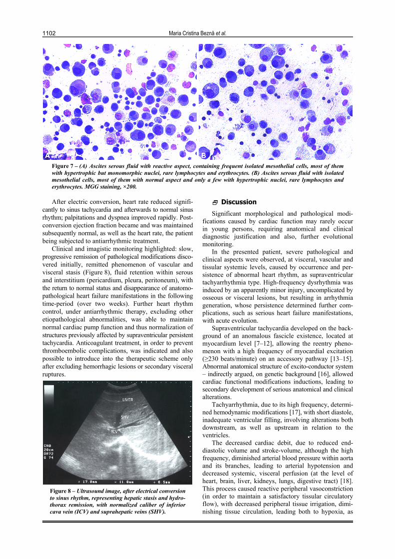
Upstream to the ventricles, the short diastole due to high heart rate, reducing ventricular filling, caused morpho-pathological left and right atrial dilatation, inferior cava vein dilatation, with visceral stasis, especially in hepatic and pulmonary territories, interstitial and tissular stasis, with lower limbs edema, stasis and transudate poly-serositis within pericardium, pleura and peritoneum. Fluids located at serous level were transudate type, caused by heart failure, in persistent tachyarrhythmia condition, without inflammatory modifications or other diverse cell-types detectable in cytology. Pathological aspect of pleural fluid was serous type (Figure 6, A and B), with isolated mesothelial cells, rare lymphocytes and erythrocytes. In the same time, cytology aspect of ascites was also serous (Figure 7, A and B), with rare cellularity, without abnormalities.
Figure 6 – (A) Pleural serous fluid with frequent isolated mesothelial cells, some with normal aspect, some with hypertrophic nuclei (reactive) and with karyorrhexis; rare lymphocytes and erythrocytes. (B) Pleural serous fluid with isolated mesothelial cells, some with intensively hypertrophic nuclei and with prominent nucleoli, but maintaining nuclei–cytoplasm balance, rare lymphocytes and erythrocytes. May–Grünwald–Giemsa (MGG) staining, ×200.
Maria Cristina Beznă et al.
1102
Figure 7 – (A) Ascites serous fluid with reactive aspect, containing frequent isolated mesothelial cells, most of them with hypertrophic but monomorphic nuclei, rare lymphocytes and erythrocytes. (B) Ascites serous fluid with isolated mesothelial cells, most of them with normal aspect and only a few with hypertrophic nuclei, rare lymphocytes and erythrocytes. MGG staining, ×200.
After electric conversion, heart rate reduced signifi-cantly to sinus tachycardia and afterwards to normal sinus rhythm; palpitations and dyspnea improved rapidly. Post-conversion ejection fraction became and was maintained subsequently normal, as well as the heart rate, the patient being subjected to antiarrhythmic treatment.
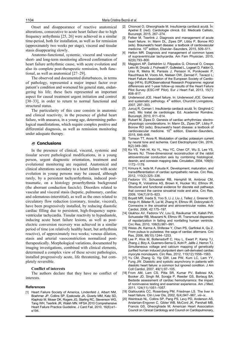
Clinical and imagistic monitoring highlighted: slow, progressive remission of pathological modifications disco-vered initially, remitted phenomenon of vascular and visceral stasis (Figure 8), fluid retention within serous and interstitium (pericardium, pleura, peritoneum), with the return to normal status and disappearance of anatomo-pathological heart failure manifestations in the following time-period (over two weeks). Further heart rhythm control, under antiarrhythmic therapy, excluding other etiopathological abnormalities, was able to maintain normal cardiac pump function and thus normalization of structures previously affected by supraventricular persistent tachycardia. Anticoagulant treatment, in order to prevent thromboembolic complications, was indicated and also possible to introduce into the therapeutic scheme only after excluding hemorrhagic lesions or secondary visceral ruptures.
Figure 8 – Ultrasound image, after electrical conversion to sinus rhythm, representing hepatic stasis and hydro-thorax remission, with normalized caliber of inferior cava vein (ICV) and suprahepatic veins (SHV).
Discussion
Significant morphological and pathological modi-fications caused by cardiac function may rarely occur in young persons, requiring anatomical and clinical diagnostic justification and also, further evolutional monitoring.
In the presented patient, severe pathological and clinical aspects were observed, at visceral, vascular and tissular systemic levels, caused by occurrence and per-sistence of abnormal heart rhythm, as supraventricular tachyarrhythmia type. High-frequency dysrhythmia was induced by an apparently minor injury, uncomplicated by osseous or visceral lesions, but resulting in arrhythmia generation, whose persistence determined further com-plications, such as serious heart failure manifestations, with acute evolution.
Supraventricular tachycardia developed on the back-ground of an anomalous fascicle existence, located at myocardium level [7–12], allowing the reentry pheno-menon with a high frequency of myocardial excitation (≥230 beats/minute) on an accessory pathway [13–15]. Abnormal anatomical structure of excito-conductor system – indirectly argued, on genetic background [16], allowed cardiac functional modifications inductions, leading to secondary development of serious anatomical and clinical alterations.
Tachyarrhythmia, due to its high frequency, determi-ned hemodynamic modifications [17], with short diastole, inadequate ventricular filling, involving alterations both downstream, as well as upstream in relation to the ventricles.
The decreased cardiac debit, due to reduced end-diastolic volume and stroke-volume, although the high frequency, diminished arterial blood pressure within aorta and its branches, leading to arterial hypotension and decreased systemic, visceral perfusion (at the level of heart, brain, liver, kidneys, lungs, digestive tract) [18]. This process caused reactive peripheral vasoconstriction (in order to maintain a satisfactory tissular circulatory flow), with decreased peripheral tissue irrigation, dimi-nishing tissue circulation, leading both to hypoxia, as

Visceral and tissular reactivity in acute heart failure due to supraventricular tachyarrhythmia in a young patient
1103
well as to biochemical, metabolic cellular and interstitial alterations, these processes providing an explanation for fatigue, fainting, dizziness symptoms.
At cardiac level, the decreased coronary diastolic filling, caused by diastole shortening, resulted in transitory ischemic non-atherosclerotic modifications, being also accentuated at subepicardial level, due to pericardial tran-sudate pressure. Developed on heart failure background, in tachyarrhythmia condition, the presence of pericardial fluid in moderate quantity contributed furthermore to hypodiastole and decreased cardiac debit processes, worsening visceral stasis.
The decreased cardiac debit, caused by hypodiastole induced by tachyarrhythmia, compensatory reduced splanchnic and peripheral blood flow.
At hepatic level [19], blood flow decrease exceeded liver’s ability to increase oxygen extraction, especially in acute situations, resulting in severe hepatocellular hypoxia, especially in acinar zone 3.
Passive hepatic congestion also increased ischemic injury risk. Increased central venous pressure (occurring in hypodiastolic heart failure) is transmitted to hepatic veins and venules that drain hepatic acini, causing atrophy of hepatocytes in liver acinar zone 3, this fact being associated to an exudation mechanism of protein-rich fluids into Disse space and also to sinus congestion. The resulted perisinusoidal edema prevented oxygen diffusion and nutrient flow towards hepatocytes. Histological aspect of combined liver pathology, such as stasis and ischemia, corresponds also to the moderate increase (×2) of serum glutamic oxaloacetic transaminase (SGOT) or aspartate aminotransferase (AST) enzyme, without assigning a proper hepatic cytolysis [GPT or alanine aminotransferase (ALT), normal level].
Morpho-pathological reactivity in visceral, vascular and tissue sectors, due to a persistent and seriously com-plicated tachyarrhythmia at a young age, sustained by recorded aspects, provided anatomo-clinical argumen-tation for positive and differential diagnosis, as well as for therapeutic control and remission monitoring [20].
Morphological altered aspects were assessed by clinical and/or paraclinical methods, some performed during emergency status and others during post-electric conver-sion evolution.
Some anatomical modifications were easy clinically detectable, such as: voluminous Vth degree hepatomegaly, with inferior edge of the liver lowered into right iliac fossa (right lobe) and mesogastrum (left lobe), rounded, with hepatojugular reflux, proofing hepatic blood stasis; presence of peritoneal fluid shown by movable abdominal dullness; subcutaneous interstitial stasis evidenced by lower limbs edema; pulmonary congestion, discovered through subcrepitant rales in the middle third of the lungs and bilateral hydrothorax through decreased vesicular murmur, with basal bilateral dullness.
Although clinically assessed, these pathological lesions required visualization and exploratory arguments [21], achieved through imagistic explorations, cardiac and thorax-abdominal ultrasound, as well as through laboratory and cytology findings.
After electric conversion, the initial ECG aspect, of wide QRS complexes paroxysmal supraventricular tachy-
cardia, turned to sinus rhythm, first with sinus tachycardia and then with normal frequency (60–80 beats/minute). Transitory ischemic subepicardial modifications were also observed in this manner, due to coronary hypoperfusion and pericardial fluid compression. ECG monitoring was necessary in order to control cardiac rhythm and rate, its decreasing allowing anatomical and clinical heart failure manifestations remission, the patient remaining under antiarrhythmic therapy until an eventual radiofrequency ablation [22].
Cardiac morphologic-functional manifestations were revealed by cardiac ultrasound exploration. It describes the presence of pericardial fluid in moderate quantity and also of bilateral pleural fluid, right and left atrial and inferior vena cava dilatation, without any anatomical modi-fications: endo-myocardial, of cavities, valves, orifices, chordae; post-resuscitation, ultrasound showed an improved ejection fraction, with 60% and an efficient decrease of heart contractions. Repeated cardiac ultrasound exami-nations allowed anatomo-functional monitoring until peri-cardial and pleural fluids disappearance, normalization of inferior vena cava caliber and remission of stasis elements.
Abdominal morphological modifications were assessed by ultrasound and computed tomography investigations. Thus, there were described: homogeneous hepatomegaly, without localized formations, infiltrative areas or post-traumatic hematoma, but with stasis aspect, due to marked dilatation of inferior cava vein and of suprahepatic veins, portal vein of normal caliber, normal configured spleen and presence of ascites within abdominal cavity.
These data are important in order to exclude other anatomical lesions causing hepatomegaly, such as lym-phoma infiltrations and leukemia (with splenomegaly, lymphadenopathy), tumors (localized), hepatic cirrhosis with portal decompensation (with an increased portal vein caliber over 15 mm, retrograde flow and congestive splenomegaly with venous dilatations within hilum), hematoma, cysts, hemangioma.
Prolonged stasis favored protein escape into tissues, favoring serum hypoproteinemia and worsening the polyserositis, requiring additional correction.
Inferior limbs peripheral edemas were consequences of prolonged stasis, fluid extravasation into interstitial subcutaneous tissue and altered capillary permeability, phenomena remitted in the following days, after anti-arrhythmic therapy initiation.
Anatomical differential diagnosis required exclusion of other pathologic conditions causing heart failure [23, 24], polyserositis, anasarca, hepatomegaly (tuberculosis, collagenosis, nephrotic syndrome, post-traumatic internal hemorrhage, protein deficiencies, neoplasia with secon-dary hepatic determinations, in a young patient.
In evolution, hepatomegaly, hydrothorax and peri-carditis remitted fully, but slowly, in about 14 days after arrhythmia cropping, maintaining normal status within the coming years.
A possible presence of an associated cardiac pathology (myocarditis, rheumatic carditis, collagenosis or cell infiltrations) would have presented adequate biological and imagistic descriptions and its remission would have required a longer period of time and a different drug therapy.
Maria Cristina Beznă et al.
1104
Onset and disappearance of reactive anatomical alterations, consecutive to acute heart failure due to high frequency arrhythmia [25, 26] were achieved in a similar time-period, both for installation, as well as for remission (approximately two weeks per stage), visceral and tissular stasis disappearing slowly.
Anatomo-functional, systemic, visceral and vascular short- and long-term monitoring allowed confirmation of heart failure arrhythmic cause, with acute evolution and also its complete post-therapeutic remission, both func-tional, as well as anatomical [27–29].
The observed and documented disturbances, in terms of pathology, represented a major impact factor over patient’s condition and worsened his general state, endan-gering his life; these facts represented an important aspect for causal treatment urgent initiation and control [30–33], in order to return to normal functional and structural status.
The particularity of this case consists in anatomic and clinical reactivity, in the presence of global heart failure, with anasarca, in a young age, determining patho-logical manifestations, which require complex positive and differential diagnosis, as well as remission monitoring under adequate therapy.
Conclusions
In the presence of clinical, visceral, systemic and tissular severe pathological modifications, in a young person, urgent diagnostic orientation, treatment and evolutional monitoring are required. Anatomical and clinical alterations secondary to a heart failure with acute evolution in young persons may be caused, although rarely, by a persistent tachyarrhythmia, induced post-traumatic, on a histology proarrhythmia background (the aberrant conduction fascicle). Disorders related to vascular and visceral stasis (hepatic, pulmonary, cardiac and edematous-interstitial), as well as the ones related to circulatory flow reduction (coronary, tissular, visceral), have been progressively installed, by reducing diastolic cardiac filling due to persistent, high-frequency supra-ventricular tachycardia. Tissular reactivity to hypodiastole, inducing acute heart failure lesions, as well as post-electric conversion recovery was achieved in a similar period of time (on relatively healthy heart, but arrhythmia reactive), of approximately two weeks; venous dilation, stasis and arterial vasoconstriction normalized post-therapeutically. Morphological variations, documented by imaging investigations, combined with clinical elements, determined a complex view of these severe pathologies, installed progressively acute, life threatening, but com-pletely reversible.
Conflict of interests The authors declare that they have no conflict of
interests.
References [1] Heart Failure Society of America, Lindenfeld J, Albert NM,
Boehmer JP, Collins SP, Ezekowitz JA, Givertz MM, Katz SD, Klapholz M, Moser DK, Rogers JG, Starling RC, Stevenson WG, Tang WH, Teerlink JR, Walsh MN. HFSA 2010 Comprehensive Heart Failure Practice Guideline. J Card Fail, 2010, 16(6):e1–e194.
[2] Chioncel O, Gheorghiade M. Insuficienţa cardiacă acută. În: Apetrei E (red). Cardiologie clinică. Ed. Medicală Callisto, Bucureşti, 2015, 267–274.
[3] Felker M, Teerlink J. Diagnosis and management of acute heart failure. In: Mann DL, Zipes DP, Libby P, Bonow RO (eds). Braunwald’s heart disease: a textbook of cardiovascular medicine. 10th edition, Elsevier–Saunders, 2015, 509–511.
[4] Helton MR. Diagnosis and management of common types of supraventricular tachycardia. Am Fam Physician, 2015, 92(9):793–800.
[5] Maggioni AP, Dahlström U, Filippatos G, Chioncel O, Crespo Leiro M, Drozdz J, Fruhwald F, Gullestad L, Logeart D, Fabbri G, Urso R, Metra M, Parissis J, Persson H, Ponikowski P, Rauchhaus M, Voors AA, Nielsen OW, Zannad F, Tavazzi L; Heart Failure Association of the European Society of Cardio-logy (HFA). EURObservational Research Programme: regional differences and 1-year follow-up results of the Heart Failure Pilot Survey (ESC-HF Pilot). Eur J Heart Fail, 2013, 15(7): 808–817.
[6] Underwood JCE. Heart failure. In: Underwood JCE. General and systematic pathology. 4th edition, Churchill Livingstone, 2007, 297–303.
[7] Jurcuţ R, Coman I. Insuficiența cardiacă acută. În: Ginghină C (red). Mic tratat de cardiologie. Ed. Academiei Române, Bucureşti, 2010, 611–614.
[8] Rubart M, Zipes D. Genesis of cardiac arrhythmias: electro-physiologic considerations. In: Mann DL, Zipes DP, Libby P, Bonow RO (eds). Braunwald’s heart disease: a textbook of cardiovascular medicine. 10th edition, Elsevier–Saunders, 2015, 646–648.
[9] Tomson TT, Arora R. Modulation of cardiac potassium current by neural tone and ischemia. Card Electrophysiol Clin, 2016, 8(2):349–360.
[10] Ko YS, Yeh HI, Ko YL, Hsu YC, Chen CF, Wu S, Lee YS, Severs NJ. Three-dimensional reconstruction of the rabbit atrioventricular conduction axis by combining histological, desmin, and connexin mapping data. Circulation, 2004, 109(9): 1172–1179.
[11] Kimura K, Ieda M, Fukuda K. Development, maturation, and transdifferentiation of cardiac sympathetic nerves. Circ Res, 2012, 110(2):325–336.
[12] Fedorov VV, Schuessler RB, Hemphill M, Ambrosi CM, Chang R, Voloshina AS, Brown K, Hucker WJ, Efimov IR. Structural and functional evidence for discrete exit pathways that connect the canine sinoatrial node and atria. Circ Res, 2009, 104(7):915–923.
[13] Boyett MR, Inada S, Yoo S, Li J, Liu J, Tellez J, Greener ID, Honjo H, Billeter R, Lei M, Zhang H, Efimov IR, Dobrzynski H. Connexins in the sinoatrial and atrioventricular nodes. Adv Cardiol, 2006, 42:175–197.
[14] Glukhov AV, Fedorov VV, Lou Q, Ravikumar VK, Kalish PW, Schuessler RB, Moazami N, Efimov IR. Transmural dispersion of repolarization in failing and nonfailing human ventricle. Circ Res, 2010, 106(5):981–991.
[15] Weiss JN, Karma A, Shiferaw Y, Chen PS, Garfinkel A, Qu Z. From pulsus to pulseless: the saga of cardiac alternans. Circ Res, 2006, 98(10):1244–1253.
[16] Lee P, Klos M, Bollensdorff C, Hou L, Ewart P, Kamp TJ, Zhang J, Bizy A, Guerrero-Serna G, Kohl P, Jalife J, Herron TJ. Simultaneous voltage and calcium mapping of genetically purified human induced pluripotent stem cell-derivated cardiac myocyte monolayers. Circ Res, 2012, 110(12):1556–1563.
[17] Yu CM, Zhang Q, Yip GW, Lee PW, Kum LC, Lam YY, Fung JW. Diastolic and systolic asynchrony in patients with diastolic heart failure: a common but ignored condition. J Am Coll Cardiol, 2007, 49(1):97–105.
[18] From AM, Lam CS, Pitta SR, Kumar PV, Balbissi KA, Booker JD, Singh IM, Sorajja P, Reeder GS, Borlaug BA. Bedside assessment of cardiac hemodynamics: the impact of noninvasive testing and examiner experience. Am J Med, 2011, 124(11):1051–1057.
[19] Giallourakis CC, Rosenberg PM, Friedman LS. The liver in heart failure. Clin Liver Dis, 2002, 6(4):947–967, viii–ix.
[20] Weintraub NL, Collins SP, Pang PS, Levy PD, Anderson AS, Arslanian-Engoren C, Gibler WB, McCord JK, Parshall MB, Francis GS, Gheorghiade M; American Heart Association Council on Clinical Cardiology and Council on Cardiopulmonary,
Visceral and tissular reactivity in acute heart failure due to supraventricular tachyarrhythmia in a young patient
1105
Critical Care, Perioperative and Resuscitation. Acute heart failure syndromes: emergency department presentation, treat-ment, and disposition: current approaches and future aims: a scientific statement from the American Heart Association. Circulation, 2010, 122(19):1975–1996.
[21] Kelder JC, Cramer MJ, van Wijngaarden J, van Tooren R, Mosterd A, Moons KG, Lammers JW, Cowie MR, Grobbee DE, Hoes AW. The diagnostic value of physical examination and additional testing in primary care patients with suspected heart failure. Circulation, 2011, 124(25):2865–2873.
[22] Isenhour JL, Craig S, Gibbs M, Littmann L, Rose G, Risch R. Wide complex tachycardia: continued evaluation of diagnostic criteria. Acad Emerg Med, 2000, 7(7):769–773.
[23] Mant J, Doust J, Roalfe A, Barton P, Cowie MR, Glasziou P, Mant D, McManus RJ, Holder R, Deeks J, Fletcher K, Qume M, Sohanpal S, Sanders S, Hobbs FD. Systematic review and individual patient data meta-analysis of diagnosis of heart failure, with modelling of implications of different diagnostic strategies in primary care. Health Technol Assess, 2009, 13(32):1–207.
[24] Little WC, Zile MR. HFpEF: cardiovascular abnormalities not just comorbidities. Circ Heart Fail, 2012, 5(6):669–671.
[25] Davie AP, Francis CM, Caruana L, Sutherland GR, McMurray JJ. Assessing diagnosis in heart failure: which features are any use? QJM, 1997, 90(5):335–339.
[26] Cruz FE, Cheriex EC, Smeets JL, Atié J, Peres AK, Penn OC, Brugada P, Wellens HJ. Reversibility of tachycardia-induced cardiomyopathy after cure of incessant supraventricular tachy-cardia. J Am Coll Cardiol, 1990, 16(3):739–744.
[27] McKelvie RS, Moe GW, Ezekowitz JA, Heckman GA, Costigan J, Ducharme A, Estrella-Holder E, Giannetti N, Grzeslo A, Harkness K, Howlett JG, Kouz S, Leblanc K, Mann E, Nigam A, O’Meara E, Rajda M, Steinhart B, Swiggum E, Le VV, Zieroth S, Arnold JM, Ashton T, D’Astous M, Dorian P, Haddad H, Isaac DL, Leblanc MH, Liu P, Rao V, Ross HJ, Sussex B. The 2012 Canadian Cardiovascular Society heart failure management guidelines update: focus on acute and chronic heart failure. Can J Cardiol, 2013, 29(2):168–181.
[28] McMurray JJ, Adamopoulos S, Anker SD, Auricchio A, Böhm M, Dickstein K, Falk V, Filippatos G, Fonseca C, Gomez-Sanchez MA, Jaarsma T, Køber L, Lip GY, Maggioni AP, Parkhomenko A, Pieske BM, Popescu BA, Rønnevik PK,
Rutten FH, Schwitter J, Seferovic P, Stepinska J, Trindade PT, Voors AA, Zannad F, Zeiher A; ESC Committee for Practice Guidelines. ESC Guidelines for the diagnosis and treatment of acute and chronic heart failure 2012: The Task Force for the Diagnosis and Treatment of Acute and Chronic Heart Failure 2012 of the European Society of Cardiology. Developed in collaboration with the Heart Failure Association (HFA) of the ESC. Eur Heart J, 2012, 33(14):1787–1847.
[29] Yancy CW, Jessup M, Bozkurt B, Butler J, Casey DE Jr, Drazner MH, Fonarow GC, Geraci SA, Horwich T, Januzzi JL, Johnson MR, Kasper EK, Levy WC, Masoudi FA, McBride PE, McMurray JJ, Mitchell JE, Peterson PN, Riegel B, Sam F, Stevenson LW, Tang WH, Tsai EJ, Wilkoff BL; American College of Cardiology Foundation; American Heart Association Task Force on Practice Guidelines. 2013 ACCF/AHA guideline for the management of heart failure: a report of the American College of Cardiology Foundation/American Heart Association Task Force on Practice Guidelines. J Am Coll Cardiol, 2013, 62(16):e147–e239.
[30] Chioncel O, Ambrosy AP, Filipescu D, Bubenek S, Vinereanu D, Petris A, Collins SP, Macarie C, Gheorghiade M; Romanian Acute Heart Failure Syndromes Study Investigators. Patterns of intensive care unit admissions in patients hospitalized for heart failure: insights from the RO-AHFS registry. J Cardiovasc Med (Hagerstown), 2015, 16(5):331–340.
[31] Mebazaa A, Pang PS, Tavares M, Collins SP, Storrow AB, Laribi S, Andre S, Mark Courtney D, Hasa J, Spinar J, Masip J, Frank Peacock W, Sliwa K, Gayat E, Filippatos G, Cleland JG, Gheorghiade M. The impact of early standard therapy on dyspnoea in patients with acute heart failure: the URGENT-dyspnoea study. Eur Heart J, 2010, 31(7):832–841.
[32] Chioncel O, Vinereanu D, Datcu M, Ionescu DD, Capalneanu R, Brukner I, Dorobantu M, Ambrosy A, Macarie C, Gheorghiade M. The Romanian Acute Heart Failure Syndromes (RO-AHFS) Registry. Am Heart J, 2011, 162(1):142–153.e1.
[33] Badar AA, Perez-Moreno AC, Hawkins NM, Brunton AP, Jhund PS, Wong CM, Solomon SD, Granger CB, Yusuf S, Pfeffer MA, Swedberg K, Gardner RS, Petrie MC, McMurray JJ. Clinical characteristics and outcomes of patients with angina and heart failure in the CHARM (Candesartan in Heart Failure Assessment of Reduction in Mortality and Morbidity) Programme. Eur J Heart Fail, 2015, 17(2):196–204.
Corresponding author Marinela Beznă, Associate Professor, MD, PhD, Department of Internal Medicine, University of Medicine and Pharmacy of Craiova, 2 Petru Rareş Street, 200349 Craiova, Dolj County, Romania; Phone +40766–100 184, e-mail: [email protected] Received: December 8, 2015
Accepted: December 3, 2016