
Rom J Morphol Embryol 2012, 53(3 Suppl):851–853 R J M E ... · Rom J Morphol Embryol 2012, 53(3...
3
Rom J Morphol Embryol 2012, 53(3 Suppl):851–853 ISSN (print) 1220–0522 ISSN (on-line) 2066–8279 CASE REPORT Granulomatous cheilitis of Miescher: the diagnostic proof for a Melkersson–Rosenthal syndrome ANDREEA LIANA RĂCHIŞAN 1) , A. HRUŞCĂ 1) , D. GHEBAN 2) , SIMONA CĂINAP 1) , T. L. POP 1) , A. BĂICAN 3) , L. FODOR 4) , N. MIU 1) , MARIANA ANDREICA 1) 1) Department of Pediatrics 2) Department of Pathology 3) Department of Dermatology 4) Department of Plastic Surgery “Iuliu Haţieganu” University of Medicine and Pharmacy, Cluj-Napoca Abstract Background: The Melkersson–Rosenthal syndrome (MRS) is a very rare clinical entity and its classical form is being characterized by the following triad: facial nerve palsy, swelling of the lips and fissured tongue. However, the monosymptomatic form is more common and the typical manifestation is facial edema and/or enlargement of the lips. Case report: We report a case of monosymptomatic MRS with a positive biopsy of granulomatous cheilitis. Conclusions: In the daily practice as a pediatrician, it is not usual to diagnose a patient as having MRS. We consider that this is partly because of misdiagnosis. We therefore believe that this case report will supply additional information, in the scope of recurrent facial paralysis and orofacial edema in both children and adults. Keywords: granulomatous cheilitis, facial nerve palsy, facial edema, fissured tongue. Introduction The Melkersson–Rosenthal syndrome is a very rare clinical entity. Its classical form is being characterized by the following triad: facial nerve palsy, swelling οf the lips and fissured tongue [1]. However, the mono- symptomatic form is more common and the typical manifestation is facial edema and/or enlargement of the lips [2]. This syndrome is very rare in childhood, thus there are only a few cases described in the pediatric age [3]. One or two manifestations in association with granulomatous cheilitis in the biopsy is sufficient for the diagnosis of Melkersson–Rosenthal syndrome [4]. Patient, Methods and Results We report a case of a 14-year-old girl presenting with non-painful swelling of the upper lip. This manifestation has occurred six months before the presentation in our department. At anamnesis, no allergic diseases, trauma or past/recent injuries were reported. With all this, from her personal antecedents we discovered a facial palsy at the age of eight years as s singular manifestation. The physical examination revealed a non-painful, non-pruritic swollen upper lip and a scrotal tongue (Figure 1). The other physical signs were within normal appearance for the age. Laboratory values, including electrolytes, liver function tests, complete blood cell count, erythrocyte sedimentation rate, total immuno- globulin E, complement (C3, C4), antinuclear antibody were all within normal limits. Figure 1 – Non-painful, swollen upper lip and scrotal tongue (before treatment with local corticosteroids). Furthermore, corroborating all three manifestations from the past until present we decided to perform a biopsy of the upper lip mucosa. The upper lip biopsy specimen showed multiple discrete, non-necrotizing granulomas composed mostly of epithelioid cells with rare multinucleated giant cells and a perivascular lymphocytic infiltration. This histo- pathological result resembled cheilitis granulomatosa (Figure 2, A–C). A three-month trial of oral corticotherapy associated with antihistaminic treatment was ineffective, similar edema of the upper lip was developed. After that, we decided to try local injections with triamcinolone with a spectacular evolution (Figure 3). R J M E Romanian Journal of Morphology & Embryology http://www.rjme.ro/
Transcript of Rom J Morphol Embryol 2012, 53(3 Suppl):851–853 R J M E ... · Rom J Morphol Embryol 2012, 53(3...
Rom J Morphol Embryol 2012, 53(3 Suppl):851–853
ISSN (print) 1220–0522 ISSN (on-line) 2066–8279
CCAASSEE RREEPPOORRTT
Granulomatous cheilitis of Miescher: the diagnostic proof for a
Melkersson–Rosenthal syndrome
ANDREEA LIANA RĂCHIŞAN1), A. HRUŞCĂ1), D. GHEBAN2), SIMONA CĂINAP1), T. L. POP1), A. BĂICAN3), L. FODOR4),
N. MIU1), MARIANA ANDREICA1)
1)Department of Pediatrics 2)Department of Pathology
3)Department of Dermatology 4)Department of Plastic Surgery
“Iuliu Haţieganu” University of Medicine and Pharmacy, Cluj-Napoca
Abstract Background: The Melkersson–Rosenthal syndrome (MRS) is a very rare clinical entity and its classical form is being characterized by the following triad: facial nerve palsy, swelling of the lips and fissured tongue. However, the monosymptomatic form is more common and the typical manifestation is facial edema and/or enlargement of the lips. Case report: We report a case of monosymptomatic MRS with a positive biopsy of granulomatous cheilitis. Conclusions: In the daily practice as a pediatrician, it is not usual to diagnose a patient as having MRS. We consider that this is partly because of misdiagnosis. We therefore believe that this case report will supply additional information, in the scope of recurrent facial paralysis and orofacial edema in both children and adults.
Keywords: granulomatous cheilitis, facial nerve palsy, facial edema, fissured tongue.
Introduction
The Melkersson–Rosenthal syndrome is a very rare clinical entity. Its classical form is being characterized by the following triad: facial nerve palsy, swelling οf the lips and fissured tongue [1]. However, the mono-symptomatic form is more common and the typical manifestation is facial edema and/or enlargement of the lips [2].
This syndrome is very rare in childhood, thus there are only a few cases described in the pediatric age [3]. One or two manifestations in association with granulomatous cheilitis in the biopsy is sufficient for the diagnosis of Melkersson–Rosenthal syndrome [4].
Patient, Methods and Results
We report a case of a 14-year-old girl presenting with non-painful swelling of the upper lip. This manifestation has occurred six months before the presentation in our department.
At anamnesis, no allergic diseases, trauma or past/recent injuries were reported. With all this, from her personal antecedents we discovered a facial palsy at the age of eight years as s singular manifestation.
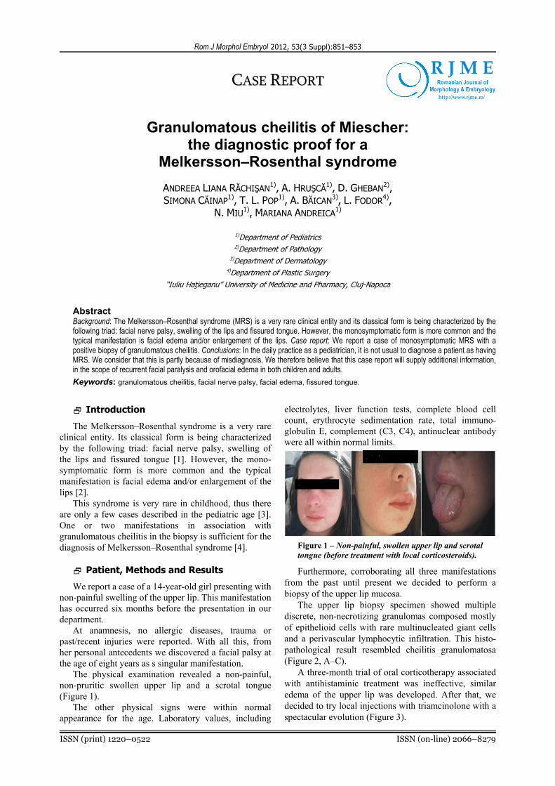
The physical examination revealed a non-painful, non-pruritic swollen upper lip and a scrotal tongue (Figure 1).
The other physical signs were within normal appearance for the age. Laboratory values, including
electrolytes, liver function tests, complete blood cell count, erythrocyte sedimentation rate, total immuno-globulin E, complement (C3, C4), antinuclear antibody were all within normal limits.
Figure 1 – Non-painful, swollen upper lip and scrotal tongue (before treatment with local corticosteroids).
Furthermore, corroborating all three manifestations from the past until present we decided to perform a biopsy of the upper lip mucosa.
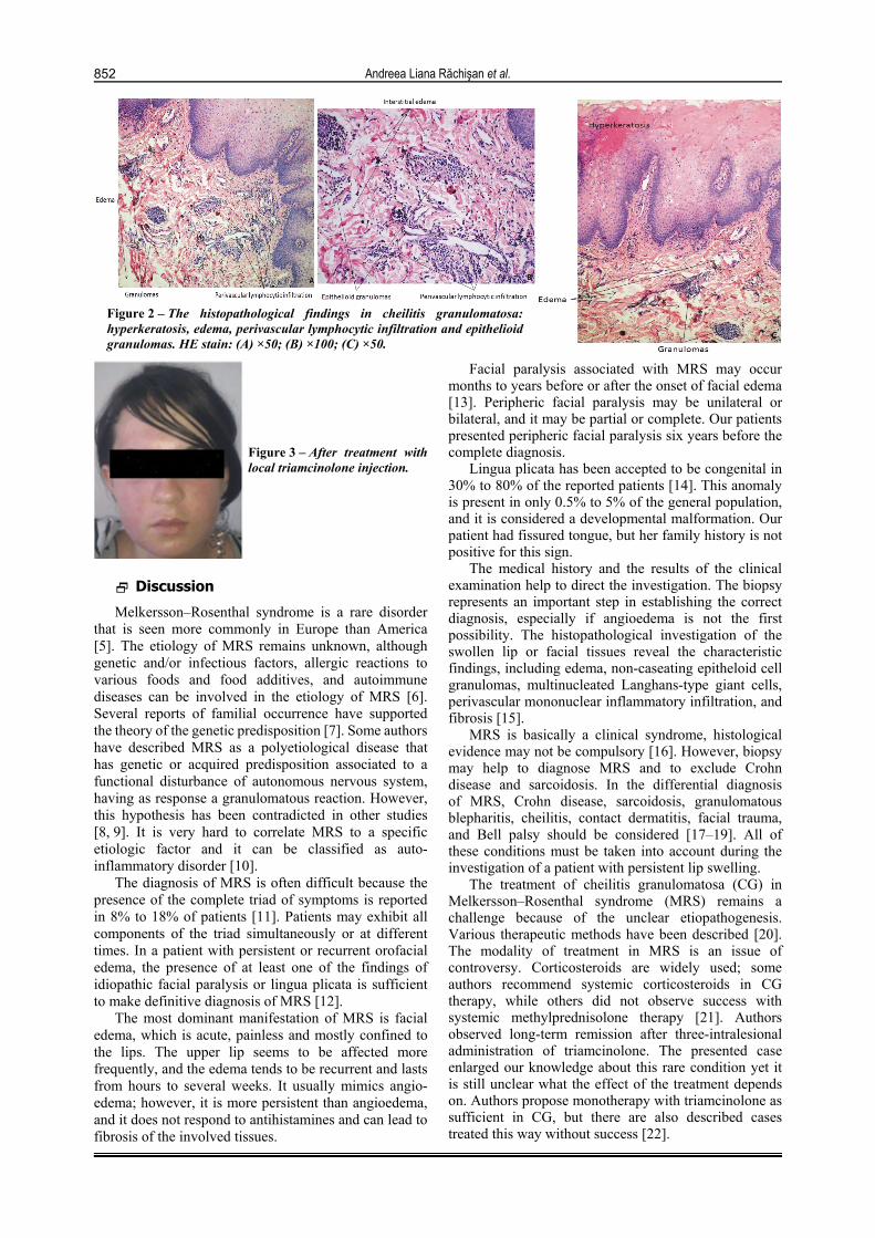
The upper lip biopsy specimen showed multiple discrete, non-necrotizing granulomas composed mostly of epithelioid cells with rare multinucleated giant cells and a perivascular lymphocytic infiltration. This histo-pathological result resembled cheilitis granulomatosa (Figure 2, A–C).
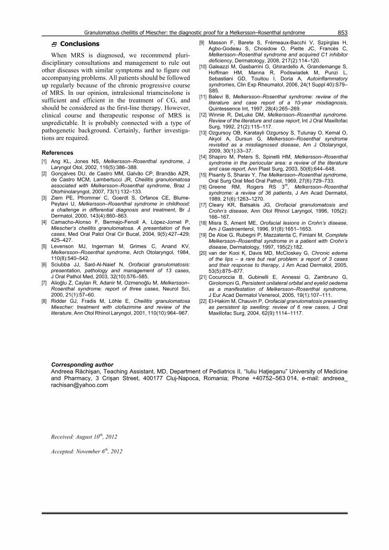
A three-month trial of oral corticotherapy associated with antihistaminic treatment was ineffective, similar edema of the upper lip was developed. After that, we decided to try local injections with triamcinolone with a spectacular evolution (Figure 3).
R J M ERomanian Journal of
Morphology & Embryologyhttp://www.rjme.ro/
Andreea Liana Răchişan et al.
852
Figure 2 – The histopathological findings in cheilitis granulomatosa: hyperkeratosis, edema, perivascular lymphocytic infiltration and epithelioid granulomas. HE stain: (A) ×50; (B) ×100; (C) ×50.
Figure 3 – After treatment withlocal triamcinolone injection.
Discussion
Melkersson–Rosenthal syndrome is a rare disorder that is seen more commonly in Europe than America [5]. The etiology of MRS remains unknown, although genetic and/or infectious factors, allergic reactions to various foods and food additives, and autoimmune diseases can be involved in the etiology of MRS [6]. Several reports of familial occurrence have supported the theory of the genetic predisposition [7]. Some authors have described MRS as a polyetiological disease that has genetic or acquired predisposition associated to a functional disturbance of autonomous nervous system, having as response a granulomatous reaction. However, this hypothesis has been contradicted in other studies [8, 9]. It is very hard to correlate MRS to a specific etiologic factor and it can be classified as auto-inflammatory disorder [10].
The diagnosis of MRS is often difficult because the presence of the complete triad of symptoms is reported in 8% to 18% of patients [11]. Patients may exhibit all components of the triad simultaneously or at different times. In a patient with persistent or recurrent orofacial edema, the presence of at least one of the findings of idiopathic facial paralysis or lingua plicata is sufficient to make definitive diagnosis of MRS [12].
The most dominant manifestation of MRS is facial edema, which is acute, painless and mostly confined to the lips. The upper lip seems to be affected more frequently, and the edema tends to be recurrent and lasts from hours to several weeks. It usually mimics angio-edema; however, it is more persistent than angioedema, and it does not respond to antihistamines and can lead to fibrosis of the involved tissues.
Facial paralysis associated with MRS may occur months to years before or after the onset of facial edema [13]. Peripheric facial paralysis may be unilateral or bilateral, and it may be partial or complete. Our patients presented peripheric facial paralysis six years before the complete diagnosis.
Lingua plicata has been accepted to be congenital in 30% to 80% of the reported patients [14]. This anomaly is present in only 0.5% to 5% of the general population, and it is considered a developmental malformation. Our patient had fissured tongue, but her family history is not positive for this sign.
The medical history and the results of the clinical examination help to direct the investigation. The biopsy represents an important step in establishing the correct diagnosis, especially if angioedema is not the first possibility. The histopathological investigation of the swollen lip or facial tissues reveal the characteristic findings, including edema, non-caseating epitheloid cell granulomas, multinucleated Langhans-type giant cells, perivascular mononuclear inflammatory infiltration, and fibrosis [15].
MRS is basically a clinical syndrome, histological evidence may not be compulsory [16]. However, biopsy may help to diagnose MRS and to exclude Crohn disease and sarcoidosis. In the differential diagnosis of MRS, Crohn disease, sarcoidosis, granulomatous blepharitis, cheilitis, contact dermatitis, facial trauma, and Bell palsy should be considered [17–19]. All of these conditions must be taken into account during the investigation of a patient with persistent lip swelling.
The treatment of cheilitis granulomatosa (CG) in Melkersson–Rosenthal syndrome (MRS) remains a challenge because of the unclear etiopathogenesis. Various therapeutic methods have been described [20]. The modality of treatment in MRS is an issue of controversy. Corticosteroids are widely used; some authors recommend systemic corticosteroids in CG therapy, while others did not observe success with systemic methylprednisolone therapy [21]. Authors observed long-term remission after three-intralesional administration of triamcinolone. The presented case enlarged our knowledge about this rare condition yet it is still unclear what the effect of the treatment depends on. Authors propose monotherapy with triamcinolone as sufficient in CG, but there are also described cases treated this way without success [22].
Granulomatous cheilitis of Miescher: the diagnostic proof for a Melkersson–Rosenthal syndrome
853
Conclusions
When MRS is diagnosed, we recommend pluri-disciplinary consultations and management to rule out other diseases with similar symptoms and to figure out accompanying problems. All patients should be followed up regularly because of the chronic progressive course of MRS. In our opinion, intralesional triamcinolone is sufficient and efficient in the treatment of CG, and should be considered as the first-line therapy. However, clinical course and therapeutic response of MRS is unpredictable. It is probably connected with a type of pathogenetic background. Certainly, further investiga-tions are required.
References
[1] Ang KL, Jones NS, Melkersson–Rosenthal syndrome, J Laryngol Otol, 2002, 116(5):386–388.
[2] Gonçalves DU, de Castro MM, Galvão CP, Brandão AZR, de Castro MCM, Lambertucci JR, Cheilitis granulomatosa associated with Melkersson–Rosenthal syndrome, Braz J Otorhinolaryngol, 2007, 73(1):132–133.
[3] Ziem PE, Pfrommer C, Goerdt S, Orfanos CE, Blume-Peytavi U, Melkersson–Rosenthal syndrome in childhood: a challenge in differential diagnosis and treatment, Br J Dermatol, 2000, 143(4):860–863.
[4] Camacho-Alonso F, Bermejo-Fenoll A, López-Jornet P, Miescher’s cheilitis granulomatosa. A presentation of five cases, Med Oral Patol Oral Cir Bucal, 2004, 9(5):427–429; 425–427.
[5] Levenson MJ, Ingerman M, Grimes C, Anand KV, Melkersson–Rosenthal syndrome, Arch Otolaryngol, 1984, 110(8):540–542.
[6] Sciubba JJ, Said-Al-Naief N, Orofacial granulomatosis: presentation, pathology and management of 13 cases, J Oral Pathol Med, 2003, 32(10):576–585.
[7] Alioğlu Z, Caylan R, Adanir M, Ozmenoğlu M, Melkersson–Rosenthal syndrome: report of three cases, Neurol Sci, 2000, 21(1):57–60.
[8] Ridder GJ, Fradis M, Löhle E, Cheilitis granulomatosa Miescher: treatment with clofazimine and review of the literature, Ann Otol Rhinol Laryngol, 2001, 110(10):964–967.
[9] Masson F, Barete S, Frémeaux-Bacchi V, Szpirglas H, Agbo-Godeau S, Chosidow O, Piette JC, Francès C, Melkersson–Rosenthal syndrome and acquired C1 inhibitor deficiency, Dermatology, 2008, 217(2):114–120.
[10] Galeazzi M, Gasbarrini G, Ghirardello A, Grandemange S, Hoffman HM, Manna R, Podswiadek M, Punzi L, Sebastiani GD, Touitou I, Doria A, Autoinflammatory syndromes, Clin Exp Rheumatol, 2006, 24(1 Suppl 40):S79–S85.
[11] Balevi B, Melkersson–Rosenthal syndrome: review of the literature and case report of a 10-year misdiagnosis, Quintessence Int, 1997, 28(4):265–269.
[12] Winnie R, DeLuke DM, Melkersson–Rosenthal syndrome. Review of the literature and case report, Int J Oral Maxillofac Surg, 1992, 21(2):115–117.
[13] Ozgursoy OB, Karatayli Ozgursoy S, Tulunay O, Kemal O, Akyol A, Dursun G, Melkersson–Rosenthal syndrome revisited as a misdiagnosed disease, Am J Otolaryngol, 2009, 30(1):33–37.
[14] Shapiro M, Peters S, Spinelli HM, Melkersson–Rosenthal syndrome in the periocular area: a review of the literature and case report, Ann Plast Surg, 2003, 50(6):644–648.
[15] Pisanty S, Sharav Y, The Melkersson–Rosenthal syndrome, Oral Surg Oral Med Oral Pathol, 1969, 27(6):729–733.
[16] Greene RM, Rogers RS 3rd, Melkersson–Rosenthal syndrome: a review of 36 patients, J Am Acad Dermatol, 1989, 21(6):1263–1270.
[17] Cleary KR, Batsakis JG, Orofacial granulomatosis and Crohn’s disease, Ann Otol Rhinol Laryngol, 1996, 105(2): 166–167.
[18] Misra S, Ament ME, Orofacial lesions in Crohn’s disease, Am J Gastroenterol, 1996, 91(8):1651–1653.
[19] De Aloe G, Rubegni P, Mazzatenta C, Fimiani M, Complete Melkersson–Rosenthal syndrome in a patient with Crohn’s disease, Dermatology, 1997, 195(2):182.
[20] van der Kooi K, Davis MD, McCloskey G, Chronic edema of the lips – a rare but real problem: a report of 3 cases and their response to therapy, J Am Acad Dermatol, 2005, 53(5):875–877.
[21] Cocuroccia B, Gubinelli E, Annessi G, Zambruno G, Girolomoni G, Persistent unilateral orbital and eyelid oedema as a manifestation of Melkersson–Rosenthal syndrome, J Eur Acad Dermatol Venereol, 2005, 19(1):107–111.
[22] El-Hakim M, Chauvin P, Orofacial granulomatosis presenting as persistent lip swelling: review of 6 new cases, J Oral Maxillofac Surg, 2004, 62(9):1114–1117.
Corresponding author Andreea Răchişan, Teaching Assistant, MD, Department of Pediatrics II, “Iuliu Haţieganu” University of Medicine and Pharmacy, 3 Crişan Street, 400177 Cluj-Napoca, Romania; Phone +40752–563 014, e-mail: andreea_ [email protected] Received: August 10th, 2012
Accepted: November 6th, 2012


![Rom J Morphol Embryol R J M E ORIGINAL … the pneumatization of the middle turbinate has often been described, pneumatized superior turbinates [1, 2], supreme turbinates, uncinate](https://static.fdocuments.us/doc/165x107/5ca9f2dc88c993c9218d71be/rom-j-morphol-embryol-r-j-m-e-original-the-pneumatization-of-the-middle-turbinate.jpg)


![Rom J Morphol Embryol 2011, 52(1):69–74 R J M E … · Rom J Morphol Embryol 2011, 52(1) ... blished by the World Health Organization (WHO) Classification [1], ... rehydrated in](https://static.fdocuments.us/doc/165x107/5b6443407f8b9a687e8d1c3f/rom-j-morphol-embryol-2011-5216974-r-j-m-e-rom-j-morphol-embryol-2011.jpg)