Rom J Morphol Embryol 2011, 52(2):669–677 R J M E · PDF fileRom J Morphol Embryol 2011,...
9
Rom J Morphol Embryol 2011, 52(2):669–677 ORIGINAL PAPER Identification of different subtypes of breast cancer using tissue microarray M. A. MUNIRAH 1) , M. A. SITI-AISHAH 1) , M. Z. REENA 1) , N. A. SHARIFAH 1) , M. ROHAIZAK 2) , A. NORLIA 2) , M. K. M. RAFIE 3) , A. ASMIATI 3) , A. HISHAM 4) , I. FUAD 5) , N. S. SHAHRUN 2) , S. DAS 6) 1) Department of Pathology 2) Department of Surgery Faculty of Medicine, Universiti Kebangsaan Malaysia Medical Centre, Kuala Lumpur, Malaysia 3) Department of Pathology 4) Department of Surgery Hospital Putrajaya, Selangor, Malaysia 5) Department of Radiotherapy & Oncology, Universiti Kebangsaan Malaysia Medical Centre, Kuala Lumpur, Malaysia 6) Department of Anatomy, Faculty of Medicine, Universiti Kebangsaan Malaysia Medical Centre, Kuala Lumpur, Malaysia Abstract Breast cancer may be classified into luminal A, luminal B, HER2+/ER-, basal-like and normal-like subtypes based on gene expression profiling or immunohistochemical (IHC) characteristics. The main aim of the present study was to classify breast cancer into molecular subtypes based on immunohistochemistry findings and correlate the subtypes with clinicopathological factors. Two hundred and seventeen primary breast carcinomas tumor tissues were immunostained for ER, PR, HER2, CK5/6, EGFR, CK8/18, p53 and Ki67 using tissue microarray technique. All subtypes were significantly associated with Malay ethnic background (p=0.035) compared to other racial origins. The most common subtypes of breast cancers were luminal A and was significantly associated with low histological grade (p<0.000) and p53 negativity (p=0.003) compared to HER2+/ER-, basal-like and normal-like subtypes with high histological grade (p<0.000) and p53 positivity (p=0.003). Luminal B subtype had the smallest mean tumor size (p=0.009) and also the highest mean number of lymph nodes positive (p=0.032) compared to other subtypes. All markers except EGFR and Ki67 were significantly associated with the subtypes. The most common histological type was infiltrating ductal carcinoma, NOS. Majority of basal-like subtype showed comedo-type necrosis (68.8%) and infiltrative margin (81.3%). Our studies suggest that IHC can be used to identify the different subtypes of breast cancer and all subtypes were significantly associated with race, mean tumor size, mean number of lymph node positive, histological grade and all immunohistochemical markers except EGFR and Ki67. Keywords: immunohistochemistry, breast cancer, estrogen receptor, progesterone receptor, HER2, keratins. Introduction Breast cancer is a heterogeneous disease such that they may have different prognoses and respond to therapy differently despite similarities in histological types, grade and stage. These differences are not well understood but are possibly due to the differences in the cell of origin. Normal human mammary gland epithelium is characterized by ductal glandular or luminal, basal myoepithelial and mammary gland stem/progenitor cells [1]. Breast cancers of glandular epithelial immuno- phenotype suggest that these neoplastic cells are derived from late stage of the glandular epithelial differentiation. Gene expression profiling in breast cancer has been successfully used to classify breast cancer into pathologically distinct subtypes [2]. For example, cDNA microarray studies on breast carcinoma have shown that breast cancers can be divided into expressing ER (ER+) luminal subtype and non expressing ER (ER-) tumors which in turn encompass three subgroups; HER2+, basal-like and normal-like [2, 3]. However, the application of cDNA microarrays in the clinical setting is still limited due to financial concerns and the few samples that it can test at one time. On the other hand, tissue microarray (TMA) and immunohistochemistry can be used to test few markers on a large number of samples [4]. Given the limitations of molecular classification of breast cancer based on gene expression profiling, an immunohistochemistry based molecular classification has been proposed [3, 5, 6]. Immunohisto- chemistry demonstrated good and acceptable surrogate of the gene analysis [3, 7, 8]. Previous immunohistochemical based molecular profiling studies on breast cancer have found that basal- like cancers tend to be associated with triple negative phenotype (immunohistochemically defined as ER, PR R J M E Romanian Journal of Morphology & Embryology http://www.rjme.ro/
Transcript of Rom J Morphol Embryol 2011, 52(2):669–677 R J M E · PDF fileRom J Morphol Embryol 2011,...

Rom J Morphol Embryol 2011, 52(2):669–677
OORRIIGGIINNAALL PPAAPPEERR
Identification of different subtypes of breast cancer using tissue microarray
M. A. MUNIRAH1), M. A. SITI-AISHAH1), M. Z. REENA1), N. A. SHARIFAH1), M. ROHAIZAK2), A. NORLIA2), M. K. M. RAFIE3), A. ASMIATI3),
A. HISHAM4), I. FUAD5), N. S. SHAHRUN2), S. DAS6)
1)Department of Pathology 2)Department of Surgery
Faculty of Medicine, Universiti Kebangsaan Malaysia Medical Centre, Kuala Lumpur, Malaysia
3)Department of Pathology 4)Department of Surgery
Hospital Putrajaya, Selangor, Malaysia 5)Department of Radiotherapy & Oncology,
Universiti Kebangsaan Malaysia Medical Centre, Kuala Lumpur, Malaysia 6)Department of Anatomy,
Faculty of Medicine, Universiti Kebangsaan Malaysia Medical Centre, Kuala Lumpur, Malaysia
Abstract Breast cancer may be classified into luminal A, luminal B, HER2+/ER-, basal-like and normal-like subtypes based on gene expression profiling or immunohistochemical (IHC) characteristics. The main aim of the present study was to classify breast cancer into molecular subtypes based on immunohistochemistry findings and correlate the subtypes with clinicopathological factors. Two hundred and seventeen primary breast carcinomas tumor tissues were immunostained for ER, PR, HER2, CK5/6, EGFR, CK8/18, p53 and Ki67 using tissue microarray technique. All subtypes were significantly associated with Malay ethnic background (p=0.035) compared to other racial origins. The most common subtypes of breast cancers were luminal A and was significantly associated with low histological grade (p<0.000) and p53 negativity (p=0.003) compared to HER2+/ER-, basal-like and normal-like subtypes with high histological grade (p<0.000) and p53 positivity (p=0.003). Luminal B subtype had the smallest mean tumor size (p=0.009) and also the highest mean number of lymph nodes positive (p=0.032) compared to other subtypes. All markers except EGFR and Ki67 were significantly associated with the subtypes. The most common histological type was infiltrating ductal carcinoma, NOS. Majority of basal-like subtype showed comedo-type necrosis (68.8%) and infiltrative margin (81.3%). Our studies suggest that IHC can be used to identify the different subtypes of breast cancer and all subtypes were significantly associated with race, mean tumor size, mean number of lymph node positive, histological grade and all immunohistochemical markers except EGFR and Ki67. Keywords: immunohistochemistry, breast cancer, estrogen receptor, progesterone receptor, HER2, keratins.
Introduction
Breast cancer is a heterogeneous disease such that they may have different prognoses and respond to therapy differently despite similarities in histological types, grade and stage. These differences are not well understood but are possibly due to the differences in the cell of origin. Normal human mammary gland epithelium is characterized by ductal glandular or luminal, basal myoepithelial and mammary gland stem/progenitor cells [1]. Breast cancers of glandular epithelial immuno-phenotype suggest that these neoplastic cells are derived from late stage of the glandular epithelial differentiation.
Gene expression profiling in breast cancer has been successfully used to classify breast cancer into pathologically distinct subtypes [2]. For example, cDNA microarray studies on breast carcinoma have shown that breast cancers can be divided into expressing
ER (ER+) luminal subtype and non expressing ER (ER-) tumors which in turn encompass three subgroups; HER2+, basal-like and normal-like [2, 3]. However, the application of cDNA microarrays in the clinical setting is still limited due to financial concerns and the few samples that it can test at one time. On the other hand, tissue microarray (TMA) and immunohistochemistry can be used to test few markers on a large number of samples [4]. Given the limitations of molecular classification of breast cancer based on gene expression profiling, an immunohistochemistry based molecular classification has been proposed [3, 5, 6]. Immunohisto-chemistry demonstrated good and acceptable surrogate of the gene analysis [3, 7, 8].
Previous immunohistochemical based molecular profiling studies on breast cancer have found that basal-like cancers tend to be associated with triple negative phenotype (immunohistochemically defined as ER, PR
R J M ERomanian Journal of
Morphology & Embryologyhttp://www.rjme.ro/

M. A. Munirah et al.
670
and HER2 negative) [6, 9]. In addition, Tan DSP et al. (2008) have found that the majority of triple negative tumor markers also expressed known basal markers such as CK5/6, CK14 and CK17, indicating the tumor heterogeneity of the triple negative phenotype tumors [10]. Basal CK5/6 and CK14 and other molecular markers such as vimentin and p63 were also used to define basal-like subtype by expressing one or a combination of these markers [5, 11, 12]. These immunohistochemical studies paralleled with gene expression studies of basal-like subtypes, which showed high gene expression of CK5, CK17, vimentin, EGFR and c-kit [2, 3, 13]. Therefore, findings combining triple negative tumors and cytokeratin expression of tumor subtypes derived from the data of gene expression profiling can be used as an approach to form an IHC based classification [3].
In 2006, breast cancer was the most common cancer in Malaysian women and was also the leading cause of cancer regardless of sex in Peninsular Malaysia [14]. The main aim of the present study was to examine and identify the different subtypes of breast carcinomas in Malaysian population by immunohistochemistry using tissue microarray technique.
Materials and Methods
Patients This study was approved by Ethical Committee
of Universiti Kebangsaan Malaysia Medical Centre. A total of two hundred and seventeen patients diagnosed with invasive breast carcinoma were recruited from two hospitals, Hospital Universiti Kebangsaan Malaysia and Hospital Putrajaya comprising of 213 (98.2%) females and four males. Clinical information was retrieved from the medical records. Breast cancers were classified according to the World Health Organization (WHO, 2003) [15] while histological grading and staging were performed according to Modified Bloom-Richardson classification [16] and American Joint Committee on Cancer (AJCC) [17], respectively.
Tissue microarray construction
A tissue microarray of 217 breast carcinomas was constructed into four blocks from which a total of 31 tissue array blocks were built. These Hematoxylin–Eosin (H&E) stained sections were made from each original block to define representative tumor regions and to determine the spots that are suitable before encircling with a marker [18].
Guided by marked H&E stained slides, cores from the selected area of the donor blocks were punched with 0.6 mm diameter needle using manual MTABooster from Alphelys (Plaisir, France) and inserted into new paraffin block (recipient block) with 1.8 mm spacing between the cores at defined array coordinates. In total, four tissue cores were sampled from the donor blocks. Two tissue cores were taken from the centre of the tumor donor block with the remaining cores taken from the periphery of the tumor. Following completion of the TMA block, the block was then heated at 600C for
approximately 5 minutes, which was able to melt the wax thus closing the small gap between the inserted cores.
Immunohistochemical staining
The immunohistochemical staining was performed on tissue microarray as described previously with minor modifications [19]. A detailed summary of primary antibodies (biomarkers) used for immunohistochemistry (IHC) including their clone, dilution and antigen retrieval were shown in Table 1.
Table 1 – The primary antibodies used for immuno-histochemistry (IHC) staining
Primary antibodies (biomarkers)
Clone / Source Dilution Antigenic
retrieval
Estrogen receptor (ER)
1D5 / DAKO 1:75
Tris/EDTA, pH 9 (water bath, 40 minutes)
Progesterone receptor (PR)
PgR 636 / DAKO 1:75
Citrate buffer, pH 6 (water bath, 40 minutes)
HER2 (c-erbB-2) SP3 / Neomarker 1:350
Citrate buffer, pH 6 (water bath, 40 minutes)
Cytokeratin 5/6 (CK5/6)
D5/16 B4 / DAKO 1:75
Tris/EDTA, pH 9 (water bath, 40 minutes)
Epidermal growth factor receptor
(EGFR)
111.6 / Thermo
Scientific 1:50
Proteanase K (room temperature, 5 minutes)
Cytokeratin 8/18 (CK8/18)
5D3 / Neomarker 1:75
Pepsin (room temperature, 5 minutes)
P53 DO7 / DAKO 1:150
Tris/EDTA, pH 9 (water bath, 40 minutes)
Ki67 MIB1 / DAKO 1:75
Citrate buffer (pt), pH 6 (water bath,
40 minutes)
Positive and negative controls were included for each immunohistochemical run. The positive control slides were prepared from tissues of breast cancer (ER, PR, HER2, and CK8/18), adenocarcinoma of the colon (p53), tonsil (CK5/6 and Ki67) and placenta (EGFR). The negative control slides were prepared from the same tissue blocks used for positive controls but TBS buffer was used instead of the primary antibody.
Evaluation of immunohistochemistry The expressions of all markers were evaluated by
two independent pathologists. ER (estrogen receptor), PR (progesterone receptor), p53 and Ki67 stains were considered positive if immunostaining was seen in more (>) than 10% of tumor nuclei [5, 11, 13].
In this study, a positive staining (3+) for HER2 corresponds to strong complete staining in >30% of tumor cells whereas weak to moderate complete staining in >10% cells was scored as equivocal (2+) and neither staining nor faint incomplete staining was scored as negative (0 and 1+) [20]. Cases scoring 2+ were subsequently assessed by fluorescence in situ hybridization (FISH) [6].
EGFR staining was considered positive if at least (≥) 10% membrane staining of tumor cells was observed [11]. A positive score for CK5/6 and CK8/18 was

Identification of different subtypes of breast cancer using tissue microarray
671
recorded if any cytoplasmic and/or membrane staining of any invasive malignant cells was present [21].
Fluorescence in situ hybridization (FISH) technique
FISH was performed using PathVysis HER2 DNA Probe kit (Abbott Molecular, Canada) according to the manufacturer’s instructions and with minor modification. Tissue whole section were cut into 3 µm thick and adhered on silanized slide. The slides were baked overnight at 600C, deparaffinized and pre-treatment with Skip-dewax (1:10, Dako, Glostrup, UK) for one hour. Then, slides were washed in distilled water for 2 minutes each before incubation in Protease (Vysis Abbott Molecular, Canada) for 50 minutes at 370C with agitation. Slides were washed twice in 2% sodium chloride/sodium citrate (SSC) at room temperature, 5 minutes each, and were then allowed to air dry. Slides were incubated with DNA probe (LSI HER2/neu SpectrumOrange/CEP17 SpectrumGreen, PathVysis, Abbott Molecular, Canada) overnight at 370C after denaturing for 15 minutes at 900C. After hybridization, the slides were washed for 2 minutes with agitation in 0.4% SSC / 0.3% NP40 at 740C followed by ×2 SSC 0.1% NP40 at room temperature. The slides were allowed to air dry before they were counterstained with diamidino-2’-phenylindole (DAPI, PathVysis HER2 DNA Probe kit, Abbott Molecular, Canada) and stored at 40C in the dark.
Evaluation of FISH
The slides were evaluated using fluorescence microscope with Applied Spectral Imaging (ASI) software and examined by an investigator and a pathologist. The number of LSI HER2/neu in orange signal and CEP17 in green signal per nucleus was recorded. For each case, 60 non-overlapping nuclei of
invasive carcinoma cells were counted and scored as amplified when the mean ratio between LSI HER2/neu and CEP17 was greater than 2 [6].
Definition of breast cancer subtypes
The tumors were classified into molecular subtypes and according to immunohistochemical expression profiles of ER, HER2, EGFR and CK5/6. The cases were classified into molecular subtypes of luminal A (ER+ and/or PR+, HER2-), luminal B (ER+ and/or PR+, HER2+), HER2+/ER- (ER-, PR-, HER2+), basal-like (ER-, PR-, HER2-, CK5/6+ and/or EGFR+) and normal-like tumors (negative for all five markers) [3, 8]. The expression of CK8/18, p53 and Ki67 were evaluated on all subtypes.
The basal-like subtypes were examined on whole paraffin sections of breast carcinomas stained by H&E for pathologic evaluation. All samples were examined by two independent pathologists.
Statistical analysis
All statistical analyses were performed using SPSS version 12. Analysis chi-square Pearson was used to investigate the association between categorical variables. The patients’ mean age, tumor size and mean numbers of lymph nodes positive were evaluated by Kruskal–Wallis. A p-value of less than 0.05 (p<0.05) was considered as statistically significant.
Results
Patients, tumor subtypes and tumor characteristics
Table 2 showed the tumor subtypes and demographic data breast cancer patients and there was a total of 217 cases.
Table 2 – Tumor subtypes and demographic data of patients with invasive breast carcinoma
Total 217 (100%)
Luminal A 125 (57.6%)
Luminal B 15 (6.9%)
HER-2+/ER-31 (14.3%)
Basal-like 16 (7.4%)
Normal-like 30 (13.8%) P-value
Age at diagnosis [years]: Mean±SD (range) <50 >50
53.1±11.5 (27–87) 100 (46.1%) 117 (53.9%)
55.1±12 (31–87) 49 (39.2%) 76 (60.8%)
48.2±10.7 (31–66) 9 (60%) 6 (40%)
52.6±9.7 (36–74) 14 (45.2%) 17 (54.8%)
48.3±11.4 (27–66) 9 (56.3%) 7 (43.7%)
50.1±9.7 (38–73) 19 (63.3%) 11 (36.7%)
0.189
Menopause status: Pre Post Missing
77 (41.8%) 107 (58.2%) 33
41 (37.6%) 68(62.4%) 16
8 (53.3%) 7 (46.7%) 0
11 (44%) 14 (56%) 6
6 (46.2%) 7 (53.8%) 3
11 (50%) 11 (50%) 8
0.669
Race: Malay Chinese Others
128 (59%) 69 (31.8%) 20 (9.2%)
62 (49.6%) 51 (40.8%) 12 (9.6%)
13 (86.7%) 1 (6.7%) 1 (6.7%)
21 (67.7%) 8 (25.8%) 2 (6.5%)
11 (68.8%) 4 (25%) 1 (6.3%)
21 (70%) 5 (16.7%) 4 (13.3%)
0.035
Tumor size [cm]: Mean±SD (range) 0.1–2 2.1–5 >5
4.2±3.1 (0.5–24) 52 (24%) 116 (53.5%) 49 (22.6%)
3.6±2.3 (0.5–15) 36 (28.8%) 69 (55.2%) 20 (16%)
3.4±2.5 (1–11) 5 (33.3%) 8 (53.3%) 2 (13.3%)
6.2±5.1 (1.4–24) 5 (16.1%) 14 (50%) 12 (38.7%)
4.7±3.3 (1.7–15) 3 (18.8%) 8 (50%) 5 (31.3%)
4.6±2.6 (0.6–10.5) 3 (10%) 17 (56.7%) 10 (33.3%)
0.009 0.069
Lymph node (LN) [cm]: Mean±SD (range) Negative Positive
3.5±5.7 (0–31) 100 (46.1%) 117 (53.9%)
2.6±4.7 (0–21) 66 (52.8%) 59 (47.2%)
5.4±6.5 (0–17) 4 (26.7%) 11 (73.3%)
4.9±6.7 (0–31) 9 (29%) 22 (71%)
4.7±7.7 (0–23) 8 (50%) 8 (50%)
4.0±6.7 (0–23) 13 (43.3%) 17 (56.7%)
0.032 0.079
M. A. Munirah et al.
672
Total 217 (100%)
Luminal A 125 (57.6%)
Luminal B 15 (6.9%)
HER-2+/ER-31 (14.3%)
Basal-like 16 (7.4%)
Normal-like 30 (13.8%) P-value
Histological types: IDC, NOS ILC Mucinous Ca Cribiform Ca Mixed Ca Metaplastic Ca IP Ca
186 (85.7%) 11 (5.1%) 6 (2.8%) 4 (1.8%) 5 (2.3%) 3 (1.4%) 2 (0.9%)
104 (83.2%) 7 (5.6%) 6 (4.8%) 4 (3.2%) 3 (2.4%) 0 1 (0.8%)
13(86.7%) 1(6.7%) 0 0 0 0 1 (6.7%)
31 (100%) 0 0 0 0 0 0
13 (81.3%) 0 0 0 1 (6.3%) 2 (12.5%) 0
25 (83.3%) 3 (10.3%) 0 0 1 (3.3%) 1 (3.3%) 0
0.067
Grade: 1 2 3
57 (26.3%) 96 (44.2%) 64 (29.5%)
46 (36.8%) 58 (46.4%) 21 (16.8%)
2 (13.3%) 9 (60%) 4 (26.7%)
3 (9.7%) 13 (41.9%) 15 (48.4%)
1 (6.3%) 6 (37.5%) 9 (56.3%)
5 (16.7%) 10 (33.3%) 15 (50%)
0.000
Recurrence: Yes No
23 (10.6%) 194 (89.4%)
13 (10.4%) 112 (89.6%)
4 (26.7%) 11 (73.3%)
1 (3.2%) 30 (96.8%)
2 (12.5%) 14 (87.5%)
3 (10.1%) 27 (90%)
0.242
Stage: I II III IV
16 (7.4%) 89 (41%) 61 (28.1%) 51 (23.5%)
12 (9.6%) 55 (44%) 34 (27.2%) 24 (19.2%)
1 (6.7%) 7 (46.7%) 4 (26.7%) 3 (20%)
0 10 (3.2.3%) 13 (41.9%) 8 (25%)
0 7 (43.8%) 3 (18.8%) 6 (37.5%)
3 (10%) 10 (33.3%) 7 (23.3%) 10 (33.3%)
0.212
IDC – infiltrating ductal carcinoma, not otherwise specified (NOS); ILC – infiltrating lobular carcinoma; Ca – carcinoma; Mixed Ca – mixed ductal and lobular carcinoma; IP Ca – invasive papillary carcinoma.
The luminal A (57.6%) was the most common IHC-subtypes, followed in descending order by HER2+/ER- (14.3%), normal-like (13.8%), basal-like (7.4%) and luminal B (6.9%) subtypes. During 93 months of follow up, only 10.6% of patients had recurrence and 13.4% were died.
Chi-square analysis showed a significant relation-ship between subtypes and race (p=0.035) and histological grade (p<0000) (Table 2). There were no significant relationships between subtypes and age (p=0.189), menopausal status (p=0.669), tumor size (p=0.069), lymph nodes (p=0.079), histological types (p=0.067), recurrence (p=0.242) and stage (p=0.212). By Kruskal–Wallis analysis, there were statistically significant different between subtypes with mean size of tumor (p=0.009) and mean number of lymph nodes positive (p=0.032).
More than 50% of patients were above 50-year-old (117/217; 53.9%) with mean age of diagnosis at 53.1 years, ranging between 27 and 87-year-old. Luminal A had the oldest mean age (55.1 years) and the highest percentage of patients (76/125; 60.8%) above 50-year-old as compared with other subtypes. The mean age of basal-like tumors was 48.3 years with the youngest being 27-year-old. Luminal A (68/109; 62.4%), HER2+/ER- (14/25; 56%) and basal-like (7/13; 53.8%) subtypes were mainly seen in the post-menopausal women.
The ethnic population of Malaysia countries mainly comprise of three groups of racial background: Malays, Chinese and Indians. In this study, the large racial distribution was Malay (128/217; 59%) followed by Chinese (69/217; 31.8%) and others including Indians (20/217; 9.2%). All subtypes were also more common in Malays followed by the Chinese and the Indians except that the incidence of luminal B subtype was similar for both Chinese and other groups (1/15; 6.7%). All subtypes occurred in more than 65% of Malays but the incidence is slightly less than 50% for luminal A. On the contrary, 40.8% (51/125) of Chinese cases were
of luminal A subtype, with other IHC-subtypes being less than 30%.
The size of the tumor ranged between 0.5 to 24 cm (mean 4.2±3.1 cm) and the largest mean size of tumor (6.2±5.1 cm; range 1.4–24 cm) was seen in HER2+/ER- subtype (p=0.009). While the smallest mean size of tumor was seen in luminal B subtype (mean 3.4±2.5 cm). The mean number of lymph node positive was significantly largest in luminal B subtype (5.4±6.5; range 0–17) and smallest in luminal A subtype (2.6±4.7; range 0–21; p=0.032).
The most common histological type was infiltrating ductal carcinomas (IDC), not otherwise specified (NOS) comprising 186 (85.7%) cases. Luminal A, luminal B, HER2+/ER-, basal-like and normal-like subtypes consisted of 83.2% (104/125), 86.7% (13/15), 100% (31/31), 81.3% (13/16) and 83.3% (25/30) of IDC, NOS of the cases respectively.
The majority of patients significantly (p<0.000) showed tumor histologic grade 2 (44.2%; 96/217) followed by grades 3 (64/217; 29.5%) and 1 (57/217; 26.3%). 46.4% (58/125) and 60.0% (9/15) of luminal A and luminal B subtypes respectively showed tumor histologic grade 2 while HER2+/ER- (15/31; 48.4%), normal-like (15/30; 50%) and basal-like subtype (9/16; 56.3%) mainly showed tumor histologic grade 3 (p<0.000). More luminal B subtype tumors (4/15; 26.7%) showed tumor histology grade 3 compared to luminal A (21/125; 16.8%).
Among the subtypes, tumor recurrence was mainly seen in luminal B subtype (4/15; 26.7%). Stage I comprised of 7.4% (16/217) of all cases and was seen in all IHC-subtypes except HER2+/ER- and basal-like but this association was not found to be statistically significant (p=0.212).
Immunohistochemical results
The frequency of immunohistochemical expression in different subtypes of invasive breast carcinoma was presented in Table 3.

Identification of different subtypes of breast cancer using tissue microarray
673
Table 3 – Immunohistochemistry results in different subtypes of invasive breast carcinomas
Total 217 (100%)
Luminal A 125 (57.6%)
Luminal B 15 (6.9%)
HER2+/ER-31 (14.3%)
Basal-like 16 (7.4%)
Normal-like 30 (13.8%) P-value
ER: Positive Negative Missing
120 (55.8%) 95 (44.2%)
2
112 (90.3%)
12 (9.7%) 1
8 (53.3%) 7 (46.7%)
0
0
31 (100%) 0
0
16 (100%) 0
0
29 (100%) 1
0.000
PR: Positive Negative
91 (41.9%)
126 (58.1%)
80 (64%) 45 (36%)
11 (73.3%) 4 (26.7%)
0
31 (100%)
0
16 (100%)
0
30 (100%) 0.000
HER2: Positive Negative
46 (21.2%)
171 (78.8%)
0
125 (100%)
15 (100%)
0
31 (100%)
0
0
16 (100%)
0
30 (100%) 0.000
CK5/6: Positive Negative Missing
38 (17.8%)
176 (82.2%) 3
15 (12.2%)
108 (87.8%) 2
1 (7.1%)
13 (92.9%) 1
6 (19.4%)
25 (80.6%) 0
16 (100%)
0 0
0
30 (100%) 0
0.000
EGFR: Positive Negative Missing
3 (1.5%)
191 (98.5%) 23
1 (0.9%)
112 (99.1%) 12
0
14 (100%) 1
1 (3.4%)
28 (96.6%) 2
1 (7.7%)
12 (92.3%) 3
0
25 (100%) 5
0.438
CK8/18: Positive Negative Missing
173 (80.5%) 42 (19.5%)
2
106 (85.5%) 18 (14.5%)
1
13 (86.7%) 2 (13.3%)
0
27 (90%) 3 (10%)
1
11 (68.7%) 5 (31.3%)
0
16 (53.3%) 14 (46.7%)
0
0.002
P53: Positive Negative
120 (55.3%) 97 (44.7%)
60 (48%) 65 (52%)
12 (80%) 3 (20%)
17 (54.8%) 14 (45.8%)
15 (93.8%)
1 (6.3%)
16 (53.3%) 14 (46.7%)
0.003
Ki67: Positive Negative Missing
89 (41.8%)
124 (58.2%) 4
47 (38.5%) 75 (61.5%)
3
10 (66.7%) 5 (33.3%)
0
13 (41.9%) 18 (58.1%)
0
9 (56.3%) 7 (43.7%)
0
10 (34.5%) 19 (65.5%)
1
0.177
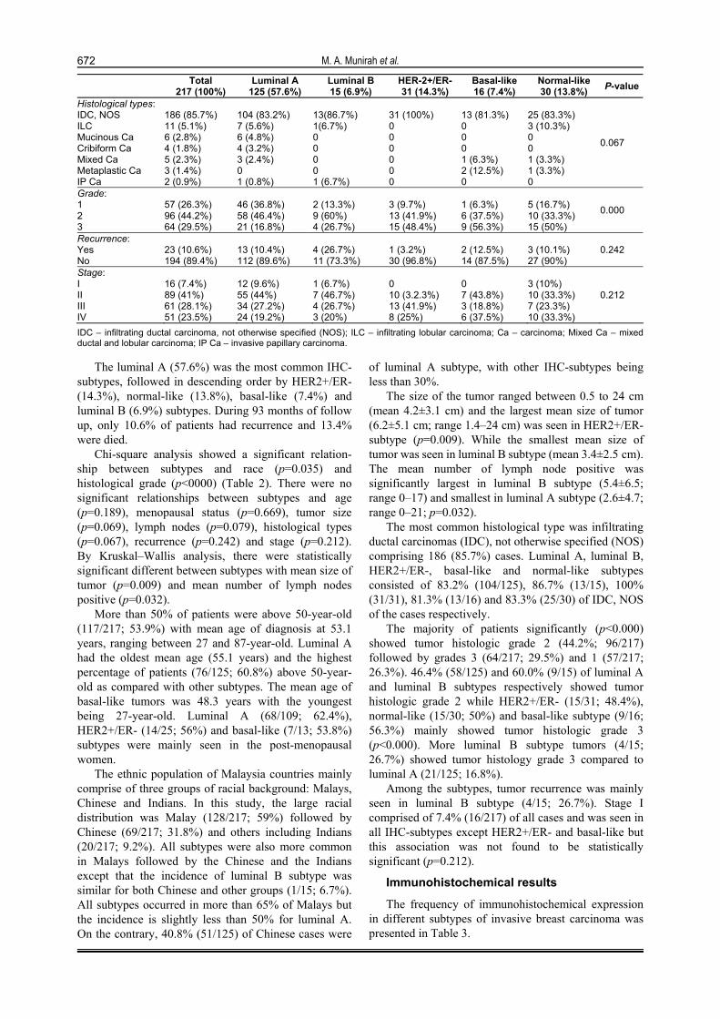
The most frequent positive immunohistochemical
expression was observed in ER (55.8%; 120/215) (Figure 1A) and the least frequent positive expression was seen in EGFR (1.5%; 3/194) (Figure 1B).
There was a significant association between ER, PR, HER2, CK5/6, CK8/18 and p53 with all of the tumor subtypes. However, no significant correlation could be found for EGFR and Ki67.
There were 25 (11.5%) cases of HER2 scoring 2+ that were subsequently assessed by fluorescence in situ hybridization (FISH) [6].
Twenty-two of 25 (88%) cases were amplified and three were not. The final total cases positive for HER2 was of 21.2% (46/217).
The ER (Figure 1A) was mainly expressed in 90.3% (112/124) in luminal A and 53.3% (8/15) in luminal B of all cases (p<0.000).
While EGFR (Figure 1B) was expressed in 7.7% (1/13) basal-like, 3.4% (1/29) HER2+/ER- and 0.9% (1/113) luminal A.
PR (Figure 1C) was expressed in 64.0% (80/125) in luminal A and 73.3% (11/15) in luminal B subtypes of all cases.
HER2 (Figure 1D) was expressed only in luminal B (100%; 15/15) and HER2+/ER- (100%; 31/31) but not in luminal A, basal-like and normal-like subtypes.
CK5/6 (Figure 1E) was positive in 100% (16/16), 19.4% (6/31), 12.2% (15/123), 7.1% (1/14) of basal-like, HER2+/ER-, luminal A and luminal B subtypes respectively (p<0.000).
CK8/18 (Figure 1F) positive was seen in all subtypes (p=0.009) whereas p53 (Figure 1G) was expressed in 93.8% (15/16), 80% (12/15), 54.8% (17/31), 53.3% (16/30) and 48% (60/125) of basal-like, luminal B, HER2+/ER-, normal-like and luminal A respectively (p=0.003).
Luminal B (Figure 1H) showed high expression of Ki67 (10/15; 66.7%) followed by basal-like (9/16; 56.3%), HER2+/ER- (13/31; 41.9%), luminal A (47/122; 38.5%) and normal-like (10/30; 34.5%) sub-types (p=0.250).
Figure 1I showed negative staining of the breast carcinoma.
Histologic features of basal-like subtype
The most common histological type was infiltrating ductal carcinoma, NOS (81.3%) and most were in grade 3 (56.3%).
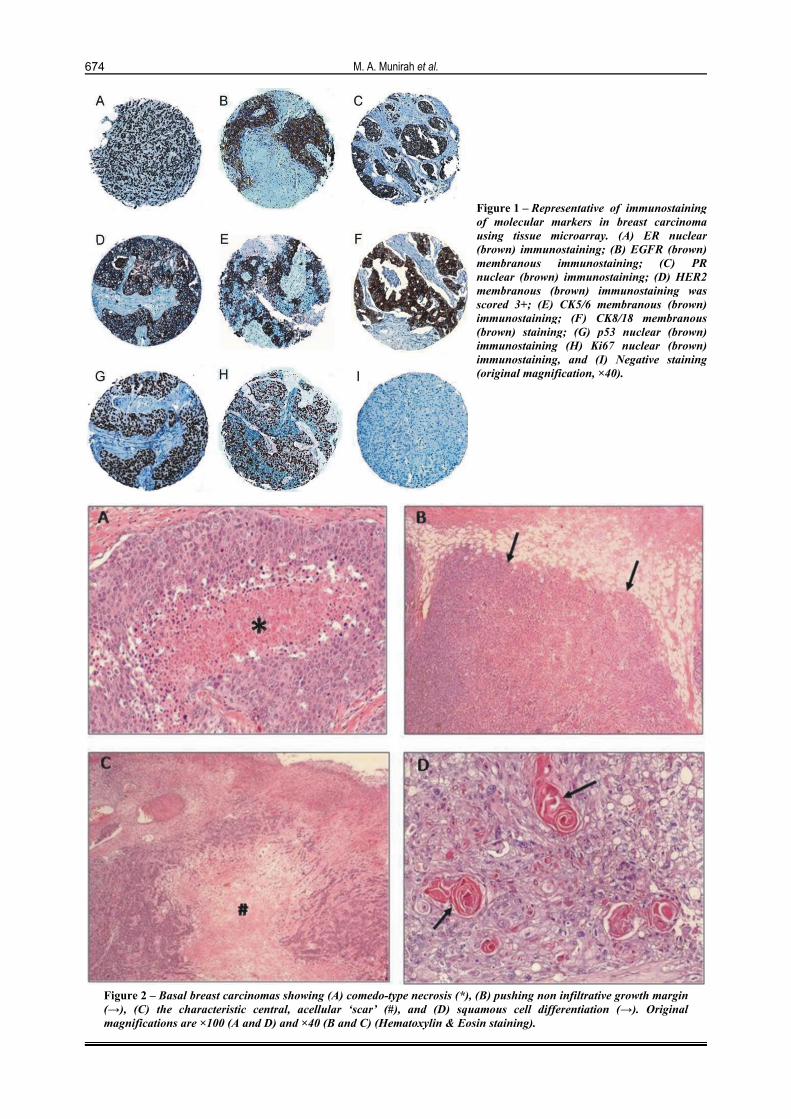
Apart from IDC, basal-like subtype tumors also consisted of metaplastic carcinoma (2/16; 12.5%) and mixed ductal and lobular carcinoma (1/16; 6.3%). Majority of basal-like subtypes showed comedo-type necrosis (10/16; 68.8%) (Figure 2A), infiltrative margin (13/16; 81.3%) and scanty to moderate inflammation (8/16; 50%).
Only a few cases demonstrated solid pattern (5/16; 31.3%), pushing margin (3/16; 18.8%) (Figure 2B), central acellular scar (1/16; 6.3%) (Figure 2C) and squamous cell differentiation (2/16; 12.5%) (Figure 2D).
M. A. Munirah et al.
674
Figure 1 – Representative of immunostaining of molecular markers in breast carcinoma using tissue microarray. (A) ER nuclear (brown) immunostaining; (B) EGFR (brown) membranous immunostaining; (C) PR nuclear (brown) immunostaining; (D) HER2 membranous (brown) immunostaining was scored 3+; (E) CK5/6 membranous (brown) immunostaining; (F) CK8/18 membranous (brown) staining; (G) p53 nuclear (brown) immunostaining (H) Ki67 nuclear (brown) immunostaining, and (I) Negative staining (original magnification, ×40).
Figure 2 – Basal breast carcinomas showing (A) comedo-type necrosis (*), (B) pushing non infiltrative growth margin (→), (C) the characteristic central, acellular ‘scar’ (#), and (D) squamous cell differentiation (→). Original magnifications are ×100 (A and D) and ×40 (B and C) (Hematoxylin & Eosin staining).

Identification of different subtypes of breast cancer using tissue microarray
675
Discussion
In this present study, luminal A (57.6%) was the most common IHC-subtypes and fall within the range of other studies from 27% up to 73.4% whereas other subtypes were between 5.2% to 32% [9, 13, 22, 23].
We have found significant correlation between breast carcinoma subtypes with ethnicity and histological grade. However, there was no significant association between breast carcinoma subtypes with age, menopausal status, tumor size, lymph nodes status and stage consistent with that previously reported [9, 24, 25].
We have also found that luminal A subtypes of breast cancer were usually observed in patients older than 50-year-old even though it was not significantly associated. On the other hand, 35.5% of patients was found to be 70 years and above [23] and had occurred in postmenopausal women [8, 26].
The Carolina Breast Cancer Study showed that the luminal A subtype was more frequent among post menopausal African American (59%) or non-African American (54%) women [8]. We, on the other hand have found that luminal B subtype was the most frequent among Malay patients regardless of menopausal status.
Our study have also found that the mean number and percentages of positive lymph nodes were high in HER2+/ER- and/or luminal B subtypes, similar to that previously reported [8, 11, 22, 25]. In keeping with previous studies [6, 8, 23, 25], we had also observed the low percentage of positive lymph nodes seen in basal-like subtype cancers. Foulkes WD et al. (2004) have found that CK5/6-positive cancers were less likely to be node positive when the tumor is large in size, suggesting a preferred route of metastatic spread for basal cancers [27].
Prior to immunohistochemistry and expression profiling, breast cancer was classified mainly by histology and grade [26]. In this study, we have shown that all cases of invasive ductal carcinoma also displayed HER2+/ER-, suggesting that breast cancer could be classified according to the expression of immunohistochemistry. The two of three (66.7%) cases of metaplastic carcinoma showed basal like subtype features in our study were also similar to previous reports by Kim MJ et al. (2006) [11].
We had shown, similar to other studies, that the majority of histological low-grade (1 and 2) tumors also displayed features of luminal A and B, and that high-grade (3) tumors were mainly of HER2+/ER- and basal-like subtypes [8, 22, 23, 25, 26]. Therefore, the features for all subtypes were in accordance with the multistep model for breast cancer progression proposed by Simpson PT et al. (2005) which was based on findings that tumors with hormone receptor-negative and frequently positive for either HER2 or basal markers usually progressed towards histological grade 3 [28].
In keeping with another study [23], we had also found that basal-like and HER2+/ER- subtypes were observed in stage III and IV tumors, although this was not found to be significant.
Based on patterns of protein expression, luminal subtypes are classified into luminal A and luminal B in
which both express ER, PR, BCL2, CK8 and CK8/18 [13, 29, 30]. Both luminal subtypes may also show low expression of basal molecular markers (CK5, CK5/6 and CK17) as well as other molecular markers (EGFR and vimentin) [13, 30].
Compared to luminal A, luminal B subtypes of breast cancer show less expression of ER and PR [13, 29, 30]. Because luminal B subtype also expresses genes associated with HER2 such as ERBB2 and GRB7 [31], HER2 has been used to distinguish luminal A and luminal B. However, HER2 positivity was only seen in 30% of luminal B subtype tumors, indicating that HER2 alone was not sufficiently sensitive in identifying this type of tumors [31]. Luminal B showed high expression of Ki67 compared to luminal A but was not significant in this study. Cheang MCU et al. (2009) found that Ki67 together with ER, PR and HER2 could be used to distinguish luminal A and luminal B [31].
HER2+/ER- and basal-like subtypes share similar protein profiles with the exception that HER2 was shown to be expressed by HER2+/ER- subtype and those basal molecular markers such as CK5, CK17, c-kit, EGFR and vimentin were expressed by basal-like subtype tumors [13, 30]. In addition, HER2+/ER- and basal-like subtypes showed either less expression or negative for ER and PR [13, 30] and this was supported by findings of cDNA-microarray studies [2, 3].
This study showed positive frequency of immuno-histochemical expression of HER2 was 21.2% (46/217) of the cases and was within the range of other studies [3, 6, 11]. Past study concluded that HER2 status
determination was most efficient by using immunohistochemistry as a method of choice, with FISH performed in cases with moderate (2+) staining [32]. In the present study, 25 (11.5%) tumors were scored 2+ by immunohisto-chemistry; of these 88% were associated with HER2/neu gene amplification by FISH. This study employed an IHC staining of 3+ (uniform, intense membrane staining of 30% of invasive tumor cells) that could explain this high percentage [20]. An earlier study had showed thirteen per cent of tumors were IHC 2+ and overall 48% of these were FISH positive but this proportion varied markedly between the centers [33].
Basal molecular markers were not only observed in basal-like subtypes [11, 12, 29, 34] but also in other subtypes with the exception of normal-like. In this study, there were more cases of HER2+/ER- subtypes with positive CK5/6 compare to luminal subtype, consistent with a recent finding [34]. Banerjee S et al. (2006) found that basal-like subtypes had expressed ER, PR and HER2 in 18.4%, 20.4% and 8.2% of cases, respectively [5]. A study by Laakso M et al. (2006) found that two subtypes, which were uniformly positive type (basal) and a partially positive type (basoluminal), could be distinguished based on basal cytokeratin expression [35]. Both basal and basoluminal subtypes showed high-grade histology and are hormone receptor negative [35]. However, expression of Ki67, vimentin and c-kit were more frequently expressed in basal tumors whereas amplification of HER2 was more characteristic of the basoluminal subgroup. Therefore,

M. A. Munirah et al.
676
in view of these previous observations, we believe that the 19.4% of HER2+/ER- subtype tumors with CK5/6 positivity identified in our study are suggestive of basoluminal subtype.
Normal-like or multiple marker negative (MMN) subtype tumors have been shown to be negative for basal markers such as CK5 and CK7 as well as negative for other molecular markers which include EGFR, c-kit, vimentin and Ki67 [13, 30]. We have found that the majority of normal-like subtype tumors express CK8/18 with absence of CK5/6 suggesting that these cells were most probably derived from luminal gland cell.
This study has also showed a significant relationship among all subtypes with p53, in keeping with a previous report [7]. Locally advanced breast cancer with TP53 gene mutations often showed resistance to either chemotherapy or radiotherapy [36]. TP53 mutations were more frequent in basal-like and HER2+/ER- subtypes compared to luminal subtype tumors [2, 8]. Overexpression of p53 was also more frequent in cancers associated with either BRCA1 or BRCA2 germline mutations [37].
In our study, overexpression of CK8/18 was significantly seen in all tumor subtypes and accordingly was deemed unsuitable for specific luminal marker and tumor classification [11]. The majority of normal-like and basal-like subtypes expressed luminal keratins (CK8/18), albeit at lower levels than those found in other subtypes [12]. The expression of CK8/18 with absence of basal markers in breast cancer indicated that the tumor originated from luminal glandular cell [1]. Foulkes WD et al. (2003) hypothesized that BRCA1 wild type may act as stem cell regulator and thus promote differentiation of gland epithelium in normal breast tissue [38]. As a consequent, tumors of BRCA1 mutation expressed less CK8/18 [39].
Russo J et al. (2001) [40] suggested that breast cells in women with BRCA1 mutation failed to differentiate to acinus breast cell resulting in increased risk of developing breast cancer [38]. Basal-like subtype tumors with BRCA1 mutation were shown to be negative for ER, PR and HER2 but positive for p53 and basal markers such as CK5/6 or CK14 [7, 38, 39]. In addition, CK5/6 positive tumors were also five times (5×) more likely to be associated with BRCA1 mutations [27].
In this study, we have also observed the morphologic features of basal-like subtype tumors, which include comedo-type necrosis, pushing invasion borders, central scar and squamous cell differentiation [12, 41].
Conclusions
Overall, all subtypes were significantly associated with race, mean size tumor, mean number of lymph nodes, histological grade and all biomarkers except EGFR and Ki67. We have found that luminal A subtype tumors are likely to be associated with low histological grade and low expression of p53. By contrast, basal-like subtype was significantly associated with Malay ethnic background, high histological grade and high expression of p53. All subtypes showed high expression of CK8/18, indicating its inappropriate role in distinguishing tumor subtypes.
Acknowledgements Financial support for this study was provided by
IRPA fund and approval for this study was obtained from Research and Ethical Committee of Universiti Kebangsaan Malaysia Medical Centre.
References [1] Böcker W, Moll R, Poremba C, Holland R, Van Diest PJ,
Dervan P, Bürger H, Wai D, Ina Diallo R, Brandt B, Herbst H, Schmidt A, Lerch MM, Buchwallow IB, Common adult stem cells in the human breast give rise to glandular and myoepithelial cell lineages: a new cell biological concept, Lab Invest, 2002, 82(6):737–746.
[2] Sørlie T, Perou CM, Tibshirani R, Aas T, Geisler S, Johnsen H, Hastie T, Eisen MB, van de Rijn M, Jeffrey SS, Thorsen T, Quist H, Matese JC, Brown PO, Botstein D, Eystein Lønning P, Børresen-Dale AL, Gene expression patterns of breast carcinomas distinguish tumor subclasses with clinical implications, Proc Natl Acad Sci U S A, 2001, 98(19):10869–10874.
[3] Nielsen TO, Hsu FD, Jensen K, Cheang M, Karaca G, Hu Z, Hernandez-Boussard T, Livasy C, Cowan D, Dressler L, Akslen LA, Ragaz J, Gown AM, Gilks CB, van de Rijn M, Perou CM, Immunohistochemical and clinical character-ization of the basal-like subtype of invasive breast carcinoma, Clin Cancer Res, 2004, 10(16):5367–5374.
[4] van de Rijn M, Perou CM, Tibshirani R, Haas P, Kallioniemi O, Kononen J, Torhorst J, Sauter G, Zuber M, Köchli OR, Mross F, Dieterich H, Seitz R, Ross D, Botstein D, Brown P, Expression of cytokeratins 17 and 5 identifies a group of breast carcinomas with poor clinical outcome, Am J Pathol, 2002, 161(6):1991–1996.
[5] Banerjee S, Reis-Filho JS, Ashley S, Steele D, Ashworth A, Lakhani SR, Smith IE, Basal-like breast carcinomas: clinical outcome and response to chemotherapy, J Clin Pathol, 2006, 59(7):729–735.
[6] Cheang MCU, Voduc D, Bajdik C, Leung S, McKinney S, Chia SK, Perou CM, Nielsen TO, Basal-like breast cancer defined by five biomarkers has superior prognostic value than triple-negative phenotype, Clin Cancer Res, 2008, 14(5):1368–1376.
[7] Bertucci F, Finetti P, Rougemont J, Charafe-Jauffret E, Cervera N, Tarpin C, Nguyen C, Xerri L, Houlgatte R, Jacquemier J, Viens P, Birnbaum D, Gene expression profiling identifies molecular subtypes of inflammatory breast cancer, Cancer Res, 2005, 65(6):2170–2178.
[8] Carey LA, Perou CM, Livasy CA, Dressler LG, Cowan D, Conway K, Karaca G, Troester MA, Tse CK, Edmiston S, Deming SL, Geradts J, Cheang MC, Nielsen TO, Moorman PG, Earp HS, Millikan RC, Race, breast cancer subtypes, and survival in the Carolina Breast Cancer Study, JAMA, 2006, 295(21):2492–2502.
[9] Carey LA, Dees E, Sawyer L, Gatti L, Moore D, Collichio F, Ollila DW, Sartor CI, Graham ML, Perou CM, The triple negative paradox: primary tumor chemosensitivity of breast cancer subtypes, Clin Cancer Res, 2007, 13(8):2329–2334.
[10] Tan DSP, Marchió C, Jones RL, Savage K, Smith IE, Dowsett M, Reis-Filho JS, Triple negative breast cancer: molecular profiling and prognostic impact in adjuvant anthracycline-treated patients, Breast Cancer Res Treat, 2008, 111(1):27–44.
[11] Kim MJ, Ro JY, Ahn SH, Kim HH, Kim SB, Gong G, Clinicopathologic significance of the basal-like subtype of breast cancer: a comparison with hormone receptor and Her2/neu-overexpressing phenotypes, Hum Pathol, 2006, 37(9):1217–1226.
[12] Livasy CA, Karaca G, Nanda R, Tretiakova MS, Olopade OI, Moore DT, Perou CM, Phenotypic evaluation of the basal-like subtype of invasive breast carcinoma, Mod Pathol, 2006, 19(2):264–271.
[13] Diallo-Danebrock R, Ting E, Gluz O, Herr A, Mohrmann S, Geddert H, Rody A, Schaefer KL, Baldus SE, Hartmann A, Wild PJ, Burson M, Gabbert HE, Nitz U, Poremba C, Protein expression profiling in high-risk breast cancer patients treated with high-dose or conventional dose-dense chemotherapy, Clin Cancer Res, 2007, 13(2 Pt 1):488–497.

Identification of different subtypes of breast cancer using tissue microarray
677[14] Zainal A, Zainudin M, Nor Saleha I, Malaysian cancer
statistics data and figure Peninsular Malaysia, National Cancer Registry, 2006, http://www.makna.org.my/PDF/ MalaysiaCancerStatistics.pdf, Last accessed 14/1/2011.
[15] Tavassoli F, Devilee P (eds), World Health Organization Classification of Tumours: Pathology and genetics of tumours of the breast and female genital organs, IARC Press, Lyon, 2003.
[16] Elston CW, Ellis IO, Pathological prognostic factors in breast cancer. I. The value of histological grade in breast cancer: experience from a large study with long-term follow-up, Histopathology, 1991, 19(5):403–410.
[17] American Joint Committee on Cancer, AJCC cancer staging handbook, 2010, www.springer.com/medicine/surgery/ cancer+staging?SGWID=0-40654-0-0-0, Last accessed on 14/1/2011.
[18] Kononen J, Bubendorf L, Kallioniemi A, Bärlund M, Schraml P, Leighton S, Torhorst J, Mihatsch MJ, Sauter G, Kallioniemi OP, Tissue microarrays for high-throughput molecular profiling of tumor specimens, Nat Med, 1998, 4(7):844–847.
[19] Noranizah W, Siti-Aishah MA, Munirah MA, Norazlin MH, Rohaizak M, Naqiyah I, Sharifah NA, Das S, Immuno-histochemical expression of vascular endothelial growth factor (VEGF) and p53 in breast lesions, Clin Ter, 2010, 161(2):129–137.
[20] Wolff AC, Hammond ME, Schwartz JN, Hagerty KL, Allred DC, Cote RJ, Dowsett M, Fitzgibbons PL, Hanna WM, Langer A, McShane LM, Paik S, Pegram MD, Perez EA, Press MF, Rhodes A, Sturgeon C, Taube SE, Tubbs R, Vance GH, van de Vijver M, Wheeler TM, Hayes DF; American Society of Clinical Oncology; College of American Pathologists, American Society of Clinical Oncology/College of American Pathologists guideline recommendations for human epidermal growth factor receptor 2 testing in breast cancer, J Clin Oncol, 2007, 25(1):118–145.
[21] Potemski P, Kusinska R, Watala C, Pluciennik E, Bednarek AK, Kordek R, Prognostic relevance of basal cytokeratin expression in operable breast cancer, Oncology, 2005, 69(6):478–485.
[22] Tamimi RM, Baer HJ, Marotti J, Galan M, Galaburda L, Fu Y, Deitz AC, Connolly JL, Schnitt SJ, Colditz GA, Collins LC, Comparison of molecular phenotypes of ductal carcinoma in situ and invasive breast cancer, Breast Cancer Res, 2008, 10(4):R67.
[23] Spitale A, Mazzola P, Soldini D, Mazzucchelli L, Bordoni A, Breast cancer classification according to immunohisto-chemical markers: clinicopathologic features and short-term survival analysis in a population-based study from the South of Switzerland, Ann Oncol, 2009, 20(4):628–635.
[24] Rouzier R, Perou CM, Symmans WF, Ibrahim N, Cristofanilli M, Anderson K, Hess KR, Stec J, Ayers M, Wagner P, Morandi P, Fan C, Rabiul I, Ross JS, Hortobagyi GN, Pusztai L, Breast cancer molecular subtypes respond differently to preoperative chemotherapy, Clin Cancer Res, 2005, 11(16):5678–5685.
[25] Yang XR, Sherman ME, Rimm DL, Lissowska J, Brinton LA, Peplonska B, Hewitt SM, Anderson WF, Szeszenia-Dabrowska N, Bardin-Mikolajczak A, Zatonski W, Cartun R, Mandich D, Rymkiewicz G, Ligaj M, Lukaszek S, Kordek R, García-Closas M, Differences in risk factors for breast cancer molecular subtypes in a population-based study, Cancer Epidemiol Biomarkers Prev, 2007, 16(3):439–443.
[26] Nofech-Mozes S, Trudeau M, Kahn HK, Dent R, Rawlinson E, Sun P, Narod SA, Hanna WM, Patterns of recurrence in the basal and non-basal subtypes of triple-negative breast cancers, Breast Cancer Res Treat, 2009, 118(1):131–137.
[27] Foulkes WD, Brunet JS, Stefansson IM, Straume O, Chappuis PO, Bégin LR, Hamel N, Goffin JR, Wong N, Trudel M, Kapusta L, Porter P, Akslen LA, The prognostic implication of the basal-like (cyclin Ehigh/p27low/p53+/ glomeruloid-microvascular-proliferation+) phenotype of BRCA1-related breast cancer, Cancer Res, 2004, 64(3):830–835.
[28] Simpson PT, Reis-Filho JS, Gale T, Lakhani SR, Molecular evolution of breast cancer, J Pathol, 2005, 205(2):248–254.
[29] Matos I, Dufloth R, Alvarenga M, Zeferino LC, Schmitt F, p63, cytokeratin 5, and P-cadherin: three molecular markers to distinguish basal phenotype in breast carcinomas, Virchows Arch, 2005, 447(4):688–694.
[30] Honrado E, Osorio A, Milne RL, Paz MF, Melchor L, Cascón A, Urioste M, Cazorla A, Díez O, Lerma E, Esteller M, Palacios J, Benítez J, Immunohistochemical classification of non-BRCA1/2 tumors identifies different groups that demonstrate the heterogeneity of BRCAX families, Mod Pathol, 2007, 20(12):1298–1306.
[31] Cheang MCU, Chia SK, Voduc D, Gao D, Leung S, Snider J, Watson M, Davies S, Bernard PS, Parker JS, Perou CM, Ellis MJ, Nielsen TO, Ki67 index, HER2 status, and prognosis of patients with luminal B breast cancer, J Natl Cancer Inst, 2009, 101(10):736–750.
[32] Yaziji H, Goldstein LC, Barry TS, Werling R, Hwang H, Ellis GK, Gralow JR, Livingston RB, Gown AM, HER-2 testing in breast cancer using parallel tissue-based methods, JAMA, 2004, 291(16):1972–1977.
[33] Dowsett M, Bartlett J, Ellis IO, Salter J, Hills M, Mallon E, Watters AD, Cooke T, Paish C, Wencyk PM, Pinder SE, Correlation between immunohistochemistry (Hercep Test) and fluorescence in situ hybridization (FISH) for HER-2 in 426 breast carcinomas from 37 centres, J Pathol, 2003, 199(4):418–423.
[34] Pintens S, Neven P, Drijkoningen M, Van Belle V, Moerman P, Christiaens MR, Smeets A, Wildiers H, Van den Bempt I, Triple negative breast cancer: a study from the point of view of basal CK5/6 and HER-1, J Clin Pathol, 2009, 62(7):624–628.
[35] Laakso M, Tanner M, Nilsson J, Wiklund T, Erikstein B, Kellokumpu-Lehtinen P, Malmström P, Wilking N, Bergh J, Isola J, Basoluminal carcinoma: a new biologically and prognostically distinct entity between basal and luminal breast cancer, Clin Cancer Res, 2006, 12(14 Pt 1):4185–4191.
[36] Geisler S, Lønning PE, Aas T, Johnsen H, Fluge O, Haugen DF, Lillehaug JR, Akslen LA, Børresen-Dale AL, Influence of TP53 gene alterations and c-erbB-2 expression on the response to treatment with doxorubicin in locally advanced breast cancer, Cancer Res, 2001, 61(6):2505–2512.
[37] Greenblatt MS, Chappuis PO, Bond JP, Hamel N, Foulkes WD, TP53 mutations in breast cancer associated with BRCA1 or BRCA2 germ-line mutations: distinctive spectrum and structural distribution, Cancer Res, 2001, 61(10):4092–4097.
[38] Foulkes WD, Stefansson IM, Chappuis PO, Bégin LR, Goffin JR, Wong N, Trudel M, Akslen LA, Germline BRCA1 mutations and a basal epithelial phenotype in breast cancer, J Natl Cancer Inst, 2003, 95(19):1482–1485.
[39] Laakso M, Loman N, Borg A, Isola J, Cytokeratin 5/14-positive breast cancer: true basal phenotype confined to BRCA1 tumors, Mod Pathol, 2005, 18(10):1321–1328.
[40] Russo J, Lynch H, Russo IH, Mammary gland architecture as a determining factor in the susceptibility of the human breast to cancer, Breast J, 2001, 7(5):278–291.
[41] Rakha EA, Reis-Filho JS, Ellis IO, Basal-like breast cancer: a critical review, J Clin Oncol, 2008, 26(15):2568–2581.
Corresponding author M. A. Siti-Aishah, Professor, Department of Pathology, Faculty of Medicine, Universiti Kebangsaan Malaysia Medical Centre, Jalan Yaacob Latiff, Bandar Tun Razak, 56000 Cheras, Kuala Lumpur, Malaysia; Phone 006–03–91455363, e-mail: [email protected] Received: January 21st, 2011 Accepted: May 30th, 2011


![Rom J Morphol Embryol 2013, 54(1):205–210 R J M E CASE ... · PDF fileRom J Morphol Embryol 2013, 54(1):205–210 ISSN ... project report CEEX 68/2006 [3] shows that 650 000 ...](https://static.fdocuments.us/doc/165x107/5aae038b7f8b9a07498b87b2/rom-j-morphol-embryol-2013-541205210-r-j-m-e-case-j-morphol-embryol-2013.jpg)