RNA Splicing
17
RNA Splicing
-
Upload
amna-jalil -
Category
Education
-
view
480 -
download
11
Transcript of RNA Splicing

RNA Splicing

Contents
Introduction
Discovery
Early Studies in Bacteria
Splicing Pathways
Spliceosomal Pathway
Introns
Formation and Activity
Major Splicesome
Minor Splicesome
Trans-splicing
Self-splicing
tRNA Splicing
Evolution
Biochemical Mechanism
Alternative Splicing
Experimental Manipulation of Splicing
Splicing Errors
Protein Splicing

RNA Splicing
Introduction
In molecular biology and genetics, splicing is a modification of the nascent pre-messenger RNA
(pre-mRNA) transcript in which introns are removed and exons are joined. For nuclear encoded
genes, splicing takes place within the nucleus after or concurrently with transcription. Splicing is
needed for the typical eukaryotic messenger RNA (mRNA) before it can be used to produce a
correct protein through translation. For many eukaryotic introns, splicing is done in a series of
reactions which are catalyzed by the spliceosome, a complex of small nuclear ribonucleoproteins
(snRNPs), but there are also self-splicing introns.
Figure: Simple illustration of exons and introns in pre-mRNA and the formation of mature mRNA by splicing

Discovery
RNA splicing was initially discovered in the 1970s, overturning years of thought in the field of
gene expression.
Early Studies in Bacteria
Gene regulation was first studied most thoroughly in relatively simple bacterial systems. Most
bacterial RNA transcripts do not undergo splicing; these transcripts are said to be colinear, with
DNA directly encoding them. In other words, there is a one-to-one correspondence of bases
between the gene and the mRNA transcribed from the gene (excepting 5′ and 3′ noncoding
regions).
However, in 1977, several groups of researchers who were working with adenoviruses that infect
and replicate in mammalian cells obtained some surprising results. These scientists identified a
series of RNA molecules that they termed "mosaics," each of which contained sequences from
noncontiguous sites in the viral genome. These mosaics were found late in viral infection.
Studies of early infection revealed long primary RNA transcripts that contained all of the
sequences from the late RNAs, as well as what came to be called the intervening sequences
(introns).
Subsequent to the adenoviral discovery, introns were found in many other viral and eukaryotic
genes, including those for hemoglobin and immunoglobulin. Splicing of RNA transcripts was
then observed in several in vitro systems derived from eukaryotic cells, including removal of
introns from transfer RNA in yeast cell-free extracts. These observations solidified the
hypothesis that splicing of large initial transcripts did, in fact, yield the mature mRNA. Other
hypotheses proposed that the DNA template in some way looped or assumed a secondary
structure that allowed transcription from noncontiguous regions.
Splicing Pathways
Several methods of RNA splicing occur in nature; the type of splicing depends on the structure
of the spliced intron and the catalysts required for splicing to occur.

Spliceosomal splicing
Self-splicing
tRNA splicing
Spliceosomal Pathway
Introns
The word intron is derived from the term intragenic region, that is, a region inside a gene. The
term intron refers to both the DNA sequence within a gene and the corresponding sequence in
the unprocessed RNA transcript. As part of the RNA processing pathway, introns are removed
by RNA splicing either shortly after or concurrent with transcription. Introns are found in the
genes of most organisms and many viruses. They can be located in a wide range of genes,
including those that generate proteins, ribosomal RNA (rRNA), and transfer RNA (tRNA).
Spliceosomal introns often reside within the sequence of eukaryotic protein-coding genes.
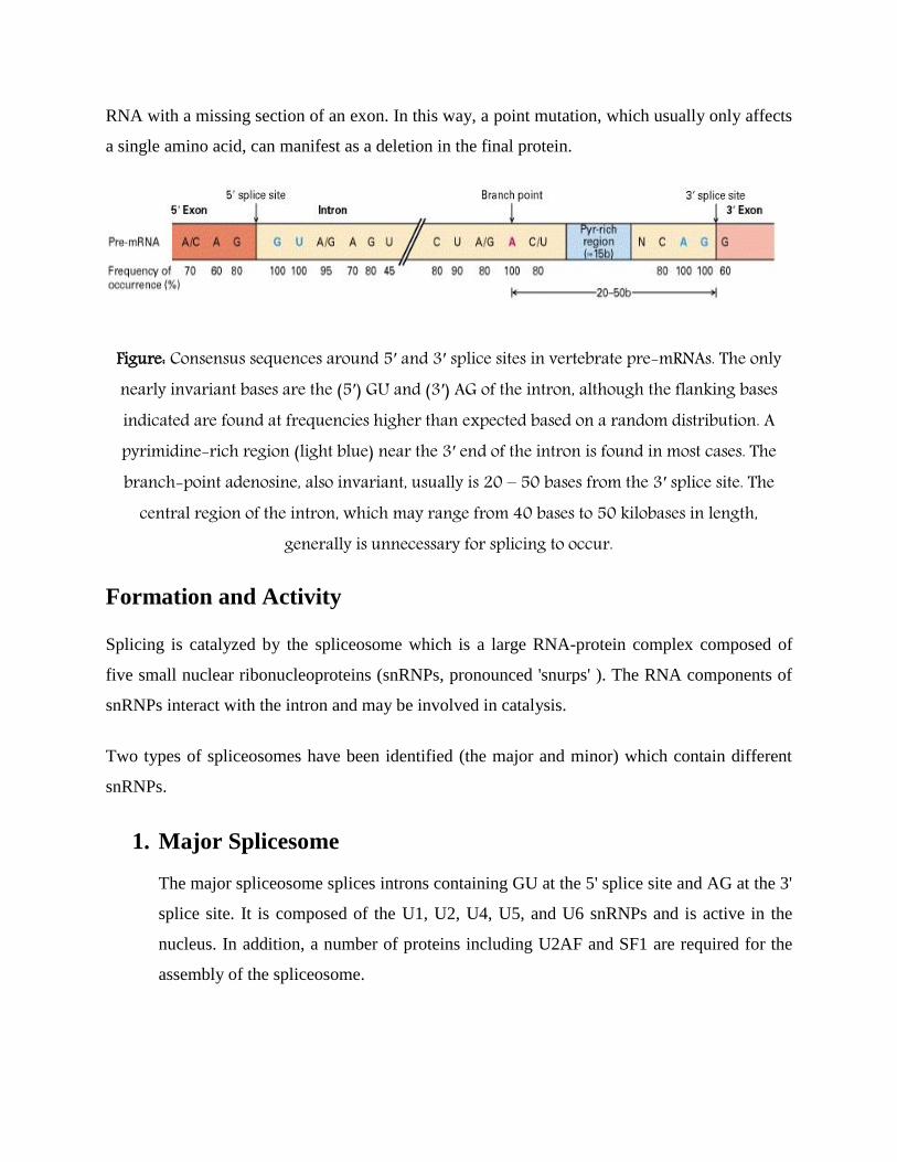
Within the intron, a donor site (5' end of the intron), a branch site (near the 3' end of the intron)
and an acceptor site (3' end of the intron) are required for splicing. The splice donor site includes
an almost invariant sequence GU at the 5' end of the intron, within a larger, less highly conserved
region. The splice acceptor site at the 3' end of the intron terminates the intron with an almost
invariant AG sequence. Upstream (5'-ward) from the AG there is a region high in pyrimidines (C
and U), or polypyrimidine tract. Upstream from the polypyrimidine tract is the branchpoint,
which includes an adenine nucleotide. The consensus sequence for an intron (in IUPAC nucleic
acid notation) is: M-A-G-[cut]-G-U-R-A-G-U (donor site) ... intron sequence ... C-U-R-[A]-Y
(branch sequence 20-50 nucleotides upstream of acceptor site) ... Y-rich-N-C-A-G-[cut]-G
(acceptor site). However, it is noted that the specific sequence of intronic splicing elements and
the number of nucleotides between the branchpoint and the nearest 3’ acceptor site affect splice
site selection.
Also, point mutations in the underlying DNA or errors during transcription can activate a cryptic
splice site in part of the transcript that usually is not spliced. This results in a mature messenger
RNA with a missing section of an exon. In this way, a point mutation, which usually only affects
a single amino acid, can manifest as a deletion in the final protein.
Figure: Consensus sequences around 5′ and 3′ splice sites in vertebrate pre-mRNAs. The only nearly invariant bases are the (5′) GU and (3′) AG of the intron, although the flanking bases indicated are found at frequencies higher than expected based on a random distribution. A pyrimidine-rich region (light blue) near the 3′ end of the intron is found in most cases. The branch-point adenosine, also invariant, usually is 20 – 50 bases from the 3′ splice site. The
central region of the intron, which may range from 40 bases to 50 kilobases in length, generally is unnecessary for splicing to occur.
Formation and Activity
Splicing is catalyzed by the spliceosome which is a large RNA-protein complex composed of
five small nuclear ribonucleoproteins (snRNPs, pronounced 'snurps' ). The RNA components of
snRNPs interact with the intron and may be involved in catalysis.
Two types of spliceosomes have been identified (the major and minor) which contain different
snRNPs.
1. Major Splicesome
The major spliceosome splices introns containing GU at the 5' splice site and AG at the 3'
splice site. It is composed of the U1, U2, U4, U5, and U6 snRNPs and is active in the
nucleus. In addition, a number of proteins including U2AF and SF1 are required for the
assembly of the spliceosome.

E Complex-U1 binds to the GU sequence at the 5' splice site, along with
accessory proteins/enzymes ASF/SF2, U2AF (binds at the Py-AG site), SF1/BBP
(BBP=Branch Binding Protein);
A Complex-U2 binds to the branch site and ATP is hydrolyzed;
B1 Complex-U5/U4/U6 trimer binds, and the U5 binds exons at the 5' site, with
U6 binding to U2;
B2 Complex-U1 is released, U5 shifts from exon to intron and the U6 binds at the
5' splice site;
C1 Complex-U4 is released, U6/U2 catalyzes transesterification, that make 5' end
of introns ligate to the A on intron and form a lariat, U5 binds exon at 3' splice
site, and the 5' site is cleaved, resulting in the formation of the lariat;
C2 Complex-U2/U5/U6 remain bound to the lariat, and the 3' site is cleaved and
exons are ligated using ATP hydrolysis. The spliced RNA is released and the
lariat debranches.
This type of splicing is termed canonical splicing or termed the lariat pathway, which accounts
for more than 99% of splicing. By contrast, when the intronic flanking sequences do not follow
the GU-AG rule, noncanonical splicing is said to occur.

Figure: The spliceosomal splicing cycle The splicing snRNPs (U1, U2, U4, U5, and U6) associate with the pre-mRNA and with each other in an ordered sequence to form the spliceosome. This large ribonucleoprotein complex then catalyzes the two transesterification reactions that result
in splicing of the exons (light and dark red) and excision of the intron (blue) as a lariat structure. Although ATP hydrolysis is not required for the transesterification reactions, it is
thought to provide the energy necessary for rearrangements of the spliceosome structure that occur during the cycle. Note that the snRNP proteins in the spliceosome are distinct from the
hnRNP proteins discussed earlier. In higher eukaryotes, the association of U2 snRNP with pre-mRNA is assisted by an hnRNP protein called U2AF, which binds to the pyrimidine-rich region
near the 3′ splice site. U2AF also probably interacts with other proteins required for splicing through a domain containing repeats of the dipeptide serine-arginine (the SR motif). The
branch-point A in pre-mRNA is indicated in boldface.

2. Minor Splicesome
The minor spliceosome is very similar to the major spliceosome, however it splices out rare
introns with different splice site sequences. While the minor and major spliceosomes contain the
same U5 snRNP, the minor spliceosome has different, but functionally analogous snRNPs for
U1, U2, U4, and U6, which are respectively called U11, U12, U4atac, and U6atac. Unike the
major spliceosome, it is found outside the nucleus, but very close to the nuclear membrane.
Trans-splicing
Trans-splicing is a special form of RNA processing in eukaryotes where exons from two
different primary RNA transcripts are joined end to end and ligated.
In contrast "normal" (cis-) splicing processes a single molecule. That is, trans-splicing results in
an RNA transcript that came from multiple RNA polymerases on the genome. This phenomenon
can be exploited for molecular therapy to address mutated gene products.
Trans-splicing can be the mechanism behind certain oncogenic fusion transcripts. Trans-splicing
is used by certain microbial organisms, notably protozoa of the Kinetoplastae class to produce
variable surface antigens and change from one life stage to another.
Figure: Trans-splicing

Self-splicing
Self-splicing occurs for rare introns that form a ribozyme, performing the functions of the
spliceosome by RNA alone. There are three kinds of self-splicing introns, Group I, Group II and
Group III. Group I and II introns perform splicing similar to the spliceosome without requiring
any protein. This similarity suggests that Group I and II introns may be evolutionarily related to
the spliceosome. Self-splicing may also be very ancient, and may have existed in an RNA world
present before protein.
Two transesterifications characterize the mechanism in which group I introns are spliced:
1. 3'OH of a free guanine nucleoside (or one located in the intron) or a nucleotide cofactor
(GMP, GDP, GTP) attacks phosphate at the 5' splice site.
2. 3'OH of the 5'exon becomes a nucleophile and the second transesterification results in the
joining of the two exons.
The mechanism in which group II introns are spliced (two transesterification reaction like group
I introns) is as follows:
1. The 2'OH of a specific adenosine in the intron attacks the 5' splice site, thereby forming
the lariat
2. The 3'OH of the 5' exon triggers the second transesterification at the 3' splice site thereby
joining the exons together.

Figure: Group I and Group II introns splicing
tRNA Splicing
tRNA (also tRNA-like) splicing is another rare form of splicing that usually occurs in tRNA. The
splicing reaction involves a different biochemistry than the spliceosomal and self-splicing
pathways. Ribonucleases cleave the RNA and ligases join the exons together.

Figure: tRNA splicing
Evolution
Splicing occurs in all the kingdoms or domains of life, however, the extent and types of splicing
can be very different between the major divisions. Eukaryotes splice many protein-coding
messenger RNAs and some non-coding RNAs. Prokaryotes, on the other hand, splice rarely and
mostly non-coding RNAs. Another important difference between these two groups of organisms
is that prokaryotes completely lack the spliceosomal pathway.
Because spliceosomal introns are not conserved in all species, there is debate concerning when
spliceosomal splicing evolved. Two models have been proposed: the intron late and intron early
models.

Splicing diversity
Eukaryotes Prokaryotes
Spliceosomal + −
Self-splicing + +
tRNA + +
Biochemical Mechanism
Spliceosomal splicing and self-splicing involves a two-step biochemical process. Both steps
involve transesterification reactions that occur between RNA nucleotides. tRNA splicing,
however, is an exception and does not occur by transesterification.
Spliceosomal and self-splicing transesterification reactions occur via two sequential
transesterification reactions.
First, the 2'OH of a specific branchpoint nucleotide within the intron that is defined
during spliceosome assembly performs a nucleophilic attack on the first nucleotide of the
intron at the 5' splice site forming the lariat intermediate.
Second, the 3'OH of the released 5' exon then performs a nucleophilic attack at the last
nucleotide of the intron at the 3' splice site thus joining the exons and releasing the intron
lariat.

Figure: Diagram illustrating the two-step biochemistry of splicing
Alternative Splicing
In many cases, the splicing process can create a range of unique proteins by varying the exon
composition of the same mRNA. This phenomenon is then called alternative splicing.
Alternative splicing can occur in many ways. Exons can be extended or skipped, or introns can
be retained. It is estimated that 95% of transcripts from multiexon genes undergo alternative
splicing, some instances of which occur in a tissue-specific manner and/or under specific cellular
conditions. Development of high throughput mRNA sequencing technology can help quantify
the expression levels of alternatively spliced isoforms. Differential regulation patterns across
tissues and cell lineages allowed computational approaches to be developed to predict the
functions of these isoforms. Given this complexity, alternative splicing of pre-mRNA transcripts
is regulated by a system of trans-acting proteins (activators and repressors) that bind to cis-acting
sites or "elements" (enhancers and silencers) on the pre-mRNA transcript itself.

These proteins and their respective binding elements promote or reduce the usage of a particular
splice site. However, adding to the complexity of alternative splicing, it is noted that the effects
of regulatory factors are many times position-dependent. For example, a splicing factor that
serves as a splicing activator when bound to an intronic enhancer element may serve as a
repressor when bound to its splicing element in the context of an exon, and vice versa.
In addition to the position-dependent effects of enhancer and silencer elements, the location of
the branchpoint (i.e., distance upstream of the nearest 3’ acceptor site) also affects splicing. The
secondary structure of the pre-mRNA transcript also plays a role in regulating splicing, such as
by bringing together splicing elements or by masking a sequence that would otherwise serve as a
binding element for a splicing factor.
Figure: Alternative splicing produces three protein isoforms.

Experimental Manipulation of Splicing
Splicing events can be experimentally altered by binding steric-blocking antisense oligos such as
Morpholinos or Peptide nucleic acids to snRNP binding sites, to the branchpoint nucleotide that
closes the lariat, or to splice-regulatory element binding sites.
Splicing Errors
Based on data current as of 2011, one-third of all hereditary diseases are thought to have a
splicing component. Common errors include:
Mutation of a splice site resulting in loss of function of that site. Results in exposure of a
premature stop codon, loss of an exon, or inclusion of an intron.
Mutation of a splice site reducing specificity. May result in variation in the splice
location, causing insertion or deletion of amino acids, or most likely, a disruption of the
reading frame.
Displacement of a splice site, leading to inclusion or exclusion of more RNA than
expected, resulting in longer or shorter exons.
Although many splicing errors are safeguarded by a cellular quality control mechanism termed
nonsense-mediated mRNA decay (NMD), a number of splicing-related diseases also exist.
Protein Splicing
In addition to RNA, proteins can undergo splicing. Although the biomolecular mechanisms are
different, the principle is the same: parts of the protein, called inteins instead of introns, are
removed. The remaining parts, called exteins instead of exons, are fused together. Protein
splicing has been observed in a wide range of organisms, including bacteria, archaea, plants,
yeast and humans.

Figure: Protein Splicing
Reference
Lodish H, Berk A, Zipursky SL, et al. Molecular Cell Biology. 4th edition. New York: W. H.
Freeman; 2000. Section 11.2, Processing of Eukaryotic mRNA.