Review Article...
19
Hindawi Publishing Corporation Journal of Allergy Volume 2012, Article ID 789232, 18 pages doi:10.1155/2012/789232 Review Article Pathogenic Mechanisms and In Vitro Diagnosis of AERD Dirk Sch¨ afer 1 and Steffen Maune 2 1 Allergie- und Intoleranzlabor, Medizinisch Klinik III, Friedrich-Alexander-Universit¨ at Erlangen-N¨ urnberg, Gl¨ uckstraße 4a, 91054 Erlangen, Germany 2 Klinik f¨ ur HNO-Heilkunde, Kopf- und Halschirurgie, Krankenhaus Holweide, Neufelder Straße 32, 51067 K¨ oln, Germany Correspondence should be addressed to Dirk Sch¨ afer, [email protected] Received 13 January 2012; Accepted 27 February 2012 Academic Editor: A. P. Sampson Copyright © 2012 D. Sch¨ afer and S. Maune. This is an open access article distributed under the Creative Commons Attribution License, which permits unrestricted use, distribution, and reproduction in any medium, provided the original work is properly cited. Aspirin-exacerbated respiratory disease (AERD) refers to chronic rhinosinusitis, nasal polyposis, bronchoconstriction, and/or eosinophilic inflammation in asthmatics following the exposure to nonsteroidal anti-inflammatory drugs (NSAIDs). A key pathogenic mechanism associated with AERD is the imbalance of eicosanoid metabolism focusing on prostanoid and leukotriene pathways in airway mucosa as well as blood cells. Genetic and functional metabolic studies on vital and non-vital cells pointed to the variability and the crucial role of lipid mediators in disease susceptibility and their response to medication. Eicosanoids, exemplified by prostaglandin E 2 (PGE 2 ) and peptidoleukotrienes (pLT), are potential metabolic biomarkers contributing to the AERD phenotype. Also other mediators are implicated in the progress of AERD. Considering the various pathogenic mechanisms of AERD, a multitude of metabolic and genetic markers is suggested to be implicated and were introduced as potential biomarkers for in vitro diagnosis during the past decades. Deduced from an eicosanoid-related pathogenic mechanism, functional tests balancing PGE 2 and pLT as well as other eicosanoids from preferentially vital leukocytes demonstrated their applicability for in vitro diagnosis of AERD. 1. Introduction Diagnostic tests assist the physician in assuring an appro- priate treatment of the symptoms and as also the disease from which a patient is suffering. In vitro diagnostic tests are widely used in the practice of modern medicine. Nons- teroidal anti-inflammatory drugs (NSAIDs) are amongst the most frequently used drugs for the treatment of a variety of symptoms and diseases. Therefore, it is unsurprising that adverse reactions to NSAIDs arise in some patients. The diagnosis of NSAID-triggered, or exacerbated symp- toms and diseases, is usually based on medical history or provocative challenge testing [1–8]. In some cases the latter is precluded on ethical grounds (e.g., pregnancy, children of young age), anatomical alterations (e.g., massive nasal polyposis), missing compliance of the patient (e.g., asthmatic experiences and therefore fear of life threatening symptoms), unavailability of specific technical and/or medical equipment (e.g., measurement of respiratory function, appropriate emergency unit), or inadequately trained staff [7, 8]. Several approaches attempted to diagnose and confirm NSAID-triggered symptoms and related diseases by in vitro diagnostic tools during the last 110 years. Some of them were discarded, others are under investigation. In vitro tests, and the results derived when they are used, frequently play a vital role in the overall diagnostic process. To ensure that each reader has the same basic knowledge, we will describe some rudimentary background information on terminology, suggested pathomechanism, test theory and test performance before discussing the in vitro test for diagnosis of NSAID- triggered symptoms and underlying diseases in more detail. To some extent there is a known discrepancy of medical history and clinical symptoms upon exposure to NSAIDs, that is, that the provocation test shows negative outcome, whereas patients’ history documented positive reaction. This may require an additional (in vitro) diagnosis to support the physician’s decision for an appropriate treatment of the patient. Unfortunately, any diagnostic procedure, clinically and in vitro, is hampered by one or more inherent as well
Transcript of Review Article...

Hindawi Publishing CorporationJournal of AllergyVolume 2012, Article ID 789232, 18 pagesdoi:10.1155/2012/789232
Review Article
Pathogenic Mechanisms and In Vitro Diagnosis of AERD
Dirk Schafer1 and Steffen Maune2
1 Allergie- und Intoleranzlabor, Medizinisch Klinik III, Friedrich-Alexander-Universitat Erlangen-Nurnberg,Gluckstraße 4a, 91054 Erlangen, Germany
2 Klinik fur HNO-Heilkunde, Kopf- und Halschirurgie, Krankenhaus Holweide, Neufelder Straße 32, 51067 Koln, Germany
Correspondence should be addressed to Dirk Schafer, [email protected]
Received 13 January 2012; Accepted 27 February 2012
Academic Editor: A. P. Sampson
Copyright © 2012 D. Schafer and S. Maune. This is an open access article distributed under the Creative Commons AttributionLicense, which permits unrestricted use, distribution, and reproduction in any medium, provided the original work is properlycited.
Aspirin-exacerbated respiratory disease (AERD) refers to chronic rhinosinusitis, nasal polyposis, bronchoconstriction, and/oreosinophilic inflammation in asthmatics following the exposure to nonsteroidal anti-inflammatory drugs (NSAIDs). A keypathogenic mechanism associated with AERD is the imbalance of eicosanoid metabolism focusing on prostanoid and leukotrienepathways in airway mucosa as well as blood cells. Genetic and functional metabolic studies on vital and non-vital cells pointedto the variability and the crucial role of lipid mediators in disease susceptibility and their response to medication. Eicosanoids,exemplified by prostaglandin E2 (PGE2) and peptidoleukotrienes (pLT), are potential metabolic biomarkers contributing to theAERD phenotype. Also other mediators are implicated in the progress of AERD. Considering the various pathogenic mechanismsof AERD, a multitude of metabolic and genetic markers is suggested to be implicated and were introduced as potential biomarkersfor in vitro diagnosis during the past decades. Deduced from an eicosanoid-related pathogenic mechanism, functional testsbalancing PGE2 and pLT as well as other eicosanoids from preferentially vital leukocytes demonstrated their applicability forin vitro diagnosis of AERD.
1. Introduction
Diagnostic tests assist the physician in assuring an appro-priate treatment of the symptoms and as also the diseasefrom which a patient is suffering. In vitro diagnostic testsare widely used in the practice of modern medicine. Nons-teroidal anti-inflammatory drugs (NSAIDs) are amongst themost frequently used drugs for the treatment of a varietyof symptoms and diseases. Therefore, it is unsurprising thatadverse reactions to NSAIDs arise in some patients.
The diagnosis of NSAID-triggered, or exacerbated symp-toms and diseases, is usually based on medical history orprovocative challenge testing [1–8]. In some cases the latteris precluded on ethical grounds (e.g., pregnancy, childrenof young age), anatomical alterations (e.g., massive nasalpolyposis), missing compliance of the patient (e.g., asthmaticexperiences and therefore fear of life threatening symptoms),unavailability of specific technical and/or medical equipment(e.g., measurement of respiratory function, appropriateemergency unit), or inadequately trained staff [7, 8].
Several approaches attempted to diagnose and confirmNSAID-triggered symptoms and related diseases by in vitrodiagnostic tools during the last 110 years. Some of themwere discarded, others are under investigation. In vitro tests,and the results derived when they are used, frequently playa vital role in the overall diagnostic process. To ensure thateach reader has the same basic knowledge, we will describesome rudimentary background information on terminology,suggested pathomechanism, test theory and test performancebefore discussing the in vitro test for diagnosis of NSAID-triggered symptoms and underlying diseases in more detail.
To some extent there is a known discrepancy of medicalhistory and clinical symptoms upon exposure to NSAIDs,that is, that the provocation test shows negative outcome,whereas patients’ history documented positive reaction. Thismay require an additional (in vitro) diagnosis to supportthe physician’s decision for an appropriate treatment of thepatient. Unfortunately, any diagnostic procedure, clinicallyand in vitro, is hampered by one or more inherent as well

2 Journal of Allergy
Table 1: Terms used for reactions of NSAID-triggered hypersensitivity. NSAID: nonsteroidal anti-inflammatory drugs; COX: cyclooxyge-nase.
Terms usedPredominant manifestation/location
of symptomsSupposed underlying pathomechanism
Syndrome de Widal AirwaysPathomechanism unknown, hyperreactivity/-sensitivity to aspirinand aspirin-like drugs
Samter’s triad AirwaysPathomechanism suspected to altered sensitivity of chemoreceptor,hyperreactivity of airway mucosa to aspirin and aspirin-like drugs
Aspirin idiosyncrasy Anywhere, ubiquitous“Peculiarity” of hypersensitive reaction to aspirin and aspirin-likedrugs which is not elicit by immunoglobulin-mediated/immunologicreactions, but by dysfunction or loss of function of enzymes
Aspirin allergy Anywhere, UbiquitousInvolvement of immunoglobulin-mediated/immunological reactionsdirected to aspirin and aspirin-like drugs
Pseudoallergic reaction toaspirin
Anywhere, ubiquitousReaction to aspirin and aspirin-like drugs, causing symptoms as seenby allergic, reactions (i.e., immunoglobulin-mediated/immunologic),but without involvement of immunological reactions
Aspirin intolerance Anywhere, ubiquitousPathomechanism unknown/not defined, but aspirin and aspirin-likedrugs are not tolerated by an individual
Aspirin sensitivity Anywhere, ubiquitousPathomechanism unknown, but hyperreactivity/-sensitivity toaspirin and aspirin-like drugs, symptomatic description
Aspirin-sensitive asthma Lower airwaysHyper-reactivity to aspirin and aspirin-like drugs causing airwayobstruction
Aspirin-induced asthma Lower airwaysPathomechanism unknown, but initiated/induced by aspirin andaspirin-like drugs
Aspirin-exacerbatedrespiratory disease (AERD)
Airways, systemic Exacerbated by NSAIDs blocking COX-1 pathway
NSAID-induced rhinitisand asthma (NIRA)
Airways Exacerbated by NSAIDs blocking COX-1 pathway
NSAID-inducedurticaria/angioedema(NIUA)
Skin, systemic Exacerbated by NSAIDs blocking COX-1 pathway
Single drug-inducedurticaria/angioedema(SDUA)
Skin Exacerbated by a single NSAID blocking COX-1 pathway
Multi-drug-inducedurticaria/angioedema(MDUA)
Skin Exacerbated by multiple NSAIDs blocking COX-1 pathway
Single drug-inducedanaphylaxis (SDA)
SystemicSensitisation to a single NSAID blocking COX pathway, suggestedimmunoglobulin-mediated/immunologic pathomechanism
NSAID-blended reaction(NBR)
Airways, skinPathomechanism unknown; not AERD, not NIRA, presumably notimmunoglobulin-mediated/immunologic
as exogenous factors. While some of them are known, mostremain unknown, leading to some uncertainty of the testoutcome.
The nomenclature for NSAID-triggered hypersensitivityreaction in medical literature might be confusing because ofthe diverse terms employed over last decades and the multi-ple clinical manifestations in humans. A list of terms used isgiven in Table 1, making no claim to be complete. Supportingthe communication we consider the proposed terminologyof “Report of the Nomenclature Review Committee of theWorld Allergy Organisation”, dating from 2003 [7]. Thisnomenclature is independent of the target organ or patientage group, but is based on the mechanisms that initiate andmediate reactions on our current knowledge, assuming thatas knowledge about basic causes and mechanisms improves,
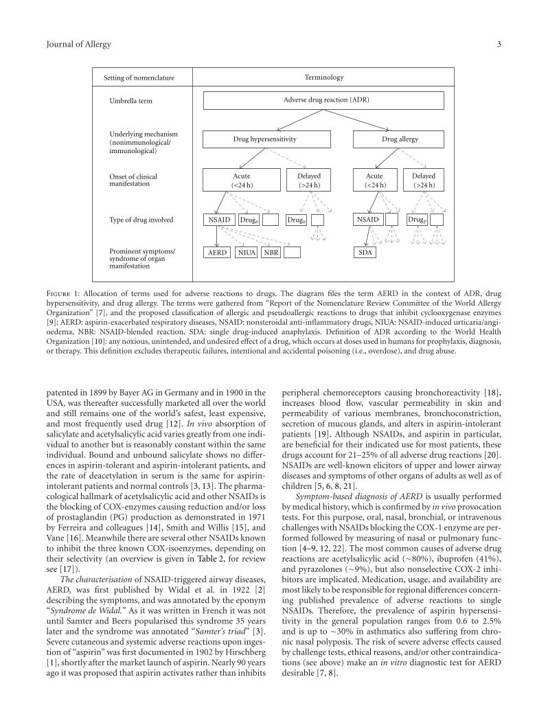
the nomenclature will need further review. In this context“hypersensitivity” describes objectively reproducible symp-toms or signs initiated by exposure to a defined stimulus at adose tolerated by normal persons. The terminology “aspirin-exacerbated respiratory disease” (AERD) characterises phys-ical reactions, underlying respiratory diseases, and inhibitorsof cyclooxygenase (COX) and refers to the clinical syndromeof chronic rhinosinusitis (CRS), nasal polyposis, bron-choconstriction in asthmatics, and/or eosinophil inflamma-tion in the upper and lower airways following the ingestionof NSAIDs blocking the COX-1 enzyme [9]. An assignmentof AERD in the context of adverse drug reactions (ADR) anddrug hypersensitivity is given in Figure 1.
NSAIDs are colloquially named “aspirin” or “aspirin-likedrugs”. Aspirin, the trade name of acetylsalicylic acid (ASA),
Journal of Allergy 3
Drug allergy
Acute DelayedDelayed
AERD NIUA SDANBR
Drugx Drugx DrugyNSAID
Umbrella term
Type of drug involved
Prominent symptoms/syndrome of organmanifestation
NSAID
Acute
Underlying mechanism(nonimmunological/immunological)
Onset of clinicalmanifestation
Setting of nomenclature
Adverse drug reaction (ADR)
Drug hypersensitivity
Terminology
(<24 h) (>24 h) (>24 h)(<24 h)
Figure 1: Allocation of terms used for adverse reactions to drugs. The diagram files the term AERD in the context of ADR, drughypersensitivity, and drug allergy. The terms were gathered from “Report of the Nomenclature Review Committee of the World AllergyOrganization” [7], and the proposed classification of allergic and pseudoallergic reactions to drugs that inhibit cyclooxygenase enzymes[9]; AERD: aspirin-exacerbated respiratory diseases, NSAID: nonsteroidal anti-inflammatory drugs, NIUA: NSAID-induced urticaria/angi-oedema, NBR: NSAID-blended reaction, SDA: single drug-induced anaphylaxis. Definition of ADR according to the World HealthOrganization [10]: any noxious, unintended, and undesired effect of a drug, which occurs at doses used in humans for prophylaxis, diagnosis,or therapy. This definition excludes therapeutic failures, intentional and accidental poisoning (i.e., overdose), and drug abuse.
patented in 1899 by Bayer AG in Germany and in 1900 in theUSA, was thereafter successfully marketed all over the worldand still remains one of the world’s safest, least expensive,and most frequently used drug [12]. In vivo absorption ofsalicylate and acetylsalicylic acid varies greatly from one indi-vidual to another but is reasonably constant within the sameindividual. Bound and unbound salicylate shows no differ-ences in aspirin-tolerant and aspirin-intolerant patients, andthe rate of deacetylation in serum is the same for aspirin-intolerant patients and normal controls [3, 13]. The pharma-cological hallmark of acetylsalicylic acid and other NSAIDs isthe blocking of COX-enzymes causing reduction and/or lossof prostaglandin (PG) production as demonstrated in 1971by Ferreira and colleagues [14], Smith and Willis [15], andVane [16]. Meanwhile there are several other NSAIDs knownto inhibit the three known COX-isoenzymes, depending ontheir selectivity (an overview is given in Table 2, for reviewsee [17]).
The characterisation of NSAID-triggered airway diseases,AERD, was first published by Widal et al. in 1922 [2]describing the symptoms, and was annotated by the eponym“Syndrome de Widal.” As it was written in French it was notuntil Samter and Beers popularised this syndrome 35 yearslater and the syndrome was annotated “Samter’s triad” [3].Severe cutaneous and systemic adverse reactions upon inges-tion of “aspirin” was first documented in 1902 by Hirschberg[1], shortly after the market launch of aspirin. Nearly 90 yearsago it was proposed that aspirin activates rather than inhibits
peripheral chemoreceptors causing bronchoreactivity [18],increases blood flow, vascular permeability in skin andpermeability of various membranes, bronchoconstriction,secretion of mucous glands, and alters in aspirin-intolerantpatients [19]. Although NSAIDs, and aspirin in particular,are beneficial for their indicated use for most patients, thesedrugs account for 21–25% of all adverse drug reactions [20].NSAIDs are well-known elicitors of upper and lower airwaydiseases and symptoms of other organs of adults as well as ofchildren [5, 6, 8, 21].
Symptom-based diagnosis of AERD is usually performedby medical history, which is confirmed by in vivo provocationtests. For this purpose, oral, nasal, bronchial, or intravenouschallenges with NSAIDs blocking the COX-1 enzyme are per-formed followed by measuring of nasal or pulmonary func-tion [4–9, 12, 22]. The most common causes of adverse drugreactions are acetylsalicylic acid (∼80%), ibuprofen (41%),and pyrazolones (∼9%), but also nonselective COX-2 inhi-bitors are implicated. Medication, usage, and availability aremost likely to be responsible for regional differences concern-ing published prevalence of adverse reactions to singleNSAIDs. Therefore, the prevalence of aspirin hypersensi-tivity in the general population ranges from 0.6 to 2.5%and is up to ∼30% in asthmatics also suffering from chro-nic nasal polyposis. The risk of severe adverse effects causedby challenge tests, ethical reasons, and/or other contraindica-tions (see above) make an in vitro diagnostic test for AERDdesirable [7, 8].

4 Journal of Allergy
Table 2: NSAIDS: classification, mechanism of action, representative structures. NSAIDs can be classified based on their chemical structureor mechanism of action; older NSAIDs were classified by chemical structure or origin, newer ones more often by their mechanism of action;COX: cyclooxygenase, 5-LO: 5-lipoxygenase.
Chemical class Example Inhibitory action Representative example
SalicylatesAcetylsalicylic acid
(Aspirin), diflunisal,mesalamine, salsalate
Nonselective; COX-1,COX-2,
Acetylsalicylic acid
OH
O
O
O
CH3
Propionic acid derivativesFenoprofen, flurbiprofen,
ibuprofen, ketoprofen,naproxen, oxaprozin
Nonselective, COX-1,COX-2
Ibuprofen
OH
O
CH3
CH3
H3C
Acetic acid derivativesDiclofenac, etodolac,
indomethacin, ketorolac,nabumetone sulindac
Nonselective, COX-1,COX-2
Diclofenac
OH
O
NH
Cl
Cl
Enolic acid (oxicam)derivatives
Droxicam, isoxicammeloxicam, piroxicam,
tenoxicam
Nonselective, preferentialCOX-2
Meloxicam
N
NS
S
N
H
OH
OO
O
Sulphonanilides NimesulideNonselective, preferential
COX-2
Nimesuleid
S
O
OO
NH
NO2
Selective COX-2 inhibitors(coxibs)
Celecoxib, parecoxib,etoricoxib
COX-2
Celecoxib
NN
S
CF3H2N
O O
H3C
p-amino phenol derivatives Paracetamol, phenacetin COX
NH
OHO
Paracetamol
Fenamic acid derivatives(fenamates)
Acid, flufenamic acid,meclofenamic, mefenamic
acidCOX
NH
OH OCH3
Cl
Flufenamic acid
Others Licofelone COX, 5-LON
OH
O
Cl
Licofelone

Journal of Allergy 5
In vitro diagnosis of AERD is discussed in literature withsome controversy, most likely based on insufficient and inpart contradicting data of earlier and recent publications, aswell as by former papers mentioning the unavailability of orinability to establish in vitro tests [4, 9]. Most clinicians havesome acquaintance of their use. However, the underlyingconcepts pertaining to diagnostic tests in general, and totheir use for diagnosis of a diseases in particular, are oftenless familiar, and perhaps less well understood. The currentconcepts point to the pathways of lipids (exemplified by eico-sanoids) and other molecules related to them (e.g., cytokines,growth factors, cell surface markers, second messengers ofcell signalling, enzymes and receptors). These will be sum-marised in brief and completed by some basic theoreticalaspects.
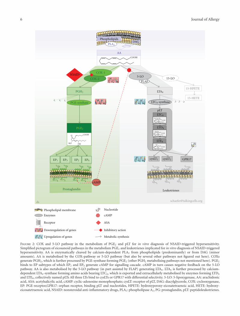
Eicosanoids (notation introduced in 1980 by Corey et al.[23], a shorthand nomenclature of eicosanoids was given in1987 by Smith and Willis [24]) are oxygenated metabolitesof the (5Z, 8Z, 11Z, 14Z)-5,8,11,14-eicosatetraenoic acid,widely known as arachidonic acid (AA). Arachidonic acid isthe main source of the eicosanoid cascade in humans involv-ing more than 50 enzymes generating a multiplicity of eico-sanoids [25, 26]. Concerning NSAID-triggered hypersensi-tivity and AERD, we selected and focused on the COX- and 5-lipoxygenase (5LO-) pathway. Both pathways are intimatelylinked to AERD and their implication is well documented(see subsequent literature). Beside these pathways and theirmetabolites, others such as those of cytokines, growth fac-tors, or second messengers of signal transduction are alsoknown to be implicated in AERD and related diseases. How-ever, it is beyond the scope of this paper to cover all of themin known detail.
Via the COX-pathway prostanoids (i.e., prostaglandins(PG), thromboxane (TX)) are generated. The COX-pathwayis blocked by NASIDs [14–16, 27] by acetylating the COXenzyme [28] and by causing inhibition of the conversionof arachidonic acid to PG [16]. COX-1 is constitutivelyexpressed in most tissues and cells and is involved in cellularhousekeeping functions. COX-2 is induced by inflammatorystimuli such as cytokines, growth factors, immunoglobulins,or bacterial toxins. Putative COX-3 mRNA is present inseveral tissues, including that from humans, but functionalprotein was still not found in humans. COX-3 is switchedon later in inflammation and is suggested for biosynthesisof endogenous anti-inflammatory mediators. Its clinicalrelevance to COX-3 remains unproven. All COX isoenzymesare modified by NSAIDs with different efficacy (for reviewsee [17, 27, 29]). The resulting metabolite PGH2 is furthermetabolised by PGE-synthase forming PGE2. The complex-ity of COX expression was demonstrated for human airways.There were no differences in the total number of cells stainedfor COX-1 and COX-2 irrespective of whether tolerantor intolerant to NSAIDs. The number and percentage ofmast cells, however, that express COX-2 was significantlyincreased in patients intolerant to NSAIDs. Furthermore, theexpression of COX-2 in epithelial and submucosal cellularwas increased in asthmatics [30]. Additionally, the expressionof COX-2 was downregulated in polypous tissue as well as inbronchial muscular cells from patients with AERD [31, 32].
PGE2 acts on at least four different seven-transmembrane-domain G-protein-coupled receptor subtypes, nominatedEP1 to EP4. Binding on the EP2 or EP4 causes bronchodilata-tive effects, whereas binding to EP1/EP3 causes opposite ef-fects [24].
The lipoxygenase pathway comprises several enzymes,generating several leukotrienes (LT). Focusing on the 5LO-pathway, LTA4 is generated from AA, which is furthermetabolised by the LTC4-synthase forming LTC4, containingthree amino acid groups, which is actively exported in theextracellular space. An overexpression of the promotor ofthe LTC4-synthase gene was observed in some patients withAERD [33]. The amino acids are degraded by subsequentenzymatic processes forming LTD4 and LTE4. These metabo-lites have been named in 1960 by Brocklehurst as slow-react-ing substances of anaphylaxis (SRS-A) [34] and were identi-fied in 1982 by Hammarstrom and Samuelsson introducingthe term leukotrienes for their occurrence in leukocytesand the characteristic chemical structure of conserved threeconjugated double bonds (see Figure 2). These LT are cha-racterised by a short half-life compared to other lipidmediators and are collectively named peptidoleukotrienes(pLT) based on their integral part of amino acids [35, 36].
The discovery of the 5-LO pathway caused an enormousinterest in this area, largely displacing the “classic” prost-aglandins. pLT are potent vaso- and bronchoconstrictors andhave several other biological activities, including an ability toincrease vascular permeability or to produce negative iono-tropic effects in cardiac contractions [37, 38]. The pLT unfoldtheir potential by currently three known seven-transmem-brane-domain G-protein-coupled receptor types, namedcysLT1 and cysLT2. A third dual orphan receptor GPR17binds uracil nucleotides and pLT [39, 40]. Increased expres-sion of cysLT1 and cysLT2 receptors is correlated to AERD[41–43].
The chemotactic metabolite LTB4, also generated fromLTA4 but formed by a separate enzymatic pathway, is 100-fold less potent concerning bronchoconstriction and acts ona separate LTB4 receptor [38, 44]. Other lipid mediators arelipoxins (LX). LXA4 is known to inhibit LTC4 response andis decreased in patients with AERD [45, 46]. Further patho-genetic aspects in AERD are extensively reviewed by Palikheet al. in this journal [47].
Attempting to condense the findings outlined above, acomplex eicosanoid-protein interaction network has beendiscovered over the past decades, comprising lipid-deriv-ed mediators, second messengers, cytokines, receptors, enzy-mes, and activation of genes. Eicosanoids have a crucial roleas mediators in inflammatory diseases like AERD. The en-zymes and receptors of the eicosanoid cascade are found tobe quite ubiquitous but also feature differences regarding dis-tribution and expression in tissue and cells in normal cir-cumstances as well as in patients with AERD. The COX-pathway can be attributed to the control of proliferativestates, the 5LO-pathway to wound healing and tissue repair.Both pathways are embedded in other metabolic pathways,for example, the network of cytokines and neuropeptides,which in turn are also interconnected [48]. Gene expressionand variability differs between AERD and NSAID-tolerant
6 Journal of Allergy
Prostaglandin
PGH2
PGE2
EP2EP1 EP4EP3
NSAID
AA
PLA2DAG
LTA4
cysLT1 cysLT2
15-HPETE
GPR17
LTE4
LTD4
LTC4
pLT
Phospholipids
15-HETELTC4-synthase
Gly–Cys–Glu
Gly–Cys
Cys
COOH
COOH
OH
COOHOH
COOHOH
cAMP
Leukotrienes
PGE-synthase
15-LOFLAP
5-LO
COOH
OHHO
O
ds
COX-1
COX-2
Phospholipid membrane
Enzymes
Receptor
Downregulation of genes
Upregulation of genes
Nucleotide
cAMP
ASA
Inhibitory action
Metabolic synthesis
Figure 2: COX and 5-LO pathway in the metabolism of PGE2 and pLT for in vitro diagnosis of NSAID-triggered hypersensitivity.Simplified pictogram of eicosanoid pathways in the metabolism PGE2 and leukotrienes implicated for in vitro diagnosis of NSAID-triggeredhypersensitivity. AA is enzymatically cleaved by calcium-dependent PLA2 from phospholipids (predominantly) or from DAG (minoramounts). AA is metabolised by the COX-pathway or 5-LO pathway (but also by several other pathways not figured out here). COXsgenerate PGH2, which is further processed by PGE-synthase forming PGE2 (other PGH2 metabolising pathways not mentioned here). PGE2
binds to EP subtypes of which EP2 and EP4 generate cAMP for signalling cascade. cAMP in turn causes negative feedback on the 5-LOpathway. AA is also metabolised by the 5-LO pathway (in part assisted by FLAP) generating LTA4. LTA4 is further processed by calcium-dependent LTA4-synthase forming amino acids bearing LTC4, which is exported and extracellularly metabolised by enzymes forming LTD4
and LTE4, collectively named pLTs All three LTs bind to cysLTs or GPR17 with differential selectivity. 5-LO: 5-lipoxygenase, AA: arachidonicacid, ASA: acetylsalicylic acid, cAMP: cyclic-adenosine monophosphate; cysLT: receptor of pLT, DAG: diacylglycerole, COX: cyclooxygenase,EP: PGE-receptor,GPR17: orphan receptor, binding pLT and nucleotides, HPETE: hydroxyperoxy-eicosatetraenoic acid, HETE: hydroxy-eicosatetraenoic acid, NSAID: nonsteroidal anti-inflammatory drugs, PLA2: phospholipase A2, PG: prostaglandin, pLT: peptidoleukotrienes.

Journal of Allergy 7
individuals with peculiarities with respect to ethnic back-ground.
Some of these elements may directly interact with intra-cellular effectors to trigger multiple signalling cascades, whileothers act extracellularly. These components control andmodulate cell migration, growth, proliferation, and activityof tissues and organs, which will result in differentiated reac-tions, unveiling symptoms like CRS, nasal polyposis, or asth-ma. A schematic overview is pictured in Figure 2.
2. Concept of Pathogenic Mechanisms
We will mention some of the known pathogenic mecha-nisms, elaborated in respect to AERD and to their supposedrelevance to AERD, but limited to in vitro diagnosis of AERDand NSAID-triggered hypersensitivity.
Since the first description of adverse reactions to aspirinin airways [2], it is common knowledge that AERD is trig-gered by NSAIDs [3–9, 11–13, 21, 22, 30, 43, 46, 47, 49–52].NSAIDS are known to modify the metabolism of unsaturatedlipids, pinpointing eicosanoids [16]. Eicosanoids comprisesa complex network of lipids essentially involved in thepathomechanisms of NSAID-triggered hypersensitivity orAERD.
NSAID hypersensitivity is characterised by an imbalanceof eicosanoid synthesis (i.e., PGE2 and pLT) prior to as wellas after exposure to aspirin. This was initially documented in1999 as a result of analysing cultured peripheral blood cells[49] as well as nasal mucosa of the same patients [50]. Theconcept of the imbalance of eicosanoid synthesis [49] wastaken up and approved recently by a theoretical study [51]and supported by former studies [52]. The genetic as well asfunctional modifications may be reasonable [33, 41–43, 46–51] but details are not fully understood, as expression ofCOX-2 is enhanced in macrophages [48] but no differencesof COX-1 or COX-2 expression in patients with AERD andNSAID-tolerant individuals is found [30].
The reduced levels of PGE2 in AERD might be one initialfactor for a diminished endogenous inhibition of the house-keeping function of PGE2, when activating the EP2 or EP4
receptor. These receptor types initiate the production ofcyclic adenosine monophosphate (cAMP), a second mes-senger, after binding of PGE2 [48]. The synthesis of pLT isreduced by a cAMP-dependent intracellular signal transduc-tion mechanism [11, 48, 52].
The reduced basal synthesis of housekeeping and inducedPGE2 [11, 49–52], as well as the postulated [49] and validatedoverexpression of LTC4-synthase [33] and cysLT receptors[41–43] give rational arguments to explain at least in part theshift toward an elevated basal synthesis of pLT. This PGE2-pLT shift will be further elevated upon exposure to NSAIDs,but also by other agents initiating the eicosanoid cascade(e.g., cytokines like interleukine-1, or bacterial antigens).Thus, the reduced housekeeping/induced PGE2 most likelyaccounts for reduced production of cAMP, which is inducedupon coupling of PGE2 on EP2 or EP4 receptors, but can beinduced by other signal transduction pathways [48].
Thus, the diminished availability of the housekeeping(basal) and induced PGE2 will cause a reduced generation
of suppressive acting endogenous cAMP upon exposure toCOX-inhibiting agents.
In this context, PGE2, pLT, NSAIDs, cAMP, and otherfactors (e.g., bacterial toxins, availability of arachidonicacid, cytokines, and others) will most likely contribute in ahighly complex manner to the multifactorial exacerbation ofNSAID-triggered symptoms and diseases.
3. Theoretical Consideration ofIn Vitro Diagnosis
Since the latter half of the 1980s enzyme immunoassay (EIA)tests are widely used to screen and diagnose a multitude ofdiseases. Results are mostly classified by a binary outcome as“positive” (“reactive”) or negative (“nonreactive”), based onthe protocols provided by the test manufacture and evalua-tion in the laboratory. The classification is the result of anordered sequence of several steps, which had been initiatedvia the testing procedure.
Measurement repeatability and reproducibility are inves-tigated during the approval process. For convenience, we willassume that the laboratory performing the test will maintainthe complex process of the measurement system, and thatthe distribution of the results of “disease-free” and “diseased”individuals are normally distributed (see Figure 4).
In an “ideal” world these two normal distributions willnot overlap. Regrettably the world of diagnostic testing israrely unequivocally ordered. Many (currently and probablyin perpetuity) unknown factors alter these distributionscausing overlap to some extent. Regardless of where the testoutcome threshold is situated on the measurement scale,some disease-free individuals and diseased (i.e., AERD) willbe incorrectly classified as “negative” (known as “false-nega-tive,” dark shaded area left-hand side of Figure 4) or “posi-tive” (known as “false-positive,” grey area right-hand side ofFigure 4), respectively. This represents one type of diagnostictest error.
Because any diagnostic test procedure has a single out-come threshold, moving the threshold to the right will reducethe false-positive results of disease-free individuals, but auto-matically will increase the false-negative error rate of the di-seased individuals. Similarly, adjusting the threshold to theleft will reduce the false-negative error rate, but automaticallyincreases the false-positive error rate (i.e., classifying disease-free individuals as patients with AERD).
Only changing the distribution of test results in one orboth groups would simultaneously reduce the rates of bothtypes of diagnostic test errors (i.e., false-positive and false-negative results). Unfortunately, in realty this will not bepracticable, due to the complex pathomechanisms underly-ing AERD, and the composition of the groups investigatedlike age, sex, medication, mentioned symptoms, interin-dividual variability of symptoms and syndromes, and ourlimited knowledge and understanding of the “plus-minus”clearly defined disease [53].
The terms sensitivity (SE), specificity (SE), and posttestprobabilities in this concern refer to probability of an (invitro) diagnostic test outcome, not to the equality of re-agent or chemicals. Tests with a high sensitivity will correctly

8 Journal of Allergy
identify virtually all patients with NSAID-triggered hyper-sensitivity with a high probability; tests with high specificityidentify all disease-free individuals correctly with a high pro-bability. This becomes obvious when referring to Figure 4:sensitivity and specificity correspond to the area under theprobability curve (i.e., the distribution) of patients withNSAID-triggered hypersensitivity (sensitivity of the test, onthe right) and disease-free individuals (specificity of the test,on the left). Unfortunately, inadequacies in the pathologicaland clinical symptoms or comorbid components and symp-tom stage, including age and sex distribution of disease-freeindividuals as well as patients with disease were described∼30 years ago [54] and continued to hamper any diagnostictest [53].
What physicians are really interested in knowing is theextent to which a positive or negative test result accuratelypredicts the true status of the patient, that is, disease-free orpatient with, for example, AERD. This is commonly referredto as the posttest probability of a disease (e.g., AERD), or pre-dictive value of a positive test result (PPV). In case of a neg-ative test result the posttest probability of being disease-free,that is, the predictive value of a negative test result (NPV)is of interest. These values depend on not only the sen-sitivity and specificity, but also on the pretest probability (orprevalence) of the disease (e.g., AERD). The mathematicalalgorithm connecting the three probabilities sensitivity,specificity, and prevalence is known as Bayes’ theorem (orig-inally published 1763 by R. Price [55] after the death of theEnglish clergyman Thomas Bayes). It might be easier to graspthe sense of this relationship more directly than lookingon the mathematical algorithm: the prevalence of AERD isarguable in respect to the supposed prevalence of 1.2 to 2.8%of a population [5, 8, 20]. However, as outlined before, thereis some uncertainty concerning the prevalence (i.e., the pre-test probability) of AERD due to the impossibility of di-agnosing this syndrome by an absolute unfailing method.This marks a further limitation for “precisely” defining theoutcome of an in vitro test by mathematical characteristics.
The probability term likelihood ratio, introduced in 1968by Lustedt and popularised in the 1980s by Sacket et al. isa ratio of the two probabilities sensitivity and 1-specificity,describing the relative probability of a positive diagnostictest result in diseased individuals compared to disease-freeindividuals which can be calculated [56, 57]. For ruling-ina disease the likelihood ratio should be at least 1, preferablymuch higher (graphically this represents the area on the rightsite of the test threshold of Figure 4). In case of ruling out adisease, the likelihood ratio of a negative test result is chosen.These values should ideally be much smaller than one.
As easily deduced from the above-mentioned aspects, thedefinition of an optimal threshold is not only a question ofstatistics but rather depends on how the test result will beused. For screening purpose the threshold will be relative-ly low, resulting in higher false-positive outcomes. This re-quires additional diagnostic testing to ensure a therapeuticregime. In case of AERD a low threshold line will captureall patients, even those without obvious symptoms. The lowthreshold also covers the risk that a patient with a poten-tial NSAID-triggered hypersensitivity but without obvious
symptoms would undergo life-threatening reactions uponexposure to NSAIDs, would not be detected. Thus, thelow threshold uncovers those patients with currently mildNSAID-triggered hypersensitivity for appropriate treatmentbefore the disease worsens in the future. The latter is vis-ualised, in part, by Figure 5, sketching schematically thecourse of NSAID-triggered hypersensitivity: The symptomsand underlying disease(s) do not relate in a uniform fashion,rather a pattern of exacerbation and remission is more like anexponentially growing sinus line. This pattern will be super-imposed on the residual changes of the underlying diseaseand is a further challenge of in vivo and in vitro diagnosis ofAERD.
4. In Vitro Diagnosis of AERD
The change in knowledge and concepts concerning thepathogenic mechanisms of AERD reflects the diversity of invitro diagnostic approaches developed during the last cen-tury.
4.1. Serum-Specific IgE against NSAIDs (SIgNT). The SIgNTexamines serum or plasma collected from patients sufferingfrom AERD and other manifestations of NSAID-triggeredsymptoms. The samples are filled into tubes coated withNSAIDs, including derivatives, or with NSAIDs/derivativescoupled to a carrier. After an incubation and washing stepan anti-IgE or anti-IgG antibody labelled with a tracer (e.g.,fluorochrome or chromogen finally converted by an enzyme)is added. Resulting values of the measurement will identifydiseased patients if the value exceeds a predefined threshold(cutoff).
Underlying this approach was the observation, that ad-verse reactions to NASIDs displayed symptoms such asallergic reactions (the term “allergy” was introduced in1906by von Pique as immunoglobulin mediated type of reaction[58]). Therefore, an immunologic reaction was assumed.Numerous attempts at detecting an antibody directed againstAspirin, derivatives thereof (e.g., anti-aspiryl antibodies), orto any other supposed NSAIDs failed to demonstrate anunequivocal antibody [3]. Even though antibodies were de-tected in 1940 by Butler et al. [59] and Zhu and colleagues[60], or propyphenazone-specific antibodies by the group ofFerreira [59], or were suspected by the group of Settipane[61]. These results have not been confirmed in the followingdecades [62, 63]. Also serum level of IgE in aspirin-intolerantpatients did not differ from non-atopic population [61].
Nevertheless, these investigations contributed some sub-stantial insights to our current understanding of AERD andto other NSAID-triggered symptoms as nonimmunologicallymediated diseases. Thus, a SIgNT for the detection of anti-bodies directed to any NSAID could not be established and isnot available for in vitro diagnosis of AERD.
4.2. Histamine Release Test (HRT). The HRT examines urinesamples from patients exposed to NSAIDs or supernatantsof cell culture medium of peripheral blood cells (PBLs)incubated in vitro with varying concentrations of differentNSAIDs.

Journal of Allergy 9
The first approach (analysing urinary samples) wouldnot be classified as an in vitro test as it affords an in vivo pro-vocation/exposure of the patient. There are some essentialdrawbacks, arguing why this procedure (in vivo challenge)might not be suitable in some cases (because of, for example,ethical reasons, age, compliance, technical; see Section 1).Using PBLs for measurement of histamine release has to bedesignated as an in vitro diagnostic test.
The known bronchoconstrictive effect of histamine sti-mulated the attempt to look for an altered histamine releasein patients with AERD [64]. Early investigations demon-strated elevated urinary excretion of a histamine metabolite[65] and elevated plasma histamine levels [66]. These meas-urements were, however, not confirmed in nasal lavageupon provocation [67, 68]. Preincubation of leucocytes withAspirin failed to alter spontaneous or calcium ionophore-induced histamine release in patients with AERD [69, 70].This was confirmed for bronchial lavage [71] and for leuco-cytes by our study performed in vivo as well as in vitro ex-posure [49, 50]. There are also some inconsistent results informer studies. Okuda and colleagues reported elevated hist-amine release induced by platelet-activating factor fromleukocytes of patients with AERD [72], Hosemann andcolleagues measured lower histamine content in polypoustissue of patients with AERD than in analgesic-tolerant pa-tients [73], and the group of Stevensson reported elevatedplasma histamine levels in only three of seventeen patients[74]. The low efficiency of histamine release by in vitro stimu-lation according to the CAST-protocol (see CAST) was alsoaffirmed by a more recent study [75].
Even though the HRT was promising, as it depicts a path-omechanistic element of AERD, and it might be suitable toconfirm AERD/NSAID sensitivity in specifically selected pa-tients (e.g., with an underlying allergic comorbidity), it isnot suggested for routine in vitro diagnosis of patients withAERD taking into consideration all data currently available.
4.3. Lymphocyte Transformation Test (LTT). The LTT (syn-onyms are lymphocyte proliferation test or lymphocyte stim-ulation test) examines the activity of lymphocytes, notably ofT-lymphocytes selected from PBLs upon exposure to vary-ing NSAIDS at different concentrations. Most widely used forquantifying the proliferation is the measurement of 3H-thy-midin uptake by dividing cells from samples of anticoagu-lated blood.
The relevance of the LTT as model system for analysingpatients with hypersensitivity to Aspirin was discussed morethan 40 decades ago [76–80]. Some NSAIDs do inhibit othersfrom enhancing the proliferation, but this was not seenconsistently [81–85].
A later study demonstrated an enhanced proliferation ofnormal lymphocytes, but a diminished 3H-thymidin uptakeby lymphocytes from patients with AERD [86]. NSAIDs areconsidered suitable for LTT investigation [87]. But the incon-sistency of results, and the more indirect relation of detectinglymphocyte proliferation to our current pathomechanisticunderstanding of AERD, often implicated unclear results.These findings questioned the clinical relevance of the LTTfor the detection of adverse reaction to NSAIDs. Therefore,
the LTT is actually not referred to be a suitable tool for invitro diagnosis for patients with AERD.
4.4. Platelet Aggregation Testing (PAT). The PAT examinessurvival and aggregation of platelets separated from venousPBLs. The platelets are exposed to varying concentrations ofthose NSAIDs which are of interest, for a defined time asvalidated by the performing laboratory.
Around 25 years ago it was suggested that plateletsmight have a pivotal role in AERD [88–93]. In a subsequentstudy, a group led by Picado, detected no differences in anyindices of platelet function studied between aspirin-tolerantand patients with AERD despite a slightly elevated aspirin-triggered PGF2α release [94]. These results are somehowunexpected, as platelets are known to be potent producer ofeicosanoids. Despite this approach and the implication of theplatelet behaviour in NSAID-triggered symptoms, the PAThas not been approved for in vitro diagnosis of AERD.
4.5. Serum-PGF2α Test (SPT). The SPT examines serumselected from peripheral blood. Upon addition of a prede-fined concentration of ASA in vitro, samples are analysedusing a radio-immunosorbent assay. Samples exceeding apredefined serum level of PGF2α indicate patients withAERD.
This approach was introduced in 1991 by Willilams andcolleagues and demonstrated no changes in PGE2 or PGD2
but lower plasma level of PGF2α in patients with AERDbefore addition of aspirin, and elevated levels of PGF2α
after addition of aspirin, when compared to aspirin-tolerantasthmatics [95]. Small concentrations of aspirin given toplatelet suspensions generated PGF2α [96]. This confirmedthe hypothesis of an NSIAD-triggered alteration of prost-anoid metabolism and altered serum protein binding capac-ities in patients with AERD. Regrettably, there are no furtherpublications documenting the routine use of this promisingapproach.
4.6. Mediators in Nasal Lavage (MNLT). The MNLT exam-ines nasal lavage collected from patients exposed in vivo tolysine aspirin. The nasal lavage is stored appropriately. Afterthawing and centrifugation the supernatant is analysed usingspecific enzyme immunoassays for two cytokines, MCP-3and RANTES [97].
It was proposed that patients with AERD are charac-terised more likely by a chronic rather than an acute overpro-duction of MCP-3 and RANTES. The MNLT increased ourpathomechanistic understanding of AERD, but an in vivoprovocation step is presupposed. Hence, this approach doesnot meet the criteria of an in vitro test. Even though, theMNLT would be suitable to confirm AERD.
4.7. Exhaled Breath Condensate Eicosanoid Testing (EBCET).The EBCET examines exhaled breath condensate of unexpos-ed patients with AERD. The condensate is stored until ana-lysis using an enzyme immunoassays specific for 8-isopro-stanes, LTB4, and PGE2. Eicosanoid values exceeding apredefined threshold characterise patients with a positive testoutcome.
10 Journal of Allergy
cAMP
Moderate-severe reaction
Mild-moderate reaction
cAMP
ds
cAMP
No-(very rare) reaction
No reaction
cAMAAAAAA P
O
O
O
OH
CH3
O
O
O
OH
CH3
ASAASA
5-LO5-LO
5-LO5-LO5-LO
Diseased
cAMP
5-LO LO5 LO
cAMP
Disease-free
cAMP
COX-1 2
EP
(A1)
(A2)
(B1)
(B2)
1 EP3 EP2 EP4 cys-LT1cys-LT2
GPR17
5-LO
cAMP
EPE
A1)
1 EP3 EP EP cys LT2GPR17
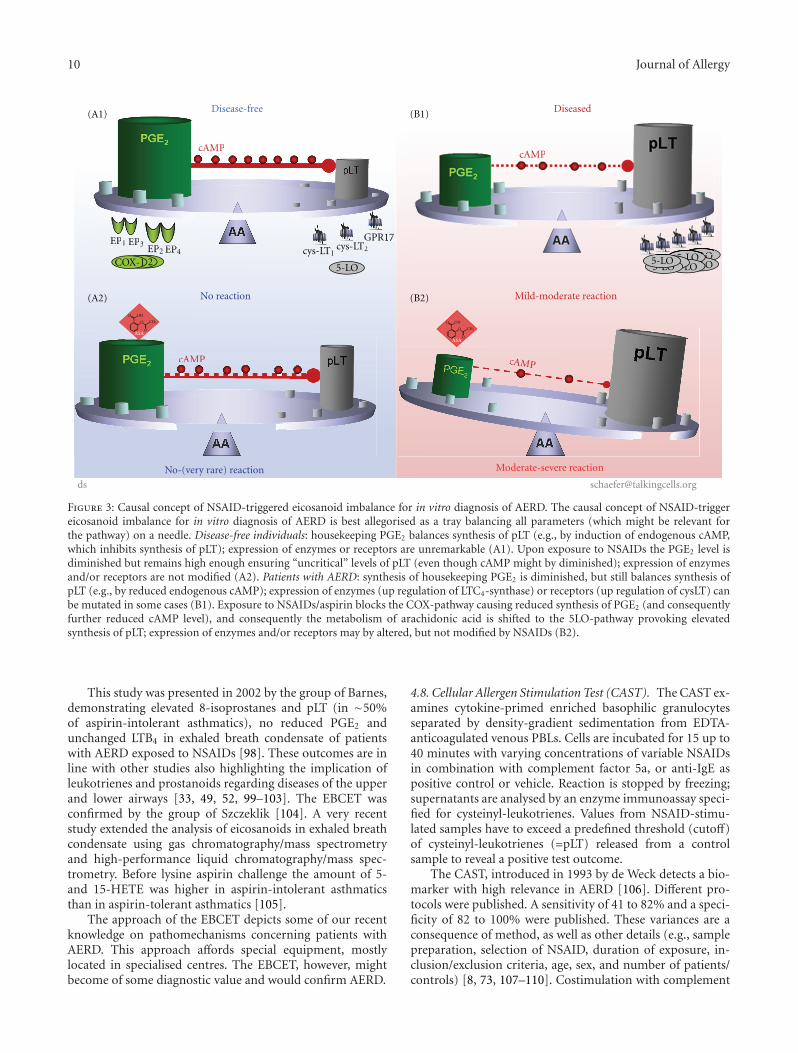
Figure 3: Causal concept of NSAID-triggered eicosanoid imbalance for in vitro diagnosis of AERD. The causal concept of NSAID-triggereicosanoid imbalance for in vitro diagnosis of AERD is best allegorised as a tray balancing all parameters (which might be relevant forthe pathway) on a needle. Disease-free individuals: housekeeping PGE2 balances synthesis of pLT (e.g., by induction of endogenous cAMP,which inhibits synthesis of pLT); expression of enzymes or receptors are unremarkable (A1). Upon exposure to NSAIDs the PGE2 level isdiminished but remains high enough ensuring “uncritical” levels of pLT (even though cAMP might by diminished); expression of enzymesand/or receptors are not modified (A2). Patients with AERD: synthesis of housekeeping PGE2 is diminished, but still balances synthesis ofpLT (e.g., by reduced endogenous cAMP); expression of enzymes (up regulation of LTC4-synthase) or receptors (up regulation of cysLT) canbe mutated in some cases (B1). Exposure to NSAIDs/aspirin blocks the COX-pathway causing reduced synthesis of PGE2 (and consequentlyfurther reduced cAMP level), and consequently the metabolism of arachidonic acid is shifted to the 5LO-pathway provoking elevatedsynthesis of pLT; expression of enzymes and/or receptors may by altered, but not modified by NSAIDs (B2).
This study was presented in 2002 by the group of Barnes,demonstrating elevated 8-isoprostanes and pLT (in ∼50%of aspirin-intolerant asthmatics), no reduced PGE2 andunchanged LTB4 in exhaled breath condensate of patientswith AERD exposed to NSAIDs [98]. These outcomes are inline with other studies also highlighting the implication ofleukotrienes and prostanoids regarding diseases of the upperand lower airways [33, 49, 52, 99–103]. The EBCET wasconfirmed by the group of Szczeklik [104]. A very recentstudy extended the analysis of eicosanoids in exhaled breathcondensate using gas chromatography/mass spectrometryand high-performance liquid chromatography/mass spec-trometry. Before lysine aspirin challenge the amount of 5-and 15-HETE was higher in aspirin-intolerant asthmaticsthan in aspirin-tolerant asthmatics [105].
The approach of the EBCET depicts some of our recentknowledge on pathomechanisms concerning patients withAERD. This approach affords special equipment, mostlylocated in specialised centres. The EBCET, however, mightbecome of some diagnostic value and would confirm AERD.
4.8. Cellular Allergen Stimulation Test (CAST). The CAST ex-amines cytokine-primed enriched basophilic granulocytesseparated by density-gradient sedimentation from EDTA-anticoagulated venous PBLs. Cells are incubated for 15 up to40 minutes with varying concentrations of variable NSAIDsin combination with complement factor 5a, or anti-IgE aspositive control or vehicle. Reaction is stopped by freezing;supernatants are analysed by an enzyme immunoassay speci-fied for cysteinyl-leukotrienes. Values from NSAID-stimu-lated samples have to exceed a predefined threshold (cutoff)of cysteinyl-leukotrienes (=pLT) released from a controlsample to reveal a positive test outcome.
The CAST, introduced in 1993 by de Weck detects a bio-marker with high relevance in AERD [106]. Different pro-tocols were published. A sensitivity of 41 to 82% and a speci-ficity of 82 to 100% were published. These variances are aconsequence of method, as well as other details (e.g., samplepreparation, selection of NSAID, duration of exposure, in-clusion/exclusion criteria, age, sex, and number of patients/controls) [8, 73, 107–110]. Costimulation with complement

Journal of Allergy 11
Disease-free Diseased
Test result scale
Threshold line
Freq
uen
cy o
f te
st m
easu
rem
ent
Real world
5 4 3 2 1 0 1 2 3 4 5
Disease-free Diseased
Test result scale
Threshold lineIdeal world
5 4 3 2 1 0 1 2 3 4 5
Freq
uen
cy o
f te
stm
easu
rem
ent
Figure 4: Framework for diagnostic test outcomes. Schema of “real” world diagnostic test outcomes; test measurement: clinical parameterslike age, sex, ethnic group, height, weight, and so forth or analytical parameters like temperature, IgE, histamine, inter-leukins, lipidmediators; shaded areas exemplify the false-positive (false-negative) measurement of disease-free (diseased) individuals, respectively; insert:pictured “ideal” world.
Time scale
Stat
us
of d
isea
se/p
atie
nt
Underlying progression
Acute disease
Potentially measurementsof a diagnostic test
Figure 5: Hypothetical progress of AERD over time.
factor 5a was claimed by the group of Weber to improve sen-sitivity [107]; they investigated patients with various under-lying diseases. Low efficiency was reported with no diagnosticutility and superiority to the HRT [75]. Nevertheless, theCAST was successfully established for diagnosis of allergies[109].
According to a more recent study, the CAST uncoversa pathway which was different from the classical IgE-med-iated pathway. CAST uses doses of ASA for in vitro stimu-lation causing nonspecific basophile activation, and therebyeliminates the usefulness of a cell based diagnostic test for
AERD. Therefore, it was suggested that the CAST would havelow value in diagnosing AERD and other diseases [108, 110].
4.9. Basophile Activation Test (BAT). The BAT, also namedFAST (Flow-cytometric Allergen Stimulation Test), examinesbasophilic granulocytes separated from EDTA-anticoagu-lated venous PBLs. Cells are incubated with varying concen-trations of different NSAIDs for up to 40 minutes. Thereafter,basophilic granulocytes are double-marked with antibodiesdirected to IgE and CD63 (or CD203). The number of positi-vely stained basophiles is measured using a fluorescence

12 Journal of Allergy
activated flow cytometer combined with appropriate soft-ware. A positive test outcome is defined by a laboratory-defined threshold (cutoff) of positively stained basophiles.
The BAT was introduced in 2000 by the group ofde Weck [111]. CD63 is a cell surface glycoprotein thatmediates signal transduction events that play a role in theregulation of cell development, (platelet) activation, growthand motility. CD203 represents a transmembrane ecto-nu-cleotide pyrophosphatase/phospho-diesterase-I enzyme (E-NPP), which cleaves phosphodiesters and phosphosulfatebonds. Both proteins are expressed on activated basophils.During the last decade follow-up studies were initiated to im-prove and ensure the technical procedures, thereby using theterm BAT [112–115].
The BAT depicts an altered appearance of granulocytes,which are known to be implicated in AERD. Variablevalues of sensitivity (∼10–64%) and specificity (∼75–100%)were published depending on the protocols used (e.g.,sample preparation, selection of NSAID, duration of expo-sure, inclusion/exclusion criteria, age, sex, and number of pa-tients/controls). The clinical use of the BAT is controversiallydiscussed [112–115], pointing to inherent factors influencingthe opportunities and limitations of an in vitro diagnostictest.
4.10. Flow Cytometric Assay and CAST (Flow-CAST). TheFlow-CAST uses two techniques, the CAST (enzyme im-munoassay) and BAT (flow cytometric assays). The outcomesof both tests are combined.
As reviewed in 2005 by the group of de Weck, thesensitivity and specificity varied depending on the NSAIDtested [116]. The global sensitivity was annotated ∼67%, thespecificity 93%. Combination of BAT with CAST elevatedsensitivity (to ∼73%) but reduced specificity (to 71%). TheFlow-CAST was proved for diagnosis of beta-lactam allergy[117]. It was proposed that in case of a negative result, aNSAID hypersensitivity cannot be excluded and a provoca-tion challenge remains necessary if clinically indicated.
This approach demonstrates the usefulness of combiningdiagnostic procedures as mentioned in the introduction part,but demonstrates also the drawbacks as explained. From apractical point of view, performing both tests makes greatdemands on laboratory equipment as well as manpower, andtherefore impacts on cost-effectiveness. The advantages ofthis procedure compared to others remain to be established.
4.11. Aspirin-Sensitive Patients Identification Test (ASPI Test).The ASPITest examines PBLs exposed in vitro to varyingconcentrations of NSAIDs. The release of 15-hydroxyeico-satetraenoic acid (15-HETE) is analysed using an enzymeimmunoassay specific for 15-HETE. Values exceeding a pre-defined amount threshold line (cutoff, ∼6% exceeding basalrelease) identify patients with AERD [118].
The report by Kowalski and colleagues in 2005 concludedthat the aspirin-triggered release of 15-HETE from PBLsdoes, to some extent, mimic the reactions observed in vivo.15-HETE was detected in epithelial cells of nasal polypoustissue as well as in PBLs from patients with AERD, but not inasthmatics without NSAID hypersensitivity [31, 119, 120].
Already in 1991 the group of Picado demonstrated thein vivo evidence of elevated release of 15-HETE in nasalsecretions of allergic patients [121]. It was demonstrated,that a PGE1 analogue (misoprostol) inhibited the aspirin-triggered 15-HETE release. A recent study investigating eightASA-intolerant patients confirmed the elevated level of 15-HETE [120]. Variable values of sensitivity (∼63–83%) andspecificity (∼50–82%) were published.
The ASPITest depicts a pathomechanistic link to AERDand obviously confirms the clinical finding in patients withAERD. Hitherto, there are only few promising publicationsand future studies will have to prove to which extent theASPITest will be applicable for routine use for in vitrodiagnosis of AERD and related diseases.
4.12. Functional Eicosanoid Testing and Typing (FET). TheFET examines PBLs of heparinised venous blood. PBLs arediluted in an appropriate buffer before exposure to ASA,neuropeptides, and arachidonic acid. The reaction is stoppedby freezing. Upon thawing and centrifugation the samplesare analysed using specific enzyme immunoassays for PGE2
and pLT. Measured data are calculated using appropriatesoftware. The resulting individualised dynamic eicosanoidpattern is classified in values ranging from 0.0 to 3.0. Thisoutcome is then more roughly classified as normal (0.0 to0.5), mild (<0.5), moderate (<1.5), and severe (<2.5 to 3.0);these values also represent a probability of severity of thesymptoms.
This approach was introduced in 1999 by Schafer andcolleagues and thereafter improved by integrating the grow-ing knowledge of pathomechanistic concepts [11, 49–52,122]. The FET depicts two biomarkers which are intimatelyinvolved in AERD and NSAID-triggered symptoms/diseases.First studies demonstrated the confirmation of clinicallydiagnosed AERD prior to, during provocation, and aftersuccessful treatment [123, 124]. Subsequent studies demon-strated the differentiation of non-airway-related but NSAID-triggered diseases [11, 125–127]. Others applied the FETfor monitoring medical treatment in patients with AERD[128, 129] or characterisation of pathophysiological aspects[130]. Values for sensitivity and specificity varied dependingon the underlying disease (airways: 96 and 89%, skin: 96 and97%, gastrointestinal tract: 64–98 and 82–89%, resp.) [8].
The FET provides context-dependent cell-based confir-mative as well as prospective information. This approachconfirms AERD, but also differentiates and/or characterisesunderlying diseases of closely related symptoms; in addition,depending on the intended diagnostic challenge (as exem-plified in Figures 6(a) and 6(b)). The FET differentiates ob-viously different symptoms of NSAID-triggered hypersensi-tivity of varying underlying disease. Future studies will haveto demonstrate whether the FET, in addition to confirmingor differentiating AERD, might provide some prognosticvalue in NSAID-triggered diseases.
5. Conclusions
During the last decades our knowledge concerning the path-ogenic mechanisms, the terminology of NSAID-triggered

Journal of Allergy 13
FET
-val
ue
Control ATA AERD
3
2.5
2
1.5
1
0.5
0
∗∗∗
∗∗
(a)
Control ATA AERD
2.5
2
1.5
1
0.5
0
∗∗
∗∗
∗
FET
-FM
D v
alu
e
NP
ns
3
(b)
Figure 6: (a): FET and NSAID-triggered eicosanoid imbalance of individuals suffering from diseases with lower airway symptoms. The FETwas performed and the FET values were calculated according to the total eicosanoid pattern score of [11] using PBLs. Patients suffering fromNSAID-triggered bronchoconstrictive symptoms were confirmed and characterised by clinical and in vitro diagnosis. Allergy was ruled outby medical history, skin test, and in vitro test for total and specific immunoglobulin. The mean FET value (solid line) of controls, ATA, andAERD was 0.7, 1.4, and 2.1, respectively. FET values > 1.0 characterise patients with lower airway symptoms. FET values ≥ 1, 8 (dashedline, potential threshold) differentiate NSAID-tolerant asthmatics and patients with AERD; ATA: patients suffering from aspirin-tolerantasthma, AERD: patients suffering from aspirin exacerbated respiratory disease; (n = 53 for each group, ∗P < 0.05, ∗∗P < 0.01). (b): FETand functional metabolic differentiation of patients with and without NSAID-triggered eicosanoid of lower and upper airway symptoms.The functional metabolic differentiation (FMD) of subgroups of patient was achieved by in vitro provocation of PBLs and calculation ofthe FET value according to the total eicosanoid pattern score of [11], but by amending the FET value by subtracting the difference of thesum of the enzymatic capacity (EC) of PG- and LT-synthesis as well as the difference of the ASA- and neuropeptide-induced eicosanoidbalances (EB) from the primary FET value (EC and EB were calculated according to [11]). The FET-FMD value takes into account twometabolites of the eicosanoid pathway and their in vitro modification by ASA and neuropeptide. The latter had been shown to be intimatelyimplicated in hyperresponsiveness of airway ([11] and ref. therein). The FET-FMD value reveals the differentiation of ATA, NP, and AERD,but without discrimination of ATA and healthy controls. The mean value of FET-FMD (solid line) was 0.4, 0.4, 1.1, and 1.7, for controls,ATA, NP, and AERD, respectively. The threshold of FET-FMD was ≥1.0 (dashed line) for NSAID-triggered lower and upper symptoms ofthe airways. In conclusion, this approach confirmed and characterised NSAID-triggered symptoms by clinical and in vitro diagnosis. ATA:patients suffering from bronchial asthma, but tolerant to NSAIDs, NP: patients suffering from nasal polyposis, AERD: patients sufferingfrom aspirin exacerbated respiratory disease with asthmatic symptoms; n = 53 for each group, ns: not significant, ∗P < 0.05, ∗∗P < 0.01.Allergy was ruled out by medical history, skin test and in vitro test of total an specific immunoglobulin.
symptoms and NSAID-exacerbated diseases (e.g., AERD)and the technical possibilities have continuously improved.This facilitated the development of new approaches forin vitro diagnosis, starting from no in vitro tests available110 years ago to twelve in vitro tests developed duringthe last decades. Some characteristics and suggestions forintended use of the in vitro tests discussed are summarised inTable 3.
Our understanding of AERD and NSAID hypersensi-tivity moved form an immunoglobulin-triggered pathome-chanism, diagnosed in the serum, to a multiplexed highlyinterconnected (eicosanoid) imbalance based on pathogenicunderstanding, diagnosing parameters from cell cultures, forexample, genes, enzymes, mediators (lipids, cytokines, pH,and others), receptors, and others. A multitude of parameterswere suggested. Surface marker of basophiles and lipidmediators remained to be the most promising biomarkers.Dynamic multiparametric approaches were favoured as
compared to static single parametric approaches. A schemat-ically simplified pictogram of the COX- and 5-LO pathwayreferred to for in vitro diagnosis is given in Figure 2.
The complexity of interacting parameters accounts forthe initial situation where NSAIDs (see Table 2) start to act.If there is an imbalance of several metabolic and/or geneticparameters, the block of the COX pathway by NSAIDs willcause an exacerbation of one or more of prestage(s) of sym-ptoms of a disease. Diagnosing the balance and imbalance ofthe eicosanoid cascade might be fundamental for diagnos-ing and treating NSAID-triggered diseases (see Figures 1and 3). These approaches might be hampered by high in-dividual variability of underlying diseases, genetics, enzy-matic/cellular function/activity, and by inclusion and exclu-sion criteria during sample collection for in vitro diagnosis.The (in vitro) test outcome has to be carefully interpreted byan appropriately trained physician and researcher concern-ing terminology, inclusion, and exclusion criteria, test theory,

14 Journal of Allergy
Table 3: Selected characteristics and suggestion for use of tests described in vitro diagnosis. ASPI Test: aspirin-sensitive patients identificationtest, BAT: basophile activation test, CAST: cellular antigen stimulation test, EBCET: exhaled breast condensate eicosanoid testing, Flow-CAST: flowcytometric assay and CAST, HRT: histamine release test, FET functional eicosanoid testing and typing, LTT: lymphocytetransformation test, MNLT: mediators of nasal lavage test, PAT: platelet aggregation test, SIgNT: serum-specific immunoglobulin E againstNSAIDs test, SPT: serum-PGF2α test; LT: leukotrienes, PG: prostaglandin, CD: cluster of differentiation, HETE: hydroy-eicosatetraenoic acid;SE: sensitivity, SP: specificity; PPV: positive predictive value, NPV: negative predictive value; n.v.d.: no values described — not suggested,(—) suggested, actually not in use, ? suggested upon further validation, (+) suggested with restrictions, + suggested.
In vitro test Test parameter Test sample SE(%) SP (%) PPV (%) NPV (%)Suggestionfor in vitrodiagnosis
SIgNT IgE, IgG serum n.v.d. n.v.d. n.v.d. n.v.d. —
HRT histamineculture medium,
PBLsn.v.d. n.v.d. n.v.d. n.v.d. —
LTT proliferation lymphocytes n.v.d. n.v.d. n.v.d. n.v.d. —
PAT aggregation platelets n.v.d. n.v.d. n.v.d. n.v.d. (—)
SPT PGF2α serum n.v.d. n.v.d. n.v.d. n.v.d. ?
MNLT MCP-3, RANTES nasal lavage n.v.d. n.v.d. n.v.d. n.v.d. —
EBCET 8-isoprostaneexhaled breast
condensaten.v.d. n.v.d. n.v.d. n.v.d. ?
CAST cysLTculture medium,
basophiles41–82 82–100 ∼96 ∼78 (—)
BAT CD63, CD203culture medium,
basophiles60–70 <90 ∼95 ∼56 (—)
Flow-CAST cysLT, CD63 basophiles ∼10–67 ∼75–100 n.v.d. n.v.d. (+)
ASPI Test 15-HETEculture medium,
PBLs63–83 >50–82 79 86 ?
FET PGE2, pLTculture medium,
PBLs96 (64–98) 83 (82–89) 90 (70–96) 93 (69–98) +
and last but not least, the most recent hypothesis and modelsof pathogenic mechanisms.
All in vitro tests, currently available, consider our currentpathogenic and clinical understanding of AERD. But theintended use by the clinician or researcher will also accountfor the selection of the most appropriate in vitro diagnosticprocedure (e.g., screening purpose, confirmation of a clinicaldiagnosis, individual risk assessment, proof of, prognosticprobability, and/or differentiation of symptomatic appear-ance, monitoring of treatment, effect of single drugs, andmany more). Considering the limitations of clinical diagnosisof AERD (see above), the “provocation” test is yet designatedas “gold standard” in clinical diagnosis, but is usually restrict-ed to confirm acute physical reactions of hyper reactive lowerairways and requires the necessity for patients’ provocation.But this “gold standard” will fail if AERD is still not thor-oughly distinctive, a prognostic goal has to be considered, orprovocation is precluded.
The relevance of the diagnostic test outcome and itsinterpretation will improve if the users of an in vitrodiagnostic procedure consider all information provided. Inthis concern, functional cellular in vitro approaches mimicsome of the complex in vivo processes seen in patients withAERD. The imbalance of eicosanoids might be a rationaldecision-making model for in vitro diagnosis of AERD as wellas NSAID-triggered hypersensitivity. Future research willdemonstrate whether and which functional in vitro approachwill prove to be the “gold standard” of in vitro diagnosis
of AERD to support treatment of patients with AERD andrelated diseases.
Acknowledgments
The authors have no financial conflicts of interests. D. Schaferhas holding in a patent on a test of eicosanoid function. Theauthors thank Dr. Ashley Cross for critical evaluation of thepaper.
References
[1] Hirschberg, “Mitteilungen uber einen Fall von Nebenwirk-ungen des Aspirin,” Dt Med Wochenschr, vol. 28, pp. 416–417,1902.
[2] F. Widal, P. Abrami, and J. Lermoyez, “Anaphylaxie et idio-syncrasie,” La Presse Medicale, vol. 30, pp. 189–193, 1922.
[3] M. Samter and R. F. Beers, “Concerning the nature of into-lerance to aspirin,” Journal of Allergy, vol. 40, no. 5, pp. 281–293, 1967.
[4] J. C. Delaney, “The diagnosis of aspirin idiosyncrasy byanalgesic challenge,” Clinical Allergy, vol. 6, no. 2, pp. 177–181, 1976.
[5] D. D. Stevenson and B. L. Zuraw, “Pathogenesis of aspirin-exacerbated respiratory disease,” Clinical Reviews in Allergyand Immunology, vol. 24, no. 2, pp. 169–187, 2003.
[6] C. Jenkins, J. Costello, and L. Hodge, “Systematic review ofprevalence of aspirin induced asthma and its implications forclinical practice,” British Medical Journal, vol. 328, no. 7437,pp. 434–437, 2004.

Journal of Allergy 15
[7] S. G. O. Johansson, T. Bieber, R. Dahl et al., “Revised nomen-clature for allergy for global use: report of the NomenclatureReview Committee of the World Allergy Organization,October 2003,” Journal of Allergy and Clinical Immunology,vol. 113, no. 5, pp. 832–836, 2004.
[8] H.-W. Baenkler, “Salicylate intolerance: pathophysiology,clinical spectrum, diagnosis and treatment,” DeutschesArzteblatt international, vol. 105, no. 8, pp. 137–142, 2008.
[9] D. D. Stevenson, M. Sanchez-Borges, and A. Szczeklik, “Clas-sification of allergic and pseudoallergic reactions to drugsthat inhibit cyclooxygenase enzymes,” Annals of Allergy, Asth-ma and Immunology, vol. 87, no. 3, pp. 177–180, 2001.
[10] Organization WH, International Drug Monitoring: The Roleof the Hospital, Geneva, Switzerland, 1996.
[11] D. Schafer, “Test and typing of eicosanoid patterns,” Journalof Physiology and Pharmacology, vol. 57, supplement 12, pp.47–64, 2006.
[12] K. B. Suresh and S. S. Sundeep, “Aspirin and asthma,” Chest,vol. 118, no. 5, pp. 1470–1476, 2000.
[13] L. A. Greenberg and M. Gross, Salicylates, A Critical Biogra-phy, Hillhouse Press, New Haven, Conn, USA, 1948.
[14] S. H. Ferreira, S. Moncada, and J. R. Vane, “Indomethacinand aspirin abolish prostaglandin release from the spleen,”Nature, vol. 231, no. 25, pp. 237–239, 1971.
[15] J. B. Smith and A. L. Willis, “Aspirin selectively inhibits pro-staglandin production in human platelets,” Nature, vol. 231,no. 25, pp. 235–237, 1971.
[16] J. R. Vane, “Inhibition of prostaglandin synthesis as a mech-anism of action for aspirin-like drugs,” Nature, vol. 231, no.25, pp. 232–235, 1971.
[17] U. Puhlmann, D. Schafer, and C. Ziemann, “Update on COX-2 inhibitor patents with a focus on optimised formulationand therapeutic scope of drug combinations making use ofCOX-2 inhibitors,” Expert Opinion on Therapeutic Patents,vol. 16, no. 4, pp. 403–430, 2006.
[18] W. S. van Leeuwn, “A possible explanation for certain cases ofsypersensitiveness to drugs in man,” Journal of Pharmacologyand Experimental Therapeutics, vol. 24, p. 25, 1924.
[19] R. W. Lamson and R. Thomas, “Some untoward effects ofacetylsalicylic acid,” Journal of the American Medical Asso-ciation, vol. 99, pp. 107–109, 1932.
[20] L. Kasper, K. Sladek, M. Duplaga et al., “Prevalence ofasthma with aspirin hypersensitivity in the adult populationof Poland,” Allergy, vol. 58, no. 10, pp. 1064–1066, 2003.
[21] J. W. Yunginger, E. J. O’Connell, and G. B. Logan, “Aspirin-induced asthma in children,” The Journal of Pediatrics, vol.82, no. 2, pp. 218–221, 1973.
[22] A. P. Hope, K. A. Woessner, R. A. Simon, and D. D. Stevenson,“Rational approach to aspirin dosing during oral challengesand desensitization of patients with aspirin-exacerbated res-piratory disease,” Journal of Allergy and Clinical Immunology,vol. 123, no. 2, pp. 406–410, 2009.
[23] E. J. Corey, H. Niwa, J. R. Falck, C. Mioskowski, Y. Arai,and A. Marfat, “Recent studies on the chemical synthesisof eicosanoids,” Advances in Prostaglandin and ThromboxaneResearch, vol. 6, pp. 19–25, 1980.
[24] D. L. Smith and A. L. Willis, “A suggested shorthandnomenclature for the eicosanoids,” Lipids, vol. 22, no. 12, pp.983–986, 1987.
[25] F. H. Chilton, A. N. Fonthe, M. E. Surette, M. Triggiani, andJ. D. Winkler, “Control of arachidonate levels within inflam-matory cells,” Biochimica et Biophysica Acta, vol. 1290, pp. 1–15, 1996.
[26] O. Laneuville, D. K. Breuer, N. Xu et al., “Fatty acid sub-strate specificities of human prostaglandin-endoperoxide
H synthase-1 and -2. Formation of 12-hydroxy-(9Z,13E/Z,15Z)-octadecatrienoic acids from α-linolenic acid,” Journal ofBiological Chemistry, vol. 270, no. 33, pp. 19330–19336, 1995.
[27] P. Brooks, P. Emery, J. F. Evans et al., “Interpreting the clinicalsignificance of the differential inhibition of cyclooxygenase-1and cyclooxygenase-2,” Rheumatology, vol. 38, no. 8, pp. 779–788, 1999.
[28] G. J. Roth, N. Stanford, and P. W. Majerus, “Acetylationof prostaglandin synthase by aspirin,” Proceedings of theNational Academy of Sciences of the United States of America,vol. 72, no. 8, pp. 3073–3076, 1975.
[29] J. A. Mitchell and T. D. Warner, “Cyclo-oxygenase-2: phar-macology, physiology, biochemistry and relevance to NSAIDtherapy,” British Journal of Pharmacology, vol. 128, no. 6, pp.1121–1132, 1999.
[30] A. R. Sousa, R. Pfister, P. E. Christie et al., “Enhanced ex-pression of cyclo-oxygenase isoenzyme 2 (COX-2) in asth-matic airways and its cellular distribution in aspirin-sensitiveasthma,” Thorax, vol. 52, no. 11, pp. 940–945, 1997.
[31] M. L. Kowalski, R. Pawliczak, J. Wozniak et al., “Differentialmetabolism of arachidonic acid in nasal polyp epithelialcells cultured from aspirin-sensitive and aspirin-tolerantpatients,” American Journal of Respiratory and Critical CareMedicine, vol. 161, no. 2 I, pp. 391–398, 2000.
[32] L. S. Chambers, J. L. Black, Q. Ge et al., “PAR-2 activation,PGE2, and COX-2 in human asthmatic and nonasthmaticairway smooth muscle cells,” American Journal of Physiology,vol. 285, no. 3, pp. L619–L627, 2003.
[33] A. S. Cowburn, K. Sladek, J. Soja et al., “Overexpressionof leukotriene C4 synthase in bronchial biopsies from pa-tients with aspirin-intolerant asthma,” Journal of ClinicalInvestigation, vol. 101, no. 4, pp. 834–846, 1998.
[34] W. E. Brocklehurst, “The release of histamine and formationof a slow-reacting substance (SRS-A) during anaphylacticshock,” The Journal of Physiology, vol. 151, pp. 416–435, 1960.
[35] S. Hammarstrom, “Leukotriene formation by mastocytomaand basophilic leukemia cells,” Progress in Lipid Research, vol.20, pp. 89–95, 1982.
[36] B. Samuelsson, “Leukotrienes: a novel group of compoundsincluding SRS-A,” Progress in Lipid Research, vol. 20, no. C,pp. 23–30, 1981.
[37] J. M. Drazen, K. F. Austen, R. A. Lewis et al., “Comparativeairway and vascular activities of leukotrienes C-1 and D invivo and in vitro,” Proceedings of the National Academy ofSciences of the United States of America , vol. 77, no. 7, pp.4354–4358, 1980.
[38] B. Samuelsson, “Leukotrienes: mediators of immediatehypersensitivity reactions and inflammation,” Science, vol.220, no. 4597, pp. 568–575, 1983.
[39] K. F. Austen, A. Maekawa, Y. Kanaoka, and J. A. Boyce,“The leukotriene E4 puzzle: finding the missing pieces andrevealing the pathobiologic implications,” Journal of Allergyand Clinical Immunology, vol. 124, no. 3, pp. 406–414, 2009.
[40] P. Ciana, M. Fumagalli, M. L. Trincavelli et al., “The orphanreceptor GPR17 identified as a new dual uracil nucleotides/cysteinyl-leukotrienes receptor,” EMBO Journal, vol. 25, no.19, pp. 4615–4627, 2006.
[41] Y. Hui and C. D. Funk, “Cysteinyl leukotriene receptors,”Biochemical Pharmacology, vol. 64, no. 11, pp. 1549–1557,2002.
[42] V. Capra, “Molecular and functional aspects of humancysteinyl leukotriene receptors,” Pharmacological Research,vol. 50, no. 1, pp. 1–11, 2004.

16 Journal of Allergy
[43] A. R. Sousa, A. Parikh, G. Scadding, C. J. Corrigan, and T. H.Lee, “Leukotriene-receptor expression on nasal mucosal in-flammatory cells in aspirin-sensitive rhinosinusitis,” New En-gland Journal of Medicine, vol. 347, no. 19, pp. 1493–1499,2002.
[44] A. W. Ford-Hutchinson, M. A. Bray, and M. V. Doig,“Leukotriene B, a potent chemokinetic and aggregating sub-stance released from polymorphonuclear leukocytes,” Na-ture, vol. 286, no. 5770, pp. 264–265, 1980.
[45] P. E. Christie, B. W. Spur, and T. H. Lee, “The effects oflipoxin A4 on airway responses in asthmatic subjects,” Ameri-can Review of Respiratory Disease, vol. 145, no. 6, pp. 1281–1284, 1992.
[46] M. Sanak, M. Pierzchalska, S. Bazan-Socha, and A. Szczeklik,“Enhanced expression of the leukotriene C4 synthase due tooveractive transcription of an allelic variant associated withaspirin-intolerant asthma,” American Journal of RespiratoryCell and Molecular Biology, vol. 23, no. 3, pp. 290–296, 2000.
[47] N. S. Palikhe, S.-H. Kim, H. J. Jin, E.-K. Hwang, Y. H. Nam,and S. H. Park, “Genetic mechanisms in aspirin-exacerbatedrespiratory disease,” Journal of Allergy, vol. 2012, Article ID794890, 6 pages, 2012.
[48] M. P. Wymann and R. Schneiter, “Lipid signalling in disease,”Nature Reviews Molecular Cell Biology, vol. 9, no. 2, pp. 162–176, 2008.
[49] D. Schafer, M. Schmid, U. C. Gode, and H. W. Baenkler,“Dynamics of eicosanoids in peripheral blood cells duringbronchial provocation in aspirin-intolerant asthmatics,” Eu-ropean Respiratory Journal, vol. 13, no. 3, pp. 638–646, 1999.
[50] M. Schmid, U. Gode, D. Schafer, and M. E. Wiegand, “Arachi-donic acid metabolism in nasal tissue and peripheral bloodcells in aspirin intolerant asthmatics,” Act Otholaryngol, vol.199, pp. 277–280, 1999.
[51] A. Dobovisek, A. Fajmut, and M. Brumen, “Role of expres-sion of prostaglandin synthase 1 and 2 and leukotriene C4synthase in in aspirin-intolernat asthma: a theoretical study,”Journal of Pharmakokin Pharmakodyn, vol. 38, pp. 261–278,2011.
[52] D. Schafer, U. Lindenthal, M. Wagner, P. L. Bolcskei, and H.W. Baenkler, “Effect of prostaglandin E2 on eicosanoid re-lease by human bronchial biopsy specimens from normal andinflamed mucosa,” Thorax, vol. 51, no. 9, pp. 919–923, 1996.
[53] M. C. Reid, M. S. Lachs, and A. R. Feinstein, “Use of metho-dological standards in diagnostic test research: getting betterbut still not good,” Journal of the American Medical Associa-tion, vol. 274, no. 8, pp. 645–651, 1995.
[54] D. F. Ransohoff and A. R. Feinstein, “Problems of spectrumand bias in evaluating the efficacy of diagnostic tests,” NewEngland Journal of Medicine, vol. 299, no. 17, pp. 926–930,1978.
[55] T. Bayes, “An essay towards solving a problem in the doctrineof chances,” Philosophical Transactions of the Royal Society ofLondon, vol. 53, pp. 370–418, 1763.
[56] L. B. Lustedt, Introduction in Medical Decision Making,Thomas, Springfield, Ohio, USA, 1968.
[57] D. L. Sacket, R. B. Haynes, G. H. Guyatt, and P. Tugwell,Clinical Epidemiology: A Basic Science for Clinical Medicine,Little Brown, London, UK, 2nd edition, 1991.
[58] C. von Piquet, “Klinische Studien uber Vakzination undvakzinale Allergie,” Munchener Medizinische Wochenschrift,vol. 53, pp. 1457–1458, 1906, Allergie, vol. 30, pp. 1457-1458,1906.
[59] G. C. Butler, C. R. Harington, and M. E. Yull, “Studies in syn-thetic immunochemistry. 5. Observation and antisera against
aspirin-protein complexes,” Biochemistry, vol. 34, pp. 838–845, 1940.
[60] D. Zhu, W. M. Becker, K. H. Schulz, K. Schubeler, and M.Schlaak, “The presence of specific IgE to salicyloyl and O-methylsalicyloyl in aspirin-sensitive patients,” Asian PacificJournal of Allergy and Immunology, vol. 10, no. 1, pp. 25–32,1992.
[61] J. K. Weltman, R. P. Szaro, and G. A. Settipane, “An analysisof the role of IgE in intolerance to aspirin and tartrazine,”Allergy, vol. 33, no. 5, pp. 273–281, 1978.
[62] B. Wuthrich, L. Fabro, and M. Walti, “’Immediate type’ re-actions to pyrazol drugs: results of skin tests and specific anti-bodies determinations,” Dermatologica, vol. 173, no. 1, pp.24–28, 1986.
[63] L. Fabro, B. Wuthrich, and M. Walti, “Acetylsalicylic acidallergy and pyrazol allergy, or pseudo-allergy? Results of skintests and antibody determination in a multicentric study,”H+G Zeitschrift fur Hautkrankheiten, vol. 62, no. 6, pp. 470–478, 1987.
[64] S. S. Braman, “Histamine receptors in the lung,” New Englandand Regional Allergy Proceedings, vol. 8, no. 2, pp. 116–120,1987.
[65] E. Oosting, S. H. Kardaun, H. M. G. Doeglas, P. Los, and J.G. R. De Monchy, “Increased urinary excretion of the his-tamine metabolite N(τ)-methylhistamine during acetylsali-cylic acid provocation in chronic urticaria patients,” Agentsand Actions, vol. 30, no. 1-2, pp. 254–257, 1990.
[66] S. I. Asad, R. Murdoch, L. J. F. Youlten, and M. H. Lessof,“Plasma level of histamine in aspirin-sensitive urticaria,”Annals of Allergy, vol. 59, no. 3, pp. 219–222, 1987.
[67] N. R. Ferreri, W. C. Howland, D. D. Stevenson, and H. L.Spiegelberg, “Release of leukotrienes, prostaglandins, andhistamine into nasal secretions of aspirin-sensitive asth-matics during reaction to aspirin,” American Review of Res-piratory Disease, vol. 137, no. 4, pp. 847–854, 1988.
[68] S. J. Lane and T. H. Lee, “Mast cell effector mechanisms,”Journal of Allergy and Clinical Immunology, vol. 98, no. 5, pp.S67–S72, 1996.
[69] B. S. Bochner, L. L. Thomas, L. Godnik, and M. Samter,“Effect of the calcium ionophore A23187 and aspirin onhistamine release in vitro from leukocytes of aspirin-into-lerant donors,” International Archives of Allergy and AppliedImmunology, vol. 74, no. 2, pp. 104–107, 1984.
[70] H. D. Schlumberger, “Pseudo-allergic reactions to drugs andchemicals,” Annals of Allergy, vol. 51, no. 2, pp. 317–324,1983.
[71] A. Szczeklik, K. Sladek, R. Dworski et al., “Bronchial aspirinchallenge causes specific eicosanoid response in aspirin-sen-sitive asthmatics,” American Journal of Respiratory and Criti-cal Care Medicine, vol. 154, no. 6, pp. 1608–1614, 1996.
[72] Y. Okuda, H. Hattori, T. Takashima et al., “Basophil hist-amine release by platelet-activating factor in aspirin-sensitivesubjects with asthma,” Journal of Allergy and Clinical Im-munology, vol. 86, no. 4 I, pp. 548–553, 1990.
[73] W. G. Hosemann, H. W. Baenkler, and F. Gunther, “ASA-induced release of histamine from nasal mucous membranesin analgesic intolerance and polyposis nasi,” Rhinology, vol.28, no. 4, pp. 231–238, 1990.
[74] J. V. Bosso, L. B. Schwartz, and D. D. Stevenson, “Tryptaseand histamine release during aspirin-induced respiratoryreactions,” Journal of Allergy and Clinical Immunology, vol.88, no. 6, pp. 830–837, 1991.
[75] B. Lebel, D. Messaad, V. Kvedariene, M. Rongier, J. Bousquet,and P. Demoly, “Cysteinyl-leukotriene release test (CAST) in

Journal of Allergy 17
the diagnosis of immediate drug reactions,” Allergy, vol. 56,no. 7, pp. 688–692, 2001.
[76] P. Holland and A. Mauer, “Drug-induced in-vitro stimula-tion of peripheral lymphocytes,” The Lancet, vol. 283, no.7347, pp. 1368–1369, 1964.
[77] A. L. DeWeck and J. R. Frey, “Immunotolerance to simplechemicals. Hypersensitivity to simple chemicals as a modelfor the study of immunological tolerance,” Monographs inallergy, vol. 1, pp. 141–142, 1966.
[78] B. Halpern, N. T. Ky, N. Amache, G. Lagrue, and J. Hazard,“Diagnosis of drug allergy ”in vitro” by use of the lym-phoblastic transformation test (LTT),” La Presse Medicale,vol. 75, no. 10, pp. 461–465, 1967.
[79] B. Halpern, N. T. Ky, N. Amache, G. Lagrue, and J. Hazard,“Drug hypersensitivity viewed from lymphocytic trans-formation,” Acquisitions Medicales Recentes, pp. 177–185,1968.
[80] D. R. Hoffman and D. H. Campbell, “Model systems for thestudy of drug hypersensitivity. II. Studies on the acetylationmechanism for aspirin intolerance,” Journal of Immunology,vol. 104, no. 3, pp. 582–584, 1970.
[81] K. E. Viken, “Effect of sodium-salicylate on the function ofcultured, human mononuclear cells,” Acta Pathologica Micro-biologica Scandinavica C, vol. 84C, no. 6, pp. 465–470, 1976.
[82] M. W. Duncan, D. A. Person, R. R. Rich, and J. T. Sharp,“Aspirin and delayed type hypersensitivity,” Arthritis andRheumatism, vol. 20, no. 6, pp. 1174–1178, 1977.
[83] P. Conti, M. G. Cifone, E. Alesse, G. Ianni, and P. U.Angeletti,, “Effect of salicylates on lymphocyte blastogenesisin vitro: association with other non-steroidal anti-inflam-matory drugs,” Clinical Rheumatology, vol. 2, no. 2, pp. 127–132, 1983.
[84] M. L. Taylor, G. A. Stewart, and P. J. Thompson, “The effect ofaspirin on mononuclear cells in aspirin- sensitive asthmaticsand control subjects,” International Archives of Allergy andImmunology, vol. 100, no. 2, pp. 156–163, 1993.
[85] G. Uhlenbruck, H. Lotzerich, J. Bernhardt, and K. Rogalla,“Acetylsalicylic acid has no effects on various isolated im-mune cells in vitro,” European Journal of Applied Physiologyand Occupational Physiology, vol. 66, no. 6, pp. 473–476,1993.
[86] S. Guez, N. Gualde, J. H. Bezian, and G. Cabanieu, “Invitro study of platelets and circulating mononuclear cells ofsubjects presenting and intolerance to aspirin,” InternationalArchives of Allergy and Immunology, vol. 97, no. 3, pp. 233–236, 1992.
[87] W. J. Pichler and J. Tilch, “The lymphocyte transformationtest in the diagnosis of drug hypersensitivity,” Allergy, vol. 59,no. 8, pp. 809–820, 2004.
[88] P. Hilgard, “Blood platelets and tumour dissemination,” inInteraction of Platelets and Tumor Cells, G. A. Jamieson andA. Scipio, Eds., pp. 143–158, Liss, New York, NY, USA, 1982.
[89] C. P. Page, W. Paul, G. S. Basran, and J. Morley, “Plateletactivation in asthma,” in Bronchial Asthma. Mechanisms andTherapeutics, E. Weiss, M. Segale, and M. Stein, Eds., pp. 24–266, Little, Brown, Boston, Mass, USA, 2nd edition, 269.
[90] B. B. Weksler, “Roles for human platelets in inflammation,” inPlatelet Membrane Receptors. Molecular Biology, Immunology,Biochemistry, and Pathology, G. A. Jamieson, Ed., pp. 611–638, Alan R Liss, New York, NY, USA, 1988.
[91] A. Taytard, H. Guenard, L. Vuillemin, J. L. Bouvot, J. Vergeret,and D. Ducassou, “Platelet kinetics in stable atopic asthmaticpatients,” American Review of Respiratory Disease, vol. 134,pp. 983–985, 1986.
[92] J. C. Ameisen, A. Capron, M. Joseph, J. Maclouf, H. Vorug,and V. Pancre, “Aspirin-sensitive asthma: abnormal plateletresponse to drugs inducing asthmatic attacks,” InternationalArchives of Allergy and Immunology, vol. 78, pp. 438–448,1985.
[93] J. C. Ameisen and A. Capron, “Aspirin-sensitive asthma,”Clinical and Experimental Allergy, vol. 20, no. 2, pp. 127–129,1990.
[94] V. Plaza, J. Prat, J. Rosello et al., “In vitro release of arachi-donic acid metabolites, glutathione peroxidase, and oxygen-free radicals from platelets of asthmatic patients with andwithout aspirin intolerance,” Thorax, vol. 50, no. 5, pp. 490–496, 1995.
[95] W. R. Williams, A. Pawlowicz, and B. H. Davies, “Aspirin-sensitive asthma: significance of the cyclooxygenase-inhibi-ting and protein-binding properties of analgesic drugs,” In-ternational Archives of Allergy and Applied Immunology, vol.95, no. 4, pp. 303–308, 1991.
[96] W. R. Williams, A. Pawlowicz, and B. H. Davies, “In vitrotests for the diagnosis of aspirin-sensitive asthma,” Journal ofAllergy and Clinical Immunology, vol. 86, no. 4 I, pp. 445–451,1990.
[97] M. Kupczyk, Z. Kurmanowska, I. Kuprys-Lipinska, M.Bochenska-Marciniak, and P. Kuna, “Mediators of inflam-mation in nasal lavage from aspirin intolerant patients afteraspirin challenge,” Respiratory Medicine, vol. 104, no. 10, pp.1404–1409, 2010.
[98] A. Antczak, P. Montuschi, S. Kharitonov, P. Gorski, and P.J. Barnes, “Increased exhaled cysteinyl-leukotrienes and 8-isoprostane in aspirin-induced asthma,” American Journal ofRespiratory and Critical Care Medicine, vol. 166, no. 3, pp.301–306, 2002.
[99] R. J. Sebaldt, J. R. Sheller, J. A. Oates, L. J. Roberts, and G. A.Fitzgerald, “Inhibition of eicosanoid biosynthesis by gluco-corticoids in humans,” Proceedings of the National Academyof Sciences of the United States of America, vol. 87, no. 18, pp.6974–6978, 1990.
[100] C. Picado, I. Ramis, J. Rosello et al., “Release of peptideleukotriene into nasal secretions after local instillationof aspirin-. aspirin-sensitive asthmatic patients,” AmericanReview of Respiratory Disease, vol. 145, pp. 65–69, 1992.
[101] K. Sladek, R. Dworski, J. Soja et al., “Eicosanoids in broncho-alveolar lavage fluid of aspirin-intolerant patients withasthma after aspirin challenge,” American Journal of Respira-tory and Critical Care Medicine, vol. 149, no. 4, pp. 940–946,1994.
[102] I. D. Pavord and A. E. Tattersfield, “Bronchoprotective rolefor endogenous prostaglandin E2,” The Lancet, vol. 345, no.8947, pp. 436–438, 1995.
[103] A. Szczeklik, M. Sanak, E. Nizankowska-Mogilnicka, andB. Kielbasa, “Aspirin intolerance and the cyclooxygenase-leukotriene pathways,” Current Opinion in PulmonaryMedicine, vol. 10, no. 1, pp. 51–56, 2004.
[104] M. Sanak, B. Kiełbasa, G. Bochenek, and A. Szczeklik,“Exhaled eicosanoids following oral aspirin challenge in asth-matic patients,” Clinical and Experimental Allergy, vol. 34, no.12, pp. 1899–1904, 2004.
[105] L. Mastalerez, M. Sanak, J. Kumik et al., “Exhaled eicosanoidsfollowing bronchial aspirin challenge in asthma patients withand without aspirin hypersensitivity,” Journal of Allergy, vol.2012, Article ID 696792, 11 pages, 2012.
[106] A. L. de Weck, B. M. Stadler, A. Urwyler, H. U. Wehner, andR. P. B uhlmann, “Cellular antigen stimulation test (CAST)—a new dimension in allergy diagnostics,” Allergy & ClinicalImmunology News, vol. 5, pp. 9–14, 1993.

18 Journal of Allergy
[107] A. May, A. Weber, H. Gall, R. Kaufmann, and T. M. Zollner,“Means of increasing sensitivity of an in vitro diagnostic testfor aspirin intolerance,” Clinical and Experimental Allergy,vol. 29, no. 10, pp. 1402–1411, 1999.
[108] S. E. Burastero, C. Paolucci, D. Breda, G. Monasterolo, R. E.Rossi, and L. Vangelista, “Unreliable measurement of baso-phil maximum leukotriene release with the BuehlmannCAST 2000 enzyme-linked immunosorbent assay kit,” Clini-cal and Vaccine Immunology, vol. 13, no. 3, pp. 420–422, 2006.
[109] A. L. de Weck and M. L. Sanz, “Cellular allergen stimulationtest (CAST),” Journal Investigational Allergology and ClinicalImmunology, vol. 14, no. 4, pp. 253–273, 2004.
[110] G. E. Celik, J. T. Schroeder, R. G. Hamilton, S. S. Saini,and N. F. Adkinson, “Effect of in vitro aspirin stimulationon basophils in patients with aspirin-exacerbated respiratorydisease,” Clinical and Experimental Allergy, vol. 39, no. 10, pp.1522–1531, 2009.
[111] C. Garcia-Aviles, G. Sanchez-Lopez, M. L. Sanz et al., “Flow-cytometric Cellular Allergen Stimulation Test (FAST) in thein vitro diagnosis of food allergy,” Allergy, vol. 55, supple-ment 63, p. 128, 2000.
[112] P. Gamboa, M. L. Sanz, M. R. Caballero et al., “The flow-cytometric determination of basophil activation induced byaspirin and other non-steroidal anti-inflammatory drugs(NSAIDs) is useful for in vitro diagnosis of the NSAID hyper-sensitivity syndrome,” Clinical and Experimental Allergy, vol.34, no. 9, pp. 1448–1457, 2004.
[113] S. Bavbek, A. Ikinciogullari, A. B. Dursun et al., “Upregu-lation of CD63 or CD203c alone or in combination is notsensitive in the diagnosis of nonsteroidal anti-inflammatorydrug intolerance,” International Archives of Allergy andImmunology, vol. 150, no. 3, pp. 261–270, 2009.
[114] A. L. de Weck, M. L. Sanz, P. M. Gamboa et al., “Nonsteroidalanti-inflammatory drug hypersensitivity syndrome: a mul-ticenter study II. Basophil activation by nonsteroidal anti-inflammatory drugs and its impact on pathogenesis,” Journalof Investigational Allergology and Clinical Immunology, vol.20, no. 1, pp. 39–57, 2010.
[115] N. Abuaf, H. Rostane, J. Barbara et al., “Comparison ofCD63 upregulation induced by NSAIDs on basophils andmonocytes in patients with NSAID hypersensitivity,” Journalof Allergy, vol. 2012, Article ID 580873, 9 pages, 2012.
[116] M. L. Sanz, P. Gamboa, and A. L. De Weck, “A new combinedtest with flowcytometric basophil activation and determina-tion of sulfidoleukotrienes is useful for in vitro diagnosisof hypersensitivity to aspirin and other nonsteroidal anti-inflammatory drugs,” International Archives of Allergy andImmunology, vol. 136, no. 1, pp. 58–72, 2005.
[117] A. L. de Weck, M. L. Sanz, P. M. Gamboa et al., “Diagnosis ofimmediate-type ß-Lactam allergy in vitro by flow-cytometricbasophil activation test and sulfidoleukotriene production: amulticenter study,” Journal of Investigational Allergology andClinical Immunology, vol. 19, no. 2, pp. 91–109, 2009.
[118] M. L. Kowalski, A. Ptasinska, M. Jedrzejczak et al., “Aspirin-triggered 15-HETE generation in peripheral blood leukocytesis a specific and sensitive aspirin-sensitive patients identifica-tion test (ASPITest),” Allergy, vol. 60, no. 9, pp. 1139–1145,2005.
[119] M. Jedrzejczak-Czechowicz, A. Lewandowska-Polak, B.Bienkiewicz, and M. L. Kowalski, “Involvement of 15-lipo-xygenase and prostaglandin EP receptors in aspirin-trig-gered 15-hydroxyeicosatetraenoic acid generation in aspirin-sensitive asthmatics,” Clinical and Experimental Allergy, vol.38, no. 7, pp. 1108–1116, 2008.
[120] M. Jedrzejczak-Czechowicz, A. Lewandowska-Polak, B.Bienkiewicz, and M. L. Kowalski, “Involvement of 15-lipo-xygenase and prostaglandin EP receptors in aspirin- Trig-gered 15-hydroxyeicosatetraenoic acid generation in aspirin-sensitive asthmatics,” Clinical and Experimental Allergy, vol.38, no. 7, pp. 1108–1116, 2008.
[121] I. Ramis, J. R. Catafau, J. Serra, O. Bulbena, C. Picado, andE. Gelpi, “In vivo release of 15-hete and other arachidonicacid metabolites in nasal secretions during early allergicreactions,” Prostaglandins, vol. 42, no. 5, pp. 411–420, 1991.
[122] D. Schafer and H.-W. Baenkler, “Functional eicosanoid testand typing (FET) of peripheral blood cells in eicosanoid re-lated diseases,” Journal of Physiology and Pharmacology, vol.56, supplement 5, pp. 103–118, 2005.
[123] J. Gosepath, F. Hoffmann, D. Schafer, R. G. Amedee, and W.J. Mann, “Aspirin intolerance in patients with chronic sinu-sitis,” Journal for Oto-Rhino-Laryngology, vol. 61, no. 3, pp.146–150, 1999.
[124] K. Hecksteden, D. Schafer, B. A. Stuck, L. Klimek, and K.Hormann, “Diagnosis of analgesic intolerance using a func-tional cell test (”analgesic intolerance test”: AIT),” Allergolo-gie, vol. 26, no. 7, pp. 263–271, 2003.
[125] H. W. Baenkler, J. Zeus, J. Schenk, and D. Schafer, “Abnormaleicosanoid pattern by blood leukocytes in gastroduodenalulcer,” Medical Science Monitor, vol. 10, no. 10, pp. CR557–CR562, 2004.
[126] H. W. Baenkler and D. Schaefer, “Abnormal eicosanoid-pattern of peripheral white-blood cells in gastro-intestinalcancer,” Journal of Physiology and Pharmacology, vol. 56,supplement 5, pp. 119–128, 2005.
[127] F. W. Velten, C. Bayerl, H. W. Baenkler, and D. Schaefer,“Functional eicosanoid test and typing (FET) in acetyl-salicylic acid intolerant patients with urticaria,” Journal ofPhysiology and Pharmacology, vol. 57, supplement 12, pp. 35–46, 2006.
[128] J. Gosepath, D. Schaefer, R. G. Amedee, and W. J. Mann,“Individual monitoring of aspirin desensitization,” Archivesof Otolaryngology, vol. 127, no. 3, pp. 316–321, 2001.
[129] J. Gosepath, D. Schafer, and W. J. Mann, “Aspirin sensitivity:long term follow-up after up to 3 years of adaptive desensiti-zation using a maintenance dose of 100 mg of aspirin a day,”Laryngo- Rhino- Otologie, vol. 81, no. 10, pp. 732–738, 2002.
[130] T. Kaldenbach, D. Schafer, J. Gosepath, F. Bittinger, L.Klimek, and W. J. Mann, “Significance of eosinophilic granu-locytes in relation to allergy and aspirin intolerance in pa-tients with sinusitis polyposa,” Laryngo-Rhino-Otologie, vol.78, pp. 429–434, 1999.

Submit your manuscripts athttp://www.hindawi.com
Stem CellsInternational
Hindawi Publishing Corporationhttp://www.hindawi.com Volume 2014
Hindawi Publishing Corporationhttp://www.hindawi.com Volume 2014
MEDIATORSINFLAMMATION
of
Hindawi Publishing Corporationhttp://www.hindawi.com Volume 2014
Behavioural Neurology
EndocrinologyInternational Journal of
Hindawi Publishing Corporationhttp://www.hindawi.com Volume 2014
Hindawi Publishing Corporationhttp://www.hindawi.com Volume 2014
Disease Markers
Hindawi Publishing Corporationhttp://www.hindawi.com Volume 2014
BioMed Research International
OncologyJournal of
Hindawi Publishing Corporationhttp://www.hindawi.com Volume 2014
Hindawi Publishing Corporationhttp://www.hindawi.com Volume 2014
Oxidative Medicine and Cellular Longevity
Hindawi Publishing Corporationhttp://www.hindawi.com Volume 2014
PPAR Research
The Scientific World JournalHindawi Publishing Corporation http://www.hindawi.com Volume 2014
Immunology ResearchHindawi Publishing Corporationhttp://www.hindawi.com Volume 2014
Journal of
ObesityJournal of
Hindawi Publishing Corporationhttp://www.hindawi.com Volume 2014
Hindawi Publishing Corporationhttp://www.hindawi.com Volume 2014
Computational and Mathematical Methods in Medicine
OphthalmologyJournal of
Hindawi Publishing Corporationhttp://www.hindawi.com Volume 2014
Diabetes ResearchJournal of
Hindawi Publishing Corporationhttp://www.hindawi.com Volume 2014
Hindawi Publishing Corporationhttp://www.hindawi.com Volume 2014
Research and TreatmentAIDS
Hindawi Publishing Corporationhttp://www.hindawi.com Volume 2014
Gastroenterology Research and Practice
Hindawi Publishing Corporationhttp://www.hindawi.com Volume 2014
Parkinson’s Disease
Evidence-Based Complementary and Alternative Medicine
Volume 2014Hindawi Publishing Corporationhttp://www.hindawi.com