Reticular formation. Consists a substantial portion of the dorsal part of the brain stem in which a...
24
Reticular formation
-
Upload
cleopatra-rosanna-robertson -
Category
Documents
-
view
217 -
download
0
Transcript of Reticular formation. Consists a substantial portion of the dorsal part of the brain stem in which a...

Reticular formation

Reticular formation
• Consists a substantial portion of the dorsal part of the brain stem in which a group of neurons and netlike fibers. The reticular formation does not include nuclei of cranial nerves, long tracts that pass through the brain stem, and those conspicuous masses.
• The function of reticular formation includes the sleep arousal cycle, perception of pain, control of movement, and the regulation of visceral activity.

Nuclei of Reticular Formation
• Nuclei of the reticular formation– Precerebellar nuclei – Raphe nuclei – The central group of nuclei – Cholinergeric and catecholamine cell groups – The lateral parvocellular reticular area – The parabrachial area – Superficial medullary neurons
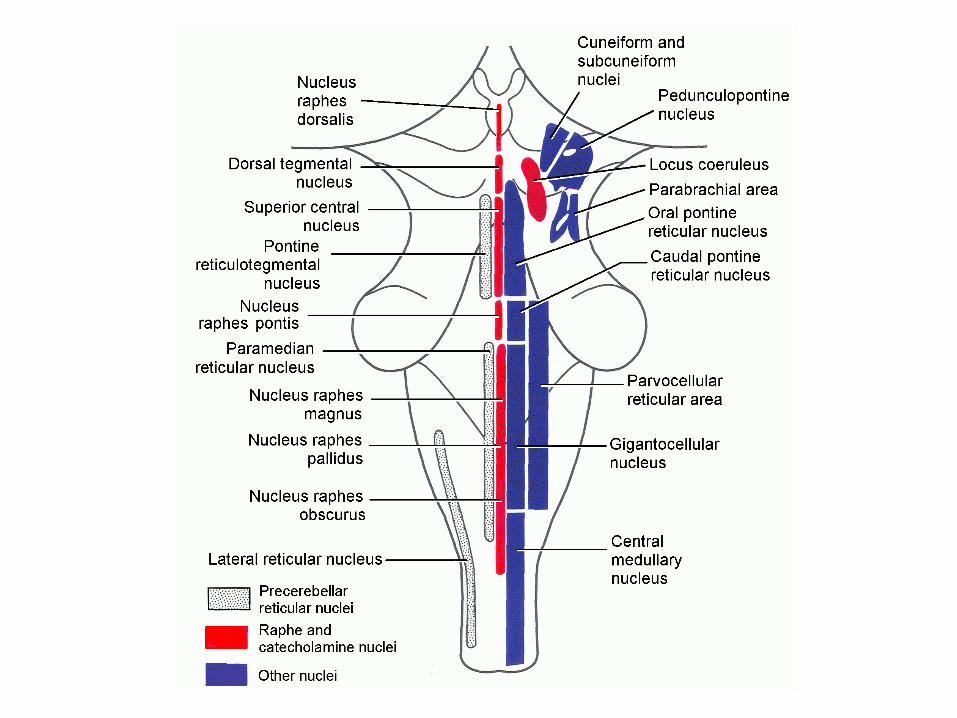

Precerebellar nuclei
• Includes lateral reticular nucleus, the paramedian reticular nucleus, and the pontine reticulotegmental nucleus. They project to the cerebellum. Their functions, associated with cerebellum are different from the rest of the reticular nuclei.


Raphe nuclei
• 1. Medullary raphe nuclei Include nucleus raphes magnus, nucleus raphes pallidus, and nucleus raphe obscurus.
• Afferent fibers are from the spinal cord, the gracile and cuneate nuclei, the trigeminal sensory nuclei, and the periaqueductal gray matter.
• Efferent fibers project to cerebellum, dorsal and ventral horns of the spinal gray matter, to the trigeminal nucleus, and to the preganglionic autonomic neurons of the brain stem and cord.
• Input from the periaqueductal gray matter and projection to spinal gray matter is responsible for suppressing the pain awareness.

Raphe nuclei
• 2. Raphe nuclei of the midbrain and rostral pons • Include nucleus raphes pontis, superior central nucleus,
nucleus raphes dorsalis, and dorsal tegmental necleus (P185)
• Afferent fibers: • Precentral cortex, and limbic system (hippocampal
formation, the hypothalamus, the interpeduncular nucleus, and the ventral tegmental area)
• Efferent fiber: • Forebrain, cerebellum, and noradrengeric nuclei of the
brain stem• Function: awareness of pain, and sleep pattern.


Central group of reticular nuclei
• Includes central medullary nucleus and the gigantocellular nucleus in the medulla, the caudal and oral pontine reticular nuclei, cuneiform and subcuneiform nuclei in the midbrain.


Central group of reticular nuclei• Afferent fibers: • From sensory system including spinoreticular tracts,
collateral tracts from spinothalamic tract and trigeminothalamic tracts, and collateral fibers form gustatory and auditory system. Visual information is provided by tectoreticular fibers originate from superior coliculus. Other afferent fibers are from fastigial nucleus of the vestibulocerebellum, reticular formation of the midbrain, cholinergic reticular nuclei, and the premotor area of the cerebral cortex.
• Efferent fibers: • To other reticular neurons (like raphe and catecholamine
neurons). Descending fibers constitute the medial (pontine) and lateral (medullary) reticulospinal tracts. Ascending fibers: to the intralaminar thalamic neuclei and the basal cholinergic nuclei of the substantia innominata. From there, fibers project to whole cortex for consciousness control and to corpus striatum for motor control.

Cholinergic and catecholamine nuclei
• Cholinergic nuclei• Includes the pedunculopontine nucleus and
nearby pontine periventricular gray matter.• Afferent fibers: • From pallidum and substantia nigra • Efferent fibers:• To central group of pontine and medullary
reticular nuclei, subthalamic nuclei, and corpus striatum.
• Function: Similar to central group, including locomotion, consciousness and arousal

Cholinergic and catecholamine nuclei
• Catecholamine nuclei
• Use NE, N and dopamine as neurotransmitter
• Includes locus coeruleus and other smaller groups of nuclei

Catecholamine nuclei• Afferent fibers:• From central group of reticular nuclei, and the nucleus
prepositus hypoglossi, the latter involves the control of eye movement. They also receive info from periaquctal gray matter and other components of the reticular formation, and from hypothalamus, amygdala, and prefrontal cortex.
• Efferent fiber:• Most of them travel in central tegmental tract. To
periaqueductal gray matter, and ascend to hypothalamus, basal cholinergic nuclei of the forebrain, and the amygdala. Some fibers end in thalamus, habenular nuclei, olfactory bulb, hippocampal formation and most of the neocortex.
• Other fibers descend to preganglionic autonomic neurons in the medulla and spinal cord
• Function: Modulator of synapses between neurons, also effects on alertness and reflexes

Parvocellular reticular area
• Afferent fibers:
• From sensory nuclei (trigeminal nuclei) and cerebral cortex
• Efferent fibers:
• To the motor nuclei of the hypoglossal, facial, and trigeminal nerves
• Function: reflexes with feeding

Parabrachial area
• includes lateral and medial parabrachial nuclei, and the Kolliker-Fuse nucleus
• Afferent fibers: • From solitary nucleus and from the cortex of
insula and adjacent parietal lobe• Efferent fibers: • To hypothalamus, preoptic area, intralaminar
thalamic nuclei, and amygdala.• Function: Relay station for visual sensation • Kolliker-Fuse nucleus is part of the pneumotaxic
center regulating the respiratory rhythm.

Superficial medullary reticular neurons
• Afferent fibers: • from spinal cord and solitary nucleus. Include fibers
sending information from chemoreceptor in the carotid and aortic sinuses. Some of these neurons can respond directly to changes of pH or carbon dioxide.
• Efferent fibers: • project to the hypothalamus and preganglionic
autonomic neurons in the medulla and spinal cord.• Function: regulates cardiovascular and respiratory
activities

Functions of the reticular formation in general
• Sleep and arousal • Varying level of consciousness is paralleled by changes
in neuronal activities, which can also be expressed in EEG. We currently believe that ascending activating system (originate from reticular nuclei to thalamus rather than to whole cerebral) increases general level of sensory stimulation thus lead to increased level of neuronal activities and increased arousal level. The noradrenergic neurons and histamine secreting neurons also stimulate arousal level.
• Sleep inducing neurons, like serotonergic neurons of the raphe nuclei could inhibit thalamus and cerebral function thus decrease arousal level.

Functions of the reticular formation
• Pain
• A descending inhibitory pathway could inhibit transmission of pain. Peptide neurotransmitters (like opiate drugs, morphine) released at synapses of periaqueductal gray matter, raphe nuclei, and dorsal horn of spinal gray matter could block the pain sensation.

Functions of the reticular formation
• Somatic motor function • reticulospinal is the major descending pathway
controls the motor movement. Most of the descending fibers run ipsilaterally to spinal cord level, some terminate ipsilaterally and others cross at white commissure of spinal cord to the contralateral side. Therefore, the reticulospinal innervation is bilaterally.
• Visceral activities• Certain regions of reticular formation regulate
visceral functions and breathing.