· Web viewbasic tissue: lymphoreticular- reticular connective tissue, reticular fibers, reticular...
15
Histology Practical 24) Lymph node 25) Thymus 26) Spleen 27) Palatine Tonsil 28) Root of Tounge General Information about lymphatic tissue: Red bone marrow Primary Lymphatic organs. All others are secondary. Thymus What is the stroma of the Network? Reticular fibers! Native lymphocytes are released from primary lymphatic organs but have to be immune competent Only secondary lymphatic Organs posses lymphatic follicles B-Cells = immunoglobines require B-Cell Receptors (BCR) therefore lymphatic follicles occur function: keeping the own antigen environment, eliminating the foreign substances/antigens lymphoid tissue lymphoid organs: primary (red bone marrow, thymus): development of lymphocytes secondrary(lymph node, spleen, tonsils, Peyer s patches, appendix): mature lymphocytes can be found here basic tissue: lymphoreticular- reticular connective tissue, reticular fibers, reticular cells; thymus: epithelial reticular cells main cell types: lymphocytes, macrophages, dendritic cells, endothelial cells Lymphoid follicles unit of the lymphoid tissue round, oval structure, many lymphocytes primary: dark, ruond, little lymphocytes, intrauterine secondary: bigger (200-400 m), originating from the primary, in the middle: light area- germinal center (centroblasts, centrocytes, FDC), peripheral: cap zone 24) Lymph node Mantle zone Lymphatic follicles Copyright by Matthias Heyner Based on the Histology practical from E. Dobo et. al at the University of Szeged videos by WashingtonDeceit (www.youtube.com/user/WashingtonDecit)
Transcript of · Web viewbasic tissue: lymphoreticular- reticular connective tissue, reticular fibers, reticular...

Histology Practical
24) Lymph node 25) Thymus 26) Spleen 27) Palatine Tonsil 28) Root of Tounge
General Information about lymphatic tissue:Red bone marrow Primary Lymphatic organs. All others are
secondary.Thymus
What is the stroma of the Network? Reticular fibers! Native lymphocytes are released from primary lymphatic organs but have to be immune competent Only secondary lymphatic Organs posses lymphatic follicles B-Cells = immunoglobines require B-Cell Receptors (BCR) therefore lymphatic follicles occur function: keeping the own antigen environment, eliminating the foreign substances/antigens lymphoid tissue lymphoid organs: primary (red bone marrow, thymus): development of lymphocytes secondrary(lymph node, spleen, tonsils, Peyer s patches, appendix): mature lymphocytes can be found here basic tissue: lymphoreticular- reticular connective tissue, reticular fibers, reticular cells; thymus: epithelial reticular cells main cell types: lymphocytes, macrophages, dendritic cells, endothelial cells
Lymphoid follicles
unit of the lymphoid tissue round, oval structure, many lymphocytes primary: dark, ruond, little lymphocytes, intrauterine secondary: bigger (200-400 m), originating from the primary, in the middle: light area- germinal center (centroblasts, centrocytes, FDC), peripheral: cap zone
24) Lymph node
Mantle zone Lymphatic follicles Cortex (inner / outer zone) Anastomosing network of lymphoids Medulla:
o Medullary cord (dark) o Medullary sinuses (pale)
Cortex: inner B-Cell Zone Medulla: B-Cell zone along the course of lymphatic vessels bean-shaped, encapsulated organs afferent lymphatic vessels efferent lymphatic vessels connective tissue capsule trabeculae hilus
Copyright by Matthias Heyner Based on the Histology practical from E. Dobo et. al at the University of Szeged videos by WashingtonDeceit (www.youtube.com/user/WashingtonDecit)
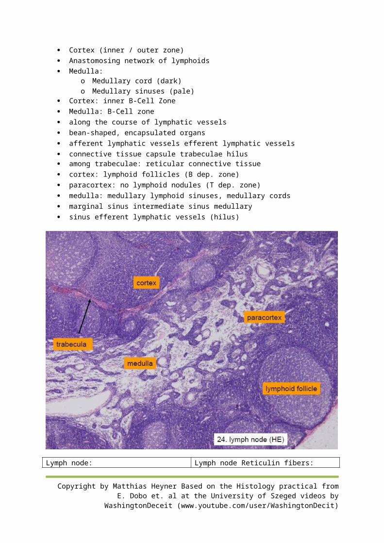
among trabeculae: reticular connective tissue cortex: lymphoid follicles (B dep. zone) paracortex: no lymphoid nodules (T dep. zone) medulla: medullary lymphoid sinuses, medullary cords marginal sinus intermediate sinus medullary sinus efferent lymphatic vessels (hilus)
Lymph node: Lymph node Reticulin fibers:
CD (cluster differentiation) marker: CD19 & CD20: B-Cells
Copyright by Matthias Heyner Based on the Histology practical from E. Dobo et. al at the University of Szeged videos by WashingtonDeceit (www.youtube.com/user/WashingtonDecit)
CD3: all CD4: helper CD8: cytotoxic
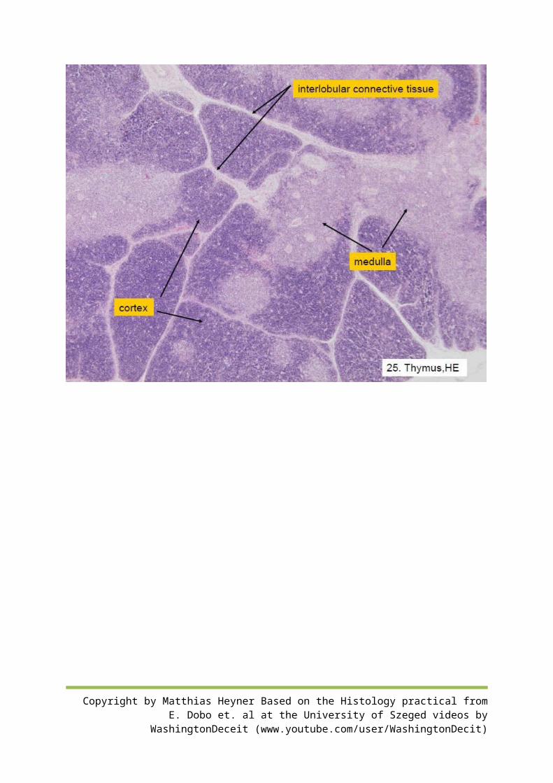
25) Thymus
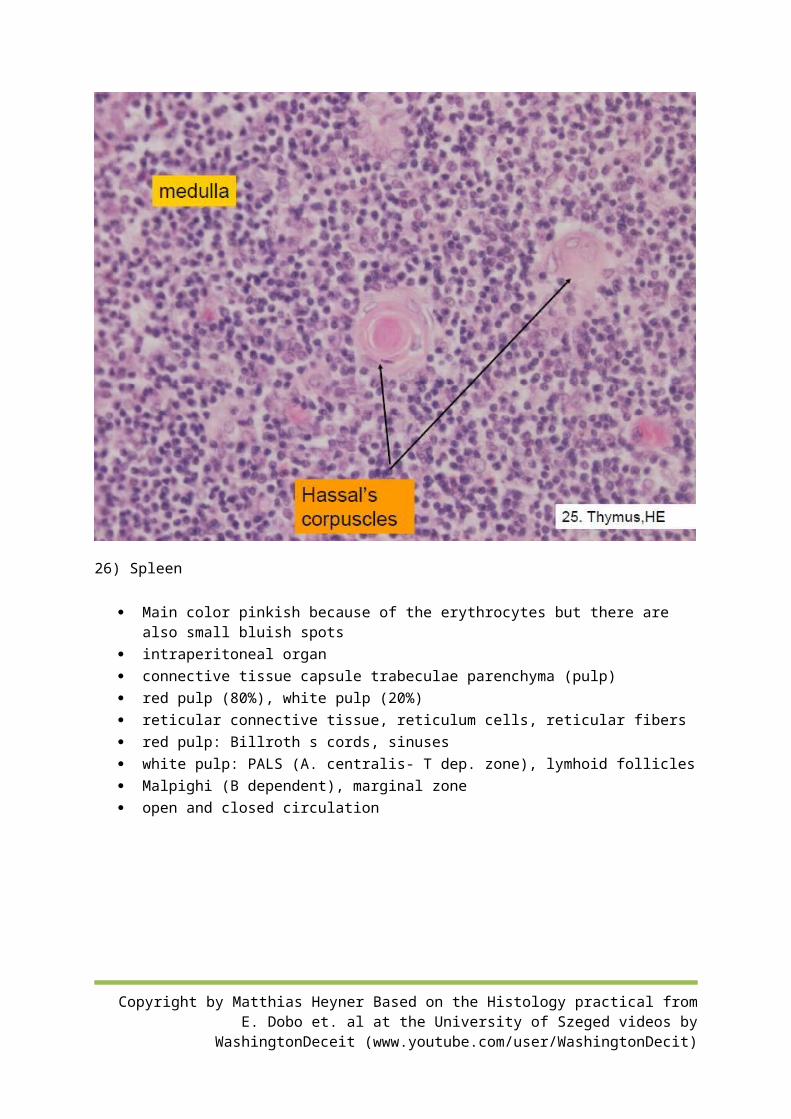
Lobulated organ, it has septa which separate the lobules Cortex is darker Medulla is paler Lymphocytes wander from cortex to medulla In the Medulla there are larger cells No incoming just outgoing. Hassals corpuscle
o Pinkish mass in center o Can only be found in Medulla o Has no function for thymus
epithelial, lymphatic, adipose form during life 2 lobes+ capsule lobules cortex: peripheral, darker, thymocytes, macrophages, epithelial reticulum cells medulla: central, lighter, more epithelial reticulum cells, Hassal-corpuscules (eosinophil,concentrically arranged,flattened epithelial cells with
keratohyalingranules), neuroendocrine cells, myoid (Hammar)cells no lymph follicles function: mature and differentiation of T lymphocytes blood-thymus barrier: pericytes, capillary basal lamina, basal lamina of the epithelial reticular
cell, cells of the endothelium, epithelial reticular cells
Copyright by Matthias Heyner Based on the Histology practical from E. Dobo et. al at the University of Szeged videos by WashingtonDeceit (www.youtube.com/user/WashingtonDecit)
Copyright by Matthias Heyner Based on the Histology practical from E. Dobo et. al at the University of Szeged videos by WashingtonDeceit (www.youtube.com/user/WashingtonDecit)
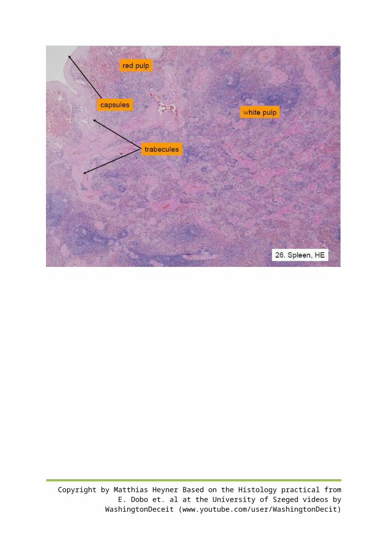
26) Spleen
Main color pinkish because of the erythrocytes but there are also small bluish spots intraperitoneal organ connective tissue capsule trabeculae parenchyma (pulp) red pulp (80%), white pulp (20%) reticular connective tissue, reticulum cells, reticular fibers red pulp: Billroth s cords, sinuses white pulp: PALS (A. centralis- T dep. zone), lymhoid follicles Malpighi (B dependent), marginal zone open and closed circulation
Copyright by Matthias Heyner Based on the Histology practical from E. Dobo et. al at the University of Szeged videos by WashingtonDeceit (www.youtube.com/user/WashingtonDecit)
Copyright by Matthias Heyner Based on the Histology practical from E. Dobo et. al at the University of Szeged videos by WashingtonDeceit (www.youtube.com/user/WashingtonDecit)
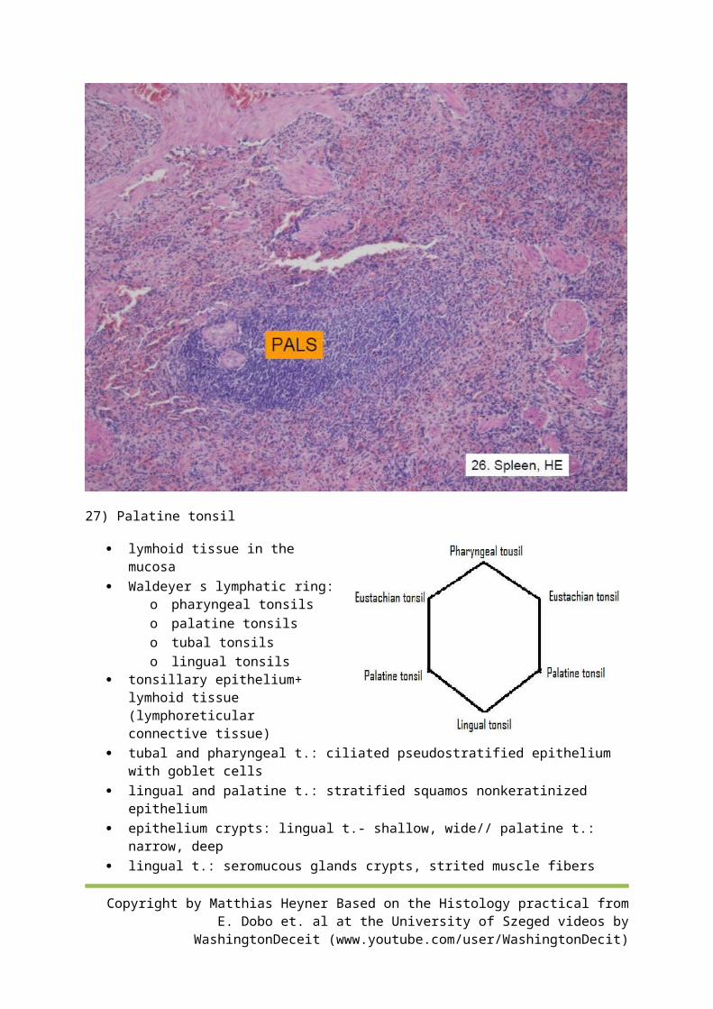
27) Palatine tonsil
lymhoid tissue in the mucosa Waldeyer s lymphatic ring:
o pharyngeal tonsils o palatine tonsils o tubal tonsils o lingual tonsils
tonsillary epithelium+ lymhoid tissue (lymphoreticular connective tissue)
tubal and pharyngeal t.: ciliated pseudostratified epithelium with goblet cells
lingual and palatine t.: stratified squamos nonkeratinized epithelium
epithelium crypts: lingual t.- shallow, wide// palatine t.: narrow, deep lingual t.: seromucous glands crypts, strited muscle fibers follicles (B dep.), interfollicular area (T dep.) epithelium is often infiltrated with lymphocytes
Palatine tonsil
Copyright by Matthias Heyner Based on the Histology practical from E. Dobo et. al at the University of Szeged videos by WashingtonDeceit (www.youtube.com/user/WashingtonDecit)
Pharyngeal tonsil
Lingual tonsil
Copyright by Matthias Heyner Based on the Histology practical from E. Dobo et. al at the University of Szeged videos by WashingtonDeceit (www.youtube.com/user/WashingtonDecit)
Copyright by Matthias Heyner Based on the Histology practical from E. Dobo et. al at the University of Szeged videos by WashingtonDeceit (www.youtube.com/user/WashingtonDecit)
Copyright by Matthias Heyner Based on the Histology practical from E. Dobo et. al at the University of Szeged videos by WashingtonDeceit (www.youtube.com/user/WashingtonDecit)