Regulation and Significance of Hepatocyte-Derived Matrix Metalloproteinases in Liver Remodeling
6
Regulation and Significance of Hepatocyte-Derived Matrix Metalloproteinases in Liver Remodeling Takahiro Haruyama, Itsuki Ajioka, Toshihiro Akaike, and Yoshifumi Watanabe 1 Department of Biomolecular Engineering, Tokyo Institute of Technology, 4259 Nagatsuda, Midori-ku, Yokohama 226-8501 Japan Received May 6, 2000 Regulation in expression and activation of protein- ases is one of the most important mechanisms in organ morphogenesis. In this study, we investigated the ex- pression of MMPs in primary hepatocytes and their roles in liver remodeling. A hepatocyte proliferation initiating cytokine, TNFa, induced MMP-9 expression in these cells while the expression of MMP-2 did not change by zymography analysis. Interestingly, both the induced MMP-9 expression and hepatocyte prolif- eration by TNFa were synergistically enhanced by HGF in vitro. The increased proliferation was sup- pressed by MMP inhibitor TIMP-1, suggesting that cytokine-induced MMP regulates proliferation. The increased expression of MMP-9 by the cytokines was inhibited by cytochalasin D or colchicine but not by PI3 kinase inhibitor wortmannin. In addition, co- stimulation by TNFa and HGF of spheroidal hepato- cytes cultured in 3-dimensional collagen gel drasti- cally induced morphological changes by cell extension and migration in the gel, which was in parallel with the induced expression of MMP-9 and was inhibited by TIMP-1 and -2. The MMP activity was also detected in vivo in the regenerating liver after partial hepatec- tomy by in situ zymography. These results suggest the roles of MMPs produced by parenchymal cells in liver remodeling. © 2000 Academic Press Organ remodeling is composed of strictly controlled multiple processes such as cell proliferation, cell death, migration and invasion, extracellular matrix (ECM) synthesis, and degradation. Liver regeneration after partial hepatectomy is one of the best-studied exam- ples of organ remodeling. The process is regulated by various molecules. Among these molecules, proteinases such as matrix metalloproteinases (MMPs) are thought to be mainly involved in the ECM degradation, which could also control cell proliferation via integrin signaling and growth factor activation. Recent studies demonstrated that proteinases play important roles in not only ECM remodeling (1) but also hepatocyte pro- liferation (2–5). MMPs are a large family of zinc requiring matrix- degrading enzymes. They have been implicated in in- vasive cell behavior and in embryonic development and organ morphogenesis. These enzymes are also sup- posed to be essential for liver remodeling after PH and the expression of some MMPs and their endogenous inhibitors, TIMP-1 and -2, were investigated in the regenerating liver (6, 7). However, the regulation and function of MMPs involved in the regenerating liver is still unclear. In terms of cell types, information has been accumulated in the MMP-2 regulation by hepatic stellate cells (8 –12). In contrast, there is little infor- mation about MMPs regulated by parenchymal cells. Therefore, in this article, we examined the expression and function of MMPs produced by primary hepato- cytes. Isolated primary hepatocytes produced constitu- tive MMP-2 and MMP-9 increased by the stimulation of liver regeneration related cytokines, TNFa and HGF. The produced MMPs increased cytokine-induced hepatocyte proliferation and induced cell invasion and migration in 3-D collagen gel. These results suggests that the hepatocyte-derived MMPs play essential roles in liver remodeling. MATERIALS AND METHODS Reagents. Recombinant human TNFa, human HGF, and human EGF were kindly provided by Asahi Chemical Inc. (Tokyo, Japan), Snow Brand Milk Products Co., Ltd. (Tokyo, Japan), and Hitachi Chemical Co. Ltd. (Tokyo, Japan), respectively. Polyclonal anti- mouse MMP-9 antibody was purchased from Santa Cruz Biotechnol- ogy (Santa Cruz, CA). Cytochalasin D, colchicine, and wortmannin were obtained from Sigma Chemical Co. (St. Louis, MO). The female ICR mice employed in the experiments in this study were purchased from Charles River Japan, Inc. (Kanagawa, Japan). All animal ex- periments were conducted in accordance with local institutional guidelines for the care and use of laboratory animals. Cell culture. Parenchymal hepatocytes were prepared as previ- ously described (13). Briefly, a liver was perfused with a 0.0125% 1 To whom correspondence should be addressed. Fax: 81-45-924- 5815. E-mail: [email protected]. Biochemical and Biophysical Research Communications 272, 681– 686 (2000) doi:10.1006/bbrc.2000.2837, available online at http://www.idealibrary.com on 681 0006-291X/00 $35.00 Copyright © 2000 by Academic Press All rights of reproduction in any form reserved.
-
Upload
takahiro-haruyama -
Category
Documents
-
view
213 -
download
1
Transcript of Regulation and Significance of Hepatocyte-Derived Matrix Metalloproteinases in Liver Remodeling
RM
TD4
R
ampriicteHpciiPsccatTvtrr
mmsppvstw
5
Biochemical and Biophysical Research Communications 272, 681–686 (2000)
doi:10.1006/bbrc.2000.2837, available online at http://www.idealibrary.com on
egulation and Significance of Hepatocyte-Derived Matrixetalloproteinases in Liver Remodeling
akahiro Haruyama, Itsuki Ajioka, Toshihiro Akaike, and Yoshifumi Watanabe1
epartment of Biomolecular Engineering, Tokyo Institute of Technology,259 Nagatsuda, Midori-ku, Yokohama 226-8501 Japan
eceived May 6, 2000
signaling and growth factor activation. Recent studiesdnl
dvoptirfsbsmTactoHhmti
M
ESCmowIfpg
o
Regulation in expression and activation of protein-ses is one of the most important mechanisms in organorphogenesis. In this study, we investigated the ex-
ression of MMPs in primary hepatocytes and theiroles in liver remodeling. A hepatocyte proliferationnitiating cytokine, TNFa, induced MMP-9 expressionn these cells while the expression of MMP-2 did nothange by zymography analysis. Interestingly, bothhe induced MMP-9 expression and hepatocyte prolif-ration by TNFa were synergistically enhanced byGF in vitro. The increased proliferation was sup-ressed by MMP inhibitor TIMP-1, suggesting thatytokine-induced MMP regulates proliferation. Thencreased expression of MMP-9 by the cytokines wasnhibited by cytochalasin D or colchicine but not byI3 kinase inhibitor wortmannin. In addition, co-timulation by TNFa and HGF of spheroidal hepato-ytes cultured in 3-dimensional collagen gel drasti-ally induced morphological changes by cell extensionnd migration in the gel, which was in parallel withhe induced expression of MMP-9 and was inhibited byIMP-1 and -2. The MMP activity was also detected inivo in the regenerating liver after partial hepatec-omy by in situ zymography. These results suggest theoles of MMPs produced by parenchymal cells in liveremodeling. © 2000 Academic Press
Organ remodeling is composed of strictly controlledultiple processes such as cell proliferation, cell death,igration and invasion, extracellular matrix (ECM)
ynthesis, and degradation. Liver regeneration afterartial hepatectomy is one of the best-studied exam-les of organ remodeling. The process is regulated byarious molecules. Among these molecules, proteinasesuch as matrix metalloproteinases (MMPs) arehought to be mainly involved in the ECM degradation,hich could also control cell proliferation via integrin
1 To whom correspondence should be addressed. Fax: 81-45-924-815. E-mail: [email protected].
681
emonstrated that proteinases play important roles inot only ECM remodeling (1) but also hepatocyte pro-
iferation (2–5).MMPs are a large family of zinc requiring matrix-
egrading enzymes. They have been implicated in in-asive cell behavior and in embryonic development andrgan morphogenesis. These enzymes are also sup-osed to be essential for liver remodeling after PH andhe expression of some MMPs and their endogenousnhibitors, TIMP-1 and -2, were investigated in theegenerating liver (6, 7). However, the regulation andunction of MMPs involved in the regenerating liver istill unclear. In terms of cell types, information haseen accumulated in the MMP-2 regulation by hepatictellate cells (8–12). In contrast, there is little infor-ation about MMPs regulated by parenchymal cells.herefore, in this article, we examined the expressionnd function of MMPs produced by primary hepato-ytes. Isolated primary hepatocytes produced constitu-ive MMP-2 and MMP-9 increased by the stimulationf liver regeneration related cytokines, TNFa andGF. The produced MMPs increased cytokine-inducedepatocyte proliferation and induced cell invasion andigration in 3-D collagen gel. These results suggests
hat the hepatocyte-derived MMPs play essential rolesn liver remodeling.
ATERIALS AND METHODS
Reagents. Recombinant human TNFa, human HGF, and humanGF were kindly provided by Asahi Chemical Inc. (Tokyo, Japan),now Brand Milk Products Co., Ltd. (Tokyo, Japan), and Hitachihemical Co. Ltd. (Tokyo, Japan), respectively. Polyclonal anti-ouse MMP-9 antibody was purchased from Santa Cruz Biotechnol-
gy (Santa Cruz, CA). Cytochalasin D, colchicine, and wortmanninere obtained from Sigma Chemical Co. (St. Louis, MO). The female
CR mice employed in the experiments in this study were purchasedrom Charles River Japan, Inc. (Kanagawa, Japan). All animal ex-eriments were conducted in accordance with local institutionaluidelines for the care and use of laboratory animals.
Cell culture. Parenchymal hepatocytes were prepared as previ-usly described (13). Briefly, a liver was perfused with a 0.0125%
0006-291X/00 $35.00Copyright © 2000 by Academic PressAll rights of reproduction in any form reserved.
collagenase solution. After the liver had been excised, parenchymalhtc(wtpctwcbfsstg
fcsaaaIsz
BlabAm
efamlk
R
eghlraaHphT1teactw
ifasIac2ipi
ipcdtdpmHvo
htwCcnTsp2
Vol. 272, No. 3, 2000 BIOCHEMICAL AND BIOPHYSICAL RESEARCH COMMUNICATIONS
epatocytes were separated from nonparenchymal cells by differen-ial centrifugation at 50g for 90 s. The dead parenchymal hepato-ytes were removed by density gradient centrifugation on PercollPharmacia, Uppsala, Sweden). The viable parenchymal hepatocytesere suspended in Williams’ E medium containing antibiotics and
hen plated at a density of 1 3 104 cells/well in flat-bottomed 96-welllates (Sumitomo Bakelite Co., Ltd., Tokyo, Japan) pre-coated withollagen. The hepatocytes were incubated at 37°C for 3 h in order forhem to adhere to the collagen-coated plates and then the mediumas changed to a new one before the experiments. For collagen gel
ulture, isolated hepatocytes were cultured on BSA-coated hydropho-ic dishes for 48 h in the presence of EGF (50 ng/ml), resulted in theormation of spheroids. These spheroids were transferred onto pre-olidified collagen gels. After the cells sedimented, upper collagenolution was overlaid and formed sandwich gels containing cells inhe interface. The cells in the gels were cultured with serum orrowth factor free medium.
Gelatin zymography. Supernatants from cultured cells or lysatesrom tissues were electrophoresed on 7.5% SDS-polyacrylamide gelontaining 0.1% gelatin. The gel was washed with 0.1% Triton X-100olution for 30 min and then incubated at 37°C for overnight in thectivation buffer (50 mM Tris–HCl, pH 7.4, 0.2 M NaCl, 5 mM CaCl2,nd 0.02% NaN3). After staining with CBB R-250, the gelatinolyticctivities were detected as clear bands against the blue background.n some cases, MMPs in supernatants were concentrated by gelatin-epharose (Pharmacia, Uppsala, Sweden). The contrast images ofymography were inverted to facilitate the band detection.In situ zymography was basically performed as described (2).riefly, freshly frozen liver sections on coverslips were directly over-
ayed with 0.7% agarose containing 0.1% gelatin or 0.1% BSA in thectivation buffer. The samples were incubated in a humidified cham-er at 37°C overnight and then counterstained with crystal violet.fter gentle washing, the samples were observed by a phase-contrasticroscopy.
Western blotting. After SDS–PAGE on a 7.5% gel, proteins werelectroblotted onto PVDF membranes (Amersham, IL) in cold trans-er buffer for 3 h at 60 V. The filters were incubated with anti-MMP-9ntibodies for 1 h at room temperature after blocking with 3% skimilk, washed with TBS, and then incubated with the peroxidase-
abeled second antibodies for 1 h. Bands were detected with an ECLit (Amersham, Illinois).
ESULTS
Expression of hepatocyte-derived MMPs. We firstxamined the effect of cytokines involved in liver re-eneration on the expression of MMPs in primaryepatocytes since these cytokines are thought to regu-
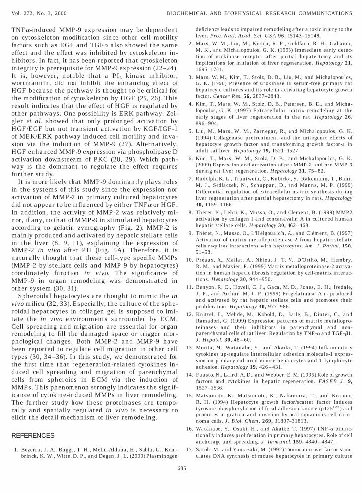
ate not only hepatocyte proliferation but also organemodeling. Liver regeneration is a very complicatednd exquisitely controlled process and many moleculesre involved in the mechanism. Among them, TNFa,GF, EGF, or TGFa are thought to contribute to theroliferation of parenchymal cells (14). When primaryepatocytes were stimulated with these cytokines, onlyNFa induced MMP-9 activity but not MMP-2 (Fig.A). Interestingly, the activity of MMP-9 was synergis-ically enhanced by either HGF, EGF, or TGFa. Theffect was synergistic since, as shown in Fig. 1B, thectivity was dependent on both TNFa and HGF at highoncentration. Western blot analysis confirmed thathe induction and increasing effect of MMP-9 activityas due to the increased expression of the correspond-
682
ng protein (Fig. 1C). We noticed that all the enhancingactors (HGF, EGF, or TGFa) of MMP-9 expression arelso known as cell motility factors. Thus, the effect wasupposed to be derived from cytoskeleton modification.n fact, both cytoskeleton inhibitors, cytochalasin Dnd colchicine dose-dependently inhibited the in-reased expression of MMP-9 by TNFa and HGF (Fig.). In contrast, it is noteworthy that PI3 kinase inhib-tor, wortmannin, did not have any effect on the ex-ression despite that the inhibitor blocked the signal-ng process from HGF receptor to cytoskeleton (15).
Roles of hepatocyte-derived MMPs in liver remodel-ng in vitro. Organ remodeling is performed by im-eccably orchestrated combination of cell proliferation,ell migration, cell death, ECM degradation, and pro-uction. MMPs are thought to play multiple roles inhese processes. In the following figures, we clearlyemonstrated that hepatocyte-derived MMPs partici-ate in the regulation of hepatocyte proliferation andigration. Hepatocyte proliferation was induced byGF, and TNFa also induced weaker proliferation in
itro as reported (16, 17). Intriguingly, the combinationf TNFa and HGF showed the synergistic positive ef-
FIG. 1. Cytokine-induced MMP-9 expression in primary mouseepatocytes. Freshly isolated mouse hepatocytes were cultured inhe absence or presence of cytokines for 48 h, then the supernatantsere subjected to zymography (A and B) or Western blotting (C). (A)ytokine induction of MMP-9 activity in primary hepatocytes. 1;ontrol, 2; HGF (100 ng/ml), 3; EGF (100 ng/ml), 4; TGF-a (100g/ml), 5; TNFa (100 ng/ml), 6; TNFa 1 HGF, 7; TNFa 1 EGF, 8;NFa 1 TGFa (B) Dose-dependent induction and increased expres-ion of MMP-9 in hepatocytes by TNFa and HGF. (C) The MMP-9rotein expression by TNFa and HGF by Western blotting. 1; control,; HGF (100 ng/ml), 3; TNFa (50 ng/ml), 4; TNFa 1 HGF.

fn(Tntrmedghiritlcsddhss(th(iiio
wcaMlaa
active forms of both MMP-2 and MMP-9 were detectedizu(t
D
ooTtalm
p(wTiD
cwtcaz
Vol. 272, No. 3, 2000 BIOCHEMICAL AND BIOPHYSICAL RESEARCH COMMUNICATIONS
ect on in vitro hepatocyte proliferation as the combi-ation showed in the induction of MMP-9 expressionFig. 3A). In addition, the increased DNA synthesis byNFa and HGF was dose-dependently suppressed by aatural MMP inhibitor, TIMP-1 (Fig. 3B), implyinghat cell proliferation induced by TNFa and HGF isegulated by autocrinally produced MMPs. Further-ore, this cytokine combination also showed a drastic
ffect on the organ morphological changes of threeimensional cultured hepatocyte spheroids in collagenel, which is considered to mimic the in vivo state ofepatocytes. TNFa slightly induced the cell extension
n or spike formation at the perimeters of the sphe-oids. In contrast, the combination of TNFa and HGFnduced the significant cell extension and migration inhese cells while HGF alone did not show any morpho-ogical effect (Fig. 4A). The spheroidal formation wasompletely collapsed and these cells were randomlycattered The drastic morphological changes were evi-ently induced by the cytokine-induced, hepatocyte-erived MMPs since the changes were completely in-ibited by TIMP-1. In addition, in this 3-D culturedystem, we also detected the cytokine-induced expres-ion of MMP-9 but not MMP-2 by gelatin zymographyFig. 4B). The different patterns of zymography be-ween monolayer (Fig. 1) and 3-D culture (Fig. 4) ofepatocytes might suggest that cell-cell interactionpossibly via E-cadherin) or 3-D interaction with ECMs also involved in the regulation of MMPs expressionn hepatocytes. Further experiments are required fordentification of the unknown bands in the zymographyf 3-D culture.
States of MMPs in regenerating liver. To examinehether MMPs function in liver remodeling in vivo, we
hecked the states of MMP activation in the regener-ting liver, a natural organ remodeling. MMP-9 andMP-2 activities were detected in zymography of the
ysates from the regenerating livers after partial hep-tectomy (Fig. 5A). These activities were observed 3 hfter PH and gradually decreased to 48 h after. The
FIG. 2. Cytokine-induced MMP-9 expression in primary hepato-ytes is regulated by cytoskeleton signaling. Primary hepatocytesere cultured with TNFa (100 ng/ml) and HGF (50 ng/ml) for 48 h in
he presence or absence of cytochalasin D (CytD) (0.2, 1, 5 mM),olchicine (Col) (0.2, 1, 5 mM), or wortmannin (Wtn) (5, 50, 500 nM)t various concentrations, then the supernatants were subjected toymography. *TNFa (50 ng/ml) 1 HGF (100 ng/ml).
683
n the regenerating liver (Fig. 5A). In addition, in situymography showed that the activity evenly distrib-ted in the histological section of the regenerating liverFig. 5B). The results directly demonstrates the activa-ion of MMPs in the regenerating liver.
ISCUSSION
Liver regeneration is a suitable model for the studyf organ remodeling. The process is regulated by vari-us types of molecules. Cytokines such as TNFa, EGF,GFa or HGF are considered to play critical roles inhe process (14). Of these cytokines, evidence has beenccumulated that TNFa is an initiation factor in theiver regeneration (14, 18, 19) although the detail
echanism is unclear. In this study, we demonstrated
FIG. 3. Involvement of hepatocyte-derived MMPs in hepatocyteroliferation. (A) Primary hepatocytes were cultured with cytokinesTNFa; 50 ng/ml, HGF; 100 ng/ml) for 48 h, then the DNA synthesisas measured as described. (B) Hepatocytes were cultured withNFa (50 ng/ml) 1 HGF (100 ng/ml) in the presence of a MMP-
nhibitor, TIMP-1 at the indicated concentrations for 48 h, and theNA synthesis was measured.

thTtTMS
teinases to trigger hepatocyte proliferation in vivo (2–5tthebcfMttMbip(
dsSfitta
Tttwtdg
ccgTAEttH
Vol. 272, No. 3, 2000 BIOCHEMICAL AND BIOPHYSICAL RESEARCH COMMUNICATIONS
hat TNFa induced MMP-9 expression in primaryepatocytes and the expression was enhanced by HGF.he same combination synergistically increased hepa-ocyte proliferation which was inhibited by TIMP-1.hese data indicated that TNFa and HGF inducedMPs critically regulated hepatocyte proliferation.
ome researchers have shown the significance of pro-
FIG. 4. Hepatocyte-derived MMPs induced morphologicalhanges in 3-D cultured spheroids. Spheroidal hepatocytes pre-ultured on BSA-coated dishes were entrapped in sandwiched colla-en gel, then cultured for 5 days in the presence of cytokines. (A)hese cells were observed under a phase contrast microscopy (380).; control, B; HGF (100 ng/ml), C; TNFa (50 ng/ml), D; TNFa 1 HGF,; TNFa 1 HGF 1 TIMP-1 (5 mg/ml). (B) The cell supernatants from
he collagen gels were subjected to zymography after being concen-rated by gelatin-sepharose. 1; control, 2; HGF, 3; TNFa, 4; TNFa 1GF. The arrow indicates MMP-9.
684
). They suggested that the proteinases may processhe proform of growth factors such as HGF, result inhe production of active form which would trigger theepatocyte proliferation. However, in our system, thexplanation is not suitable since we employed recom-inant active HGF. It is more plausible that theytokine-induced function as a growth factor or co-actor to hepatocytes. In fact, it has been reported that
MP-2 might be a growth factor in another liver cellype (11). However, the possibility can not be ruled outhat the disruption of ECM by the cytokine-inducedMPs which would modify the integrin signaling may
e involved in the mechanism. Taken together, TNFas thought to function as an initiator of hepatocyteroliferation by directly increasing the proliferation16) and inducing MMPs in hepatocytes.
The detail mechanism in which TNFa and HGF in-uces MMP-9 expression in hepatocytes remains to beolved. However, some mechanisms can be speculated.ince the activation of NF-kB signaling is important
or the expression of MMP-9 (20), TNFa may directlynduce MMP-9 via the activation of NF-kB. Alterna-ively, TNFa might indirectly induce MMP-9 in hepa-ocytes via the induction of ceramide as reported innother cell type (21). The enhancement by HGF of
FIG. 5. Activities of MMPs in the regenerating liver in vivo. (A)he regenerating livers were excised out at the indicated times andhe homogenized lysates were subjected to gelatin zymography afterhe protein concentrations were adjusted. (B) The sections (10 mm)ere prepared from the regenerating liver 48 h after partial hepa-
ectomy (PH), then in situ gelatin zymography was performed asescribed. A; normal liver with gelatin zymography, B; PH liver withelatin zymography, C; PH liver with BSA zymography.
TNFa-induced MMP-9 expression may be dependentofehiIwHtrogHosHawf
iadInamiMn(cMo
vrtCrpbttdcMiTre
R
deficiency leads to impaired remodeling after a toxic injury to the
1
1
1
1
1
1
1
1
Vol. 272, No. 3, 2000 BIOCHEMICAL AND BIOPHYSICAL RESEARCH COMMUNICATIONS
n cytoskeleton modification since other cell motilityactors such as EGF and TGFa also showed the sameffect and the effect was inhibited by cytoskeleton in-ibitors. In fact, it has been reported that cytoskeleton
ntegrity is prerequisite for MMP-9 expression (22–24).t is, however, notable that a PI3 kinase inhibitor,ortmannin, did not inhibit the enhancing effect ofGF because the pathway is thought to be critical for
he modification of cytoskeleton by HGF (25, 26). Thisesult indicates that the effect of HGF is regulated byther pathways. One possibility is ERK pathway. Zei-ler et al. showed that only prolonged activation byGF/EGF but not transient activation by KGF/IGF-1
f MEK/ERK pathway induced cell motility and inva-ion via the induction of MMP-9 (27). Alternatively,GF enhanced MMP-9 expression via phospholipase Dctivation downstream of PKC (28, 29). Which path-ay is the dominant to regulate the effect requires
urther study.It is more likely that MMP-9 dominantly plays roles
n the systems of this study since the expression norctivation of MMP-2 in primary cultured hepatocytesid not appear to be influenced by either TNFa or HGF.n addition, the activity of MMP-2 was relatively mi-or, if any, to that of MMP-9 in stimulated hepatocytesccording to gelatin zymography (Fig. 2). MMP-2 isainly produced and activated by hepatic stellate cells
n the liver (8, 9, 11), explaining the expression ofMP-2 in vivo after PH (Fig. 5A). Therefore, it is
aturally thought that these cell-type specific MMPsMMP-2 by stellate cells and MMP-9 by hepatocytes)oordinately function in vivo. The significance ofMP-9 in organ remodeling was demonstrated in
ther system (30, 31).Spheroidal hepatocytes are thought to mimic the in
ivo milieu (32, 33). Especially, the culture of the sphe-oidal hepatocytes in collagen gel is supposed to imi-ate the in vivo environments surrounded by ECM.ell spreading and migration are essential for organ
emodeling to fill the damaged space or trigger mor-hological changes. Both MMP-2 and MMP-9 haveeen reported to regulate cell migration in other cellypes (30, 34–36). In this study, we demonstrated forhe first time that regeneration-related cytokines in-uced cell spreading and migration of parenchymalells from spheroids in ECM via the induction ofMPs. This phenomenon strongly indicates the signif-
cance of cytokine-induced MMPs in liver remodeling.he further study how these proteinases are tempo-ally and spatially regulated in vivo is necessary tolicit the detail mechanism of liver remodeling.
EFERENCES
1. Bezerra, J. A., Bugge, T. H., Melin-Aldana, H., Sabla, G., Kom-brinck, K. W., Witte, D. P., and Degen, J. L. (2000) Plasminogen
685
liver. Proc. Natl. Acad. Sci. USA 96, 15143–15148.2. Mars, W. M., Liu, M., Kitson, R. P., Goldfarb, R. H., Gabauer,
M. K., and Michalopoulos, G. K. (1995) Immediate early detec-tion of urokinase receptor after partial hepatectomy and itsimplications for initiation of liver regeneration. Hepatology 21,1695–1701.
3. Mars, W. M., Kim, T., Stolz, D. B., Liu, M., and Michalopoulos,G. K. (1996) Presence of urokinase in serum-free primary rathepatocyte cultures and its role in activating hepatocyte growthfactor. Cancer Res. 56, 2837–2843.
4. Kim, T., Mars, W. M., Stolz, D. B., Petersen, B. E., and Micha-lopoulos, G. K. (1997) Extracellular matrix remodeling at theearly stages of liver regeneration in the rat. Hepatology 26,896–904.
5. Liu, M., Mars, W. M., Zarnegar, R., and Michalopoulos, G. K.(1994) Collagenase pretreatment and the mitogenic effects ofhepatocyte growth factor and transforming growth factor-a inadult rat liver. Hepatology 19, 1521–1527.
6. Kim, T., Mars, W. M., Stolz, D. B., and Michalopoulos, G. K.(2000) Expression and activation of pro-MMP-2 and pro-MMP-9during rat liver regeneration. Hepatology 31, 75–82.
7. Rudolph, K. L., Trautwein, C., Kubicka, S., Rakemann, T., Bahr,M. J., Sedlaczek, N., Schuppan, D., and Manns, M. P. (1999)Differential regulation of extracellular matrix synthesis duringliver regeneration after partial hepatectomy in rats. Hepatology30, 1159–1166.
8. Theret, N., Lehti, K., Musso, O., and Clement, B. (1999) MMP2activation by collagen I and concanavalin A in cultured humanhepatic stellate cells. Hepatology 30, 462–468.
9. Theret, N., Musso, O., L’Helgoualc’h, A., and Clement, B. (1997)Activation of matrix metalloproteinase-2 from hepatic stellatecells requires interactions with hepatocytes. Am. J. Pathol. 150,51–58.
0. Preaux, A., Mallat, A., Nhieu, J. T. V., D’Ortho, M., Hembry,R. M., and Mavier, P. (1999) Matrix metalloproteinase-2 activa-tion in human hepatic fibrosis regulation by cell-matrix interac-tions. Hepatology 30, 944–950.
1. Benyon, R. C., Hovell, C. J., Gaca, M. D., Jones, E. H., Iredale,J. P., and Arthur, M. J. P. (1999) Progelatinase A is producedand activated by rat hepatic stellate cells and promotes theirproliferation. Hepatology 30, 977–986.
2. Knittel, T., Mehde, M., Kobold, D., Saile, B., Dinter, C., andRamadori, G. (1999) Expression patterns of matrix metallopro-teinases and their inhibitors in parenchymal and non-parenchymal cells of rat liver: Regulation by TNF-a and TGF-b1.J. Hepatol. 30, 48–60.
3. Morita, M., Watanabe, Y., and Akaike, T. (1994) Inflammatorycytokines up-regulate intercellular adhesion molecule-1 expres-sion on primary cultured mouse hepatocytes and T-lymphocyteadhesion. Hepatology 19, 426–431.
4. Fausto, N., Laird, A. D., and Webber, E. M. (1995) Role of growthfactors and cytokines in hepatic regeneration. FASEB J. 9,1527–1536.
5. Matsumoto, K., Matsumoto, K., Nakamura, T., and Kramer,R. H. (1994) Hepatocyte growth factor/scatter factor inducestyrosine phosphorylation of focal adhesion kinase (p125FAK) andpromotes migration and invasion by oral squamous cell carci-noma cells. J. Biol. Chem. 269, 31807–31813.
6. Watanabe, Y., Osaki, H., and Akaike, T. (1997) TNF-a bifunc-tionally induces proliferation in primary hepatocytes. Role of cellanchorage and spreading. J. Immunol. 159, 4840–4847.
7. Satoh, M., and Yamazaki, M. (1992) Tumor necrosis factor stim-ulates DNA synthesis of mouse hepatocytes in primary culture
and is suppressed by transforming growth factor b and interleu-
1
1
2
2
2
2
2
2
2
2
metalloproteinase 9 production in growth factor-stimulated hu-
2
2
3
3
3
3
3
3
3
Vol. 272, No. 3, 2000 BIOCHEMICAL AND BIOPHYSICAL RESEARCH COMMUNICATIONS
kin 6. J. Cell. Physiol. 150, 134–139.8. Yamada, Y., Kirillova, I., Peschon, J. J., and Fausto, N. (1997)
Initiation of liver growth by tumor necrosis factor: Deficient liverregeneration in mice lacking type I tumor necrosis factor recep-tor. Proc. Natl. Acad. Sci. USA 94, 1441–1446.
9. Webber, E. M., Bruix, J., Pierce, R. H., and Fausto, N. (1998)Tumor necrosis factor primes hepatocytes for DNA replication inthe rat. Hepatology 28, 1226–1234.
0. Sato, H., and Seike, M. (1993) Regulatory mechanism of 92 kDatype IV collagenase gene expression which is associated withinvasiveness of tumor cells. Oncogene 8, 395–405.
1. Buisson-Legendre, N., Bernard, P., Bobichon, H., Emonard, H.,Schneider, C., Maquart, F., Haye, B., and Hornebeck, W. (1999)Involvement of the 92-kDa gelatinase (matrix metalloproteinase-9)in the ceramide-mediated inhibition of human keratinocyte growth.Biochem. Biophys. Res. Commun. 260, 634–640.
2. Chintala, S. K., Sawaya, R., Aggarwal, B. B., Majumder, S., Giri,D. K., Kyritsis, A. P., Gokaslan, Z. L., and Rao, J. S. (1998)Induction of matrix metalloproteinase-9 requires a polymerizedactin cytoskeleton in human malignant glioma cells. J. Biol.Chem. 273, 13545–13551.
3. MacDougalland, J. R., and Kerbel, R. S. (1995) Constitutiveproduction of 92-kDa gelatinase B can be suppressed by alter-ations in cell shape. Exp. Cell Res. 218, 508–515.
4. Xie, B., Laouar, A., and Huberman, E. (1998) Fibronectin-mediated cell adhesion is required for induction of 92-kDa typeIV collagenase/gelatinase (MMP-9) gene expression during mac-rophase differentiation. The signaling role of protein kinase C-b.J. Biol. Chem. 273, 11576–11582.
5. Boccaccio, C., Ando, M., Tamagnone, L., Bardelli, A., Michieli, P.,Battistini, C., and Comoglio, P. M. (1998) Induction of epithelialtubules by growth factor HGF depends on the STAT pathway.Nature 391, 285–288.
6. Royal, I., and Park, M. (1995) Hepatocyte growth factor-inducedscatter of madin-darby canine kidney cells requires phosphati-dylinositol 3-kinase. J. Biol. Chem. 270, 27780–27787.
7. Zeigler, M. E., Chi, Y., Schmidt, T., and Varani, J. (1999) Role ofERK and JNK pathways in regulating cell motility and matrix
686
man epidermal keratinocytes. J. Cell. Physiol. 180, 271–284.8. Adachi, T., Nakashima, S., Saji, S., Nakamura, T., and Nozawa,
Y. (1996) Phospholipase D activation in hepatocyte growthfactor-stimulated rat hepatocytes mediates the expression ofc-jun and c-fos: Involvement of protein tyrosine kinase, proteinkinase C, and Ca21. Hepatology 24, 1274–1281.
9. Williger, B., Ho, W., and Exton, J. H. (1999) Phospholipase Dmediates matrix metalloproteinase-9 secretion in phorbol ester-stimulated human fibrosarcoma cells. J. Biol. Chem. 274, 735–738.
0. Legrand, C., Gilles, C., Zahm, J., Polette, M., Buisson, A.,Kaplan, H., Birembaut, P., and Tournier, J. (1999) Airway epi-thelial cell migration dynamics: MMP-9 role in cell-extracellularmatrix remodeling. J. Cell Biol. 146, 517–529.
1. Lelongt, B., Trugnan, G., Murphy, G., and Ronco, P. M. (1997)Matrix metalloproteinases MMP2 and MMP9 are produced inearly stages of kidney morphogenesis but only MMP9 is requiredfor renal organogenesis in vitro. J. Cell Biol. 136, 1363–1373.
2. Yuasa, C., Tomita, Y., Shono, M., Ishimura, K., and Ichihara, A.(1993) Importance of cell aggregation for expression of liverfunctions and regeneration demonstrated with primary culturedhepatocytes. J. Cell. Physiol. 156, 522–530.
3. Landry, J., Bernier, D., Ouellet, C., Goyette, R., and Marceau, N.(1985) Spheroidal aggregate culture of rat liver cells: Histotypicreorganization, biomatrix deposition, and maintenance of func-tional activities. J. Cell Biol. 101, 914–923.
4. McCawley, L. J., O’Brien, P., and Hudson, L. G. (1998) Epider-mal growth factor (EGF)- and scatter factor/hepatocyte growthfactor (SF/HGF)-mediated keratinocyte migration is coincidentwith induction of matrix metalloproteinase (MMP)-9. J. Cell.Physiol. 176, 255–265.
5. Ray, J. M., and Stetler-Stevenson, W. G. (1995) Gelatinase Aactivity directly modulates melanoma cell adhesion and spread-ing. EMBO J. 14, 908–917.
6. Giannelli, G., Falk-Marzillier, J., Schiraldi, O., Stetler-Stevenson, W. G., and Quaranta, V. (1997) Induction of cellmigration by matrix metalloprotease-2 cleavage of laminin-5.Science 277, 225–228.