Therapeutic role of human hepatocyte growth factor (HGF ... · Subjects Biochemistry, Cell Biology,...
14
Therapeutic role of human hepatocyte growth factor (HGF) in treating hair loss Yonghao Qi*, Miao Li*, Lian Xu*, Zhijing Chang*, Xiong Shu* and Lijun Zhou Tianjin Key Laboratory for Modern Drug Delivery & High-Efficiency, School of Pharmaceutical Science and Technology, Tianjin University, Tianjin, People’s Republic of China * These authors contributed equally to this work. ABSTRACT Hepatocyte growth factor (HGF) is a paracrine hormone that plays an important role in epithelial-mesenchymal transition. HGF secreted by mesenchymal cells affects many properties of epithelial cells, such as proliferation, motility, and morphology. HGF has been reported to promote follicular growth. The purpose of the present study is to investigate the therapeutic role of HGF in hair loss treatment. A recombinant vector containing the human HGF (hHGF) gene (pTARGET-hHGF) was constructed, and the expression of hHGF in vitro was quantitatively and qualitatively evaluated. The effect of hHGF on hair growth was tested in mice, and results demonstrated that pTARGET-hHGF was successfully delivered into fibroblasts in vitro leading to a high expression of hHGF. Local injections of the pTARGET-hHGF recombinant vector into mice resulted in multiple beneficial effects compared to placebo, including faster hair regeneration, improved follicle development, and significantly increased HGF receptor (HGF-R). In conclusion, we have established a nonviral vector of hHGF which could be utilized to manipulate the sheath fibroblasts surrounding hair follicles (HF), thereby stimulating hair regeneration. Subjects Biochemistry, Cell Biology, Molecular Biology, Toxicology Keywords Hepatocyte growth factor, Transfection, Fibroblasts, Animal model, Hair follicles INTRODUCTION Alopecia is a common disease that is extremely difficult to treat in clinical dermatology. An increasing number of people are suffering from alopecia, particularly among the younger population. Some topical medicines have been utilized clinically to treat alopecia; however the therapeutic effects have been marginal or inconsistent. Therefore, development of new therapeutic approaches for treating alopecia represents a significant endeavor in clinical dermatology. Hair follicles (HF) play an important role in controlling the process of hair growth. HF are composed of epidermis and dermis. Epithelia have an inner root sheath (IRS) and an outer root sheath (ORS). Dermis consists of dermal papilla (DP) and dermal sheath (DS) (Peus & Pittelkow, 1996; Jahoda, Horne & Oliver, 1984; Jahoda & Reynolds, 1996). The growth cycle of mammalian HF, or the hair follicle growth cycle (HFGC), can be divided into three stages: anagen, catagen, and telogen (Fig. 6C). In the anagen phase, the entire HF is buried in the How to cite this article Qi et al. (2016), Therapeutic role of human hepatocyte growth factor (HGF) in treating hair loss. PeerJ 4:e2624; DOI 10.7717/peerj.2624 Submitted 4 June 2016 Accepted 27 September 2016 Published 1 November 2016 Corresponding author Lijun Zhou, [email protected] Academic editor Martin Poenie Additional Information and Declarations can be found on page 11 DOI 10.7717/peerj.2624 Copyright 2016 Qi et al. Distributed under Creative Commons CC-BY 4.0
Transcript of Therapeutic role of human hepatocyte growth factor (HGF ... · Subjects Biochemistry, Cell Biology,...
Therapeutic role of human hepatocytegrowth factor (HGF) in treating hair loss
Yonghao Qi*, Miao Li*, Lian Xu*, Zhijing Chang*, Xiong Shu* andLijun Zhou
Tianjin Key Laboratory for Modern Drug Delivery & High-Efficiency, School of Pharmaceutical
Science and Technology, Tianjin University, Tianjin, People’s Republic of China
* These authors contributed equally to this work.
ABSTRACTHepatocyte growth factor (HGF) is a paracrine hormone that plays an important
role in epithelial-mesenchymal transition. HGF secreted by mesenchymal cells
affects many properties of epithelial cells, such as proliferation, motility, and
morphology. HGF has been reported to promote follicular growth. The purpose of
the present study is to investigate the therapeutic role of HGF in hair loss treatment.
A recombinant vector containing the human HGF (hHGF) gene (pTARGET-hHGF)
was constructed, and the expression of hHGF in vitro was quantitatively and
qualitatively evaluated. The effect of hHGF on hair growth was tested in mice,
and results demonstrated that pTARGET-hHGF was successfully delivered into
fibroblasts in vitro leading to a high expression of hHGF. Local injections of the
pTARGET-hHGF recombinant vector into mice resulted in multiple beneficial
effects compared to placebo, including faster hair regeneration, improved follicle
development, and significantly increased HGF receptor (HGF-R). In conclusion, we
have established a nonviral vector of hHGF which could be utilized to manipulate
the sheath fibroblasts surrounding hair follicles (HF), thereby stimulating hair
regeneration.
Subjects Biochemistry, Cell Biology, Molecular Biology, Toxicology
Keywords Hepatocyte growth factor, Transfection, Fibroblasts, Animal model, Hair follicles
INTRODUCTIONAlopecia is a common disease that is extremely difficult to treat in clinical dermatology.
An increasing number of people are suffering from alopecia, particularly among the
younger population. Some topical medicines have been utilized clinically to treat alopecia;
however the therapeutic effects have been marginal or inconsistent. Therefore,
development of new therapeutic approaches for treating alopecia represents a significant
endeavor in clinical dermatology. Hair follicles (HF) play an important role in
controlling the process of hair growth. HF are composed of epidermis and dermis.
Epithelia have an inner root sheath (IRS) and an outer root sheath (ORS). Dermis consists
of dermal papilla (DP) and dermal sheath (DS) (Peus & Pittelkow, 1996; Jahoda,
Horne & Oliver, 1984; Jahoda & Reynolds, 1996). The growth cycle of mammalian HF,
or the hair follicle growth cycle (HFGC), can be divided into three stages: anagen,
catagen, and telogen (Fig. 6C). In the anagen phase, the entire HF is buried in the
How to cite this article Qi et al. (2016), Therapeutic role of human hepatocyte growth factor (HGF) in treating hair loss. PeerJ 4:e2624;
DOI 10.7717/peerj.2624
Submitted 4 June 2016Accepted 27 September 2016Published 1 November 2016
Corresponding authorLijun Zhou, [email protected]
Academic editorMartin Poenie
Additional Information andDeclarations can be found onpage 11
DOI 10.7717/peerj.2624
Copyright2016 Qi et al.
Distributed underCreative Commons CC-BY 4.0
dermal layer of the skin to sustain the growth of the hair strand, and the DP produces
the growth factors needed for hair fibers. The longer the hair stays in the anagen
phase, the longer it grows. In the catagen phase, HF shrink due to disintegration and
detachment of DP. During this phase, the hair is cut off from blood and other
nutrition supply, and HF begins to enter the resting or the telogen phase in which
they become dormant, leading to the normal process of hair loss known as shedding
(Braun-Falco et al., 2000).
The hair generation process is dictated through changes in HFGC (Stenn et al., 1996;
Randall, 1996). Extended HFGC could enhance the development of HF, leading to the
stimulation of hair generation. Different factors, such as growth factors, cell factors, and
cortical hormones during the development of HF, directly impact the progression of
HFGC. Most significantly, dermal papilla cells (DPCs), the primary mesenchymal cells of
DP, are quintessential in regulating HFGC. All DPCs from furs and whiskers exhibit the
characteristics of aggregation growth. The most important feature of DPCs is that they
can stimulate the regeneration of HF. Reynolds demonstrated that injection of DPCs
cultured in vitro into the layer between dermis and epidermis of rat palm skin leads to
more follicle and hair growth after the injected skin was transplanted elsewhere on the
same animal (Chiu et al., 1996; Francz et al., 1993). In addition, a variety of cytokines
and growth factors, as well as their receptors, are expressed in DPCs (Hibberts, Messenger
& Randall, 1996; Lachgar et al., 1996; Shimaoka et al., 1995; Philpott, Sanders & Kealey,
1994; Reynolds & Jahoda, 1992; Xu & Zhou, 2014). Among those, human HGF (hHGF) has
shown to be secreted by DPCs and exerts multiple biological effects on various epithelia
cells (Matsumoto & Nakamura, 1996; Shimaoka et al., 1995; Xu & Zhou, 2014). hHGF is
expressed in the mesenchyme and plays an important role in cell motility, proliferation,
and stimulation of branching morphogenesis in the fetal lung (Ohmichi et al., 1998);
however, hHGF’s role in hair regeneration remains unknown.
Our previous studies demonstrated that some Chinese herbal medicines could
stimulate HF growth by promoting the proliferation of DPCs and enhancing the secretion
of Hepatocyte growth factor (HGF) (Xu & Zhou, 2014). Given that hHGF could be
secreted by DPCs, in the present studies, we constructed a plasmid containing hHGF gene
(pTARGET-hHGF) and then transfected it into human fibroblasts. After injecting the
transfected fibroblasts into mice, expression of pTARGET-hHGF and its effect in vivo
were evaluated. We envisioned that the transfected hHGF could stimulate hair
regeneration by directly regulating HFGC and not DPCs, thereby providing clinical
evidence to support intradermal injection of plasmid and its potential application as a
gene therapy for treatment of alopecia.
MATERIALS AND METHODSMaterialsE. coli DH5a, E. coli JM109 (Promega, WI, USA), BamHI and SalI (TaKaRa, Japan),
pTARGET vector (Promega, Fitchburg, WI, USA), T4 DNA ligase (Promega, Fitchburg,
WI, USA), TIAN quick Midi Purification Kit, TIANgel Midi Purification Kit, EndoFree
Maxi Plasmid Kit (TIANGEN BIOTECH Co., Ltd.), Polymerase Chain Reaction (PCR)
Qi et al. (2016), PeerJ, DOI 10.7717/peerj.2624 2/14

primer (Shanghai Sango Biotech Co., Ltd.), human skin fibroblasts (Academy of Military
Medical Sciences), EntransterTM-in vivo Transfection Reagent (Engreen),
LipofectamineTM 2000 (Invitrogen), hHGF enzyme-linked immunosorbent assay (ELISA)
Kit (R&D), rabbit anti-HGF monoclonal antibody, rabbit anti-glyceraldehyde-3-
phosphate dehydrogenase (GAPDH) monoclonal antibody, rabbit anti-b-cateninmonoclonal antibody, Strept Avidin-biotin complex (SABC) kit and goat anti-rabbit IgG
horseradish peroxidase (Wuhan Boster Biological Technology Co., Ltd.), b-catenin, HGF
receptor (HGF-R) (Beijing Biosynthesis Biotechnology Co., Ltd.), Diaminobenzidine
(DAB) and SP-9001 (ZSGB-BIO), Dulbecco’s modified eagle medium (DMEM) (Gibco),
fetal bovine serum (FBS) (Hyclone), streptomycin sulfate and penicillin G sodium
(Amresco), Boehringer DIG Luminescent Detection Kit (Boehringer–Mannheim), and
Kodak X-Omat AR film (Kodak, Rochester) were commercially available and utilized
according to manufacture’s protocol. The pEGFP-N1-hHGF was previously constructed
in our laboratory (Fig. S1; Xu et al., 2015). Institute of Cancer Research (ICR) male
and female mice (Institute of Radiation Medicine, Chinese Academy of Medical Sciences,
SPF grade) were used in this study.
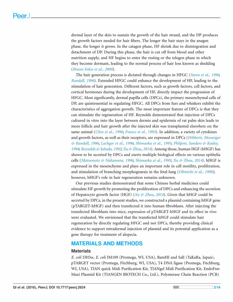
Construction and identification of recombinant vectorof pTARGET-hHGFAccording to the hHGF cDNA sequence (NM-000601) in GenBank, the hHGF cDNA
was amplified with primers (hHGF-BamHI-F: CGCGGATCCATGTGGGTGACCAAACTCC
and hHGF-SalI-R: ACGCGTCGACTGTGGTACCTTATATGTTAAAATAATT). The length
of the amplified segment was 2,200 bp. After purification (Endo-Free Maxi Plasmid Kit),
and qualitative and quantitative analysis of hHGF with 1% agarose gel electrophoresis,
hHGF cDNA and pTARGET were ligated using T4 DNA ligase as shown in Fig. 1.
The ligation mixture was transformed into E. coli JM109, and the positive clones on
LB plates containing Kanamycin-50 were then put into liquid medium and cultured at
37 �C on a shaking table. Plasmid was extracted with Endo-free Plasmid Mini Kit
(OMEGA, USA). Subsequently, the extracted plasmid was digested with SalI and BamHI
and separated with 1% agarose gel electrophoresis as well as colony sequencing.
Cell cultureHuman skin fibroblasts were generously provided by the Chinese Academy of Military
Medical Sciences (Beijing, China). Cells were cultured in DMEM supplemented with 10%
FBS. All cells were incubated at 37 �C in a humidified atmosphere with 5% CO2.
hHGF plasmid transfected fibroblastsTransfection rates of pTARGET and/or pEGFP-N1-hHGF plasmid in different
concentrations of cells, DNA and/or liposome were optimized to the highest level. At a
concentration of plasmid DNA: 0.8 mg/mL/well, liposome concentration of 2 mg/mL/well,
and inoculated cell density in 24 well plate: 0.5 � 105, 1.0 � 105, 1.5 � 105, 2.0 � 105,
2.5 � 105, 3.0 � 105, 3.5 � 105, and 4.0 � 105 cells/mL/well. The cells were cultured in
serum-free DMEM for 6 h, then replaced with DMEM supplemented with 10% FBS, and
Qi et al. (2016), PeerJ, DOI 10.7717/peerj.2624 3/14
the transfection efficiency was determined 48 h later. The fluorescence emitted by the
positive cells was observed with an inverted fluorescence microscope to assess transfection
efficiency and expression levels of pEGFP-N1-hHGF and pTARGET-hHGF.
Detection of hHGF protein levels in target cellsThe hHGF protein expression levels in the fibroblasts were assessed with western blot
analysis. Non-transfected fibroblasts and fibroblasts transfected with pTARGET-hHGF
plasmid (0.8 mg/mL) were harvested, and ice-cold lysis buffer (�2) was applied to extract
the protein. Samples containing equal amounts of protein were resolved by sodium
dodecyl sulfate polyacrylamide gel electrophoresis (SDS-PAGE), transferred to a
Polyvinylidene Fluoride (PVDF) membrane (Millipore, Billerica, MA, USA) and
incubated with corresponding primary antibody (1:200 dilution) (monoclonal rabbit
anti-hHGF antibody or monoclonal mouse anti-GAPDH antibody) at room temperature
for 2 h, washed three times with TBST (3 � 10 min), incubated with secondary antibody
(1:10,000 dilution) (anti-rabbit IgG horseradish peroxidase conjugated antibody) at
room temperature for 2 h, and then washed again. The blots were developed with an
enhanced chemiluminescence detection system (CWBIO). GAPDH was used as a control
to assess the relative expression levels of the proteins in each group.
Cell culture supernatant was collected from cultures of fibroblasts transfected with
pTARGET-hHGF plasmid at 0, 0.6, 0.8 and 1.0 mg at 48 h after transfection. ELISA was
used to detect the expression of hHGF protein according to the manufacturer’s protocols.
Readings from ST-360 Microplate Reader (Shanghai Kehua Bio-engineering Co., Ltd.,
Figure 1 The flow chart of recombinant vector construction.
Qi et al. (2016), PeerJ, DOI 10.7717/peerj.2624 4/14

Shanghai, China) were taken as the accurate values of experimental results, and were
not corrected.
Animal experimentsICR male and female mice (Specific Pathogen Free: SPF) at 4 weeks of age were used
in these studies. Animals were maintained in the Institute of Radiation Medicine, Chinese
Academy of Medical Science. All experimental procedures using these mice were
performed in accordance with relevant requirements of the guiding opinions on the
treatment of experimental animals approved by the National Institutes for Food and
Drug Control of China. All animal experiments have been approved by Ethics Committee
on Animal Experiments of Institute of Radiation Medicine, Chinese Academy of
Medical Science (The approval number is: (2016) 1-008).
Dorsal hairs were carefully removed with hair depilatory cream. Forty-eight ICR
mice from which back hair was removed were randomly divided into 8 groups with
each group comprised of 6 mice: pEGFP-N1-hHGF group (20, 40, and 60 mg/kg),
pTARGET-hHGF group (20, 40, and 60 mg/kg), empty pTARGET plasmid control group
(60 mg/kg), and negative control group. The transfection system in vivo is shown in
Table 1. Mice were dosed via intradermal injection once a day for 20 d. Hair growth
was recorded and photos were taken daily. Local skin samples were taken for Hematoxylin
and Eosin (H/E) staining at day 8.
Statistical analysisStatistical analysis was performed with SPSS software and the values expressed as the
mean ± standard error of the mean. The statistical significance of differences between
the control and treated groups were determined by Student’s t-test. Values �P < 0.05 and��P < 0.01 are considered significant.
RESULTSIdentification and sequencing of the hHGF PCR amplification andpEGFP-N1-hHGF productsAs predicted, the hHGF product was amplified with the two primers using the
pEGFP-N1-hHGF plasmid at approximately 2,200 bp on a gel (Fig. 2). After the
pTARGET-hHGF clone was double digested with SalI and BamHI, both hHGF band
(approximately 2.2 kb) and vector band (approximately 5.67 kb) were observed (Fig. 3).
The clone was submitted for sequence analysis. The base sequence of the inserted gene
Table 1 The transfection system in vivo.
400 mL Transfectioncomplexes
Plasmid dilution 100 mL Plasmid
solution
100 mL Injection
water
50 mg DNA
100 mL 10% Glucose solution
Transfection reagent
diluent
100 mL Transfection reagent diluent
100 mL 10% Glucose solution
Qi et al. (2016), PeerJ, DOI 10.7717/peerj.2624 5/14

in the pTARGET plasmid matched the genomic sequence of hHGF in GenBank.
The final plasmid was termed pTARGET-hHGF.
Production and titration of the recombinant lentiviral vectorAfter transfection of fibroblasts with the pEGFP-N1-hHGF and pTARGET-hHGF
plasmids, Green Fluorescent Protein (GFP) expression from pEGFP-N1-hHGF was
assessed using a fluorescence microscope to ensure that the plasmids were properly
generated (Fig. 4A). As analysis indicates that when the concentration of plasmid DNA is
0.8 mg/well and liposome concentration is 2 mg/well, the optimal cell seeding density is
3.0 � 105 cells/mL/well (Fig. 4B).
Figure 2 The hHGF analysis and amplification product (PCR). Lane M: Marker; Lanes 1 and 2: hHGF.
The purpose gene is 2,200 bp.
Figure 3 Results of double digestion. pTARGET plasmid (Lane 1) and double digested with SalI and
BamHI (Lane 2).
Qi et al. (2016), PeerJ, DOI 10.7717/peerj.2624 6/14

hHGF protein expression in fibroblastsProtein lanes corresponding to the non-transfected fibroblasts and the fibroblasts
transfected with pTARGET-hHGF plasmids are shown in Fig. 5A. The GAPDH protein
ran at 37 kDa and the hHGF protein at 83 kDa. The non-transfected fibroblasts did not
express hHGF, while the hHGF-transfected group expressed hHGF. ELISA showed that
pTARGET-hHGF infected fibroblasts produced hHGF protein, and that the concentration
was higher in the hHGF infected group at DNA concentration at 0.8 mg/well (P < 0.01)
(Fig. 5B).
Figure 4 Transfection results. Green fluorescent protein (GFP) was observed to determine the
expression of pEGFP-N1-hHGF plasmid in fibroblasts after transfection for 48 h (100�) ((A) brightfield; (B) fluorescence). (C) Cell seeding density was optimized through examining the transfection ratio
at a concentration of plasmid DNA: 0.8 g/well, liposome concentration: 2 µg/well (24 well plate).
Figure 5 The hHGF protein expression in fibroblasts after transfection with the pTARGE T-hHGF
plasmid. (A) Expression of hHGF as assessed by Western blot. (B) ELISA of hHGF in the fibroblasts
supernatants after the transfection at different DNA concentrations.
Qi et al. (2016), PeerJ, DOI 10.7717/peerj.2624 7/14
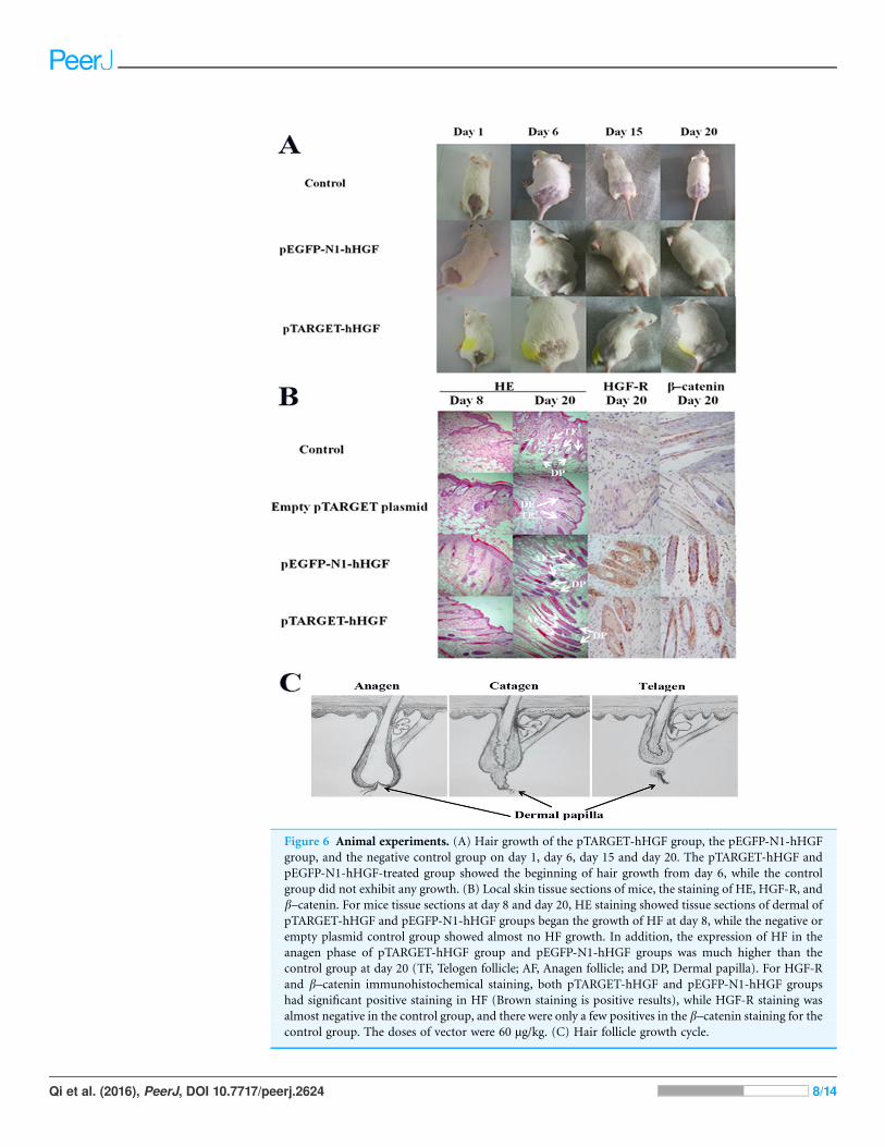
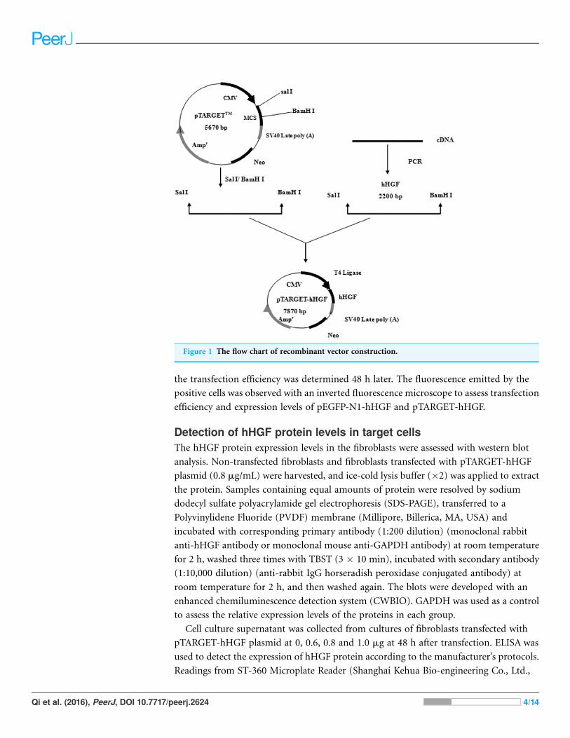
Figure 6 Animal experiments. (A) Hair growth of the pTARGET-hHGF group, the pEGFP-N1-hHGF
group, and the negative control group on day 1, day 6, day 15 and day 20. The pTARGET-hHGF and
pEGFP-N1-hHGF-treated group showed the beginning of hair growth from day 6, while the control
group did not exhibit any growth. (B) Local skin tissue sections of mice, the staining of HE, HGF-R, and
b–catenin. For mice tissue sections at day 8 and day 20, HE staining showed tissue sections of dermal of
pTARGET-hHGF and pEGFP-N1-hHGF groups began the growth of HF at day 8, while the negative or
empty plasmid control group showed almost no HF growth. In addition, the expression of HF in the
anagen phase of pTARGET-hHGF group and pEGFP-N1-hHGF groups was much higher than the
control group at day 20 (TF, Telogen follicle; AF, Anagen follicle; and DP, Dermal papilla). For HGF-R
and b–catenin immunohistochemical staining, both pTARGET-hHGF and pEGFP-N1-hHGF groups
had significant positive staining in HF (Brown staining is positive results), while HGF-R staining was
almost negative in the control group, and there were only a few positives in the b–catenin staining for thecontrol group. The doses of vector were 60 µg/kg. (C) Hair follicle growth cycle.
Qi et al. (2016), PeerJ, DOI 10.7717/peerj.2624 8/14

Animal experimentsICR mice were used to investigate the effects of pTARGET-hHGF and pEGFP-N1-hHGF on
the hair growth. The dorsal hair of ICR mice was removed and then the resulting mice were
injected with various concentrations of pTARGET-hHGF or pEGFP-N1-hHGF. Results
demonstrated that the hair growth rate in the dorsal skin of the ICR mice treated with
pTARGET-hHGF or pEGFP-N1-hHGF (Injection concentration was 60 mg/kg) was higher
than that of the negative or empty plasmid control group (Fig. 6A). H/E staining showed
that at the beginning of day 8, pTARGET-hHGF and pEGFP-N1-hHGF groups experienced
a significant increase in total number of HF when compared to the control or empty
plasmid group (Fig. 6B; Table 2). Furthermore, immunohistochemical staining showed a
significant increase of HGF-R and b-catenin in HF for the pTARGET-hHGF and
pEGFP-N1-hHGF groups, when compared with the control or empty plasmid group
(Fig. 6B).
DISCUSSIONMany physiological events are involved in the growth and development of HF, such as
interaction between dermis and epidermis, as well as development and differentiation of
the cells. HF play an important role in skin self-repair, wound healing, and onset of
carcinoma. The dermal parts of HF consist of DPCs and DS fibroblasts which have the
ability to induce the formation of HF through the interaction between epithelial cells and
the mesenchyme cells of HF. DPCs are a composite of very unique fibroblast cells that
produce an extracellular matrix (ECM) and show great significance in the interaction
between epithelial cells and stromal cells (Matsuzaki & Yoshizato, 1998; Almond-Roesler
et al., 1997). The cause for hair loss is the imbalance between the anagen and telogen
phases of HFGC. In the telogen phase, the ECM of DPCs is reduced, and epithelial cells of
the hair bulb are narrowed and separated from the DPCs. In the catagen phase, DP
remains close to the base of HF to form cells that are closely bunched and embedded in the
ECM, leading to an unrecognizable ultra-structure (Stenn & Paus, 2001; Pans & Cotsarelis,
1999). Thus, the DP plays an important regulatory role in HFGC, and hair loss is
associated with DP dysfunctions.
Our previous work illustrated that certain Chinese herbal medicines can stimulate
HF growth by promoting the proliferation of DPCs and enhancing the secretion of hHGF
(Xu & Zhou, 2014; Xu et al., 2015), thereby stimulating HF growth. In addition to our
study showing hHGF’s role in promoting growth of HF, other recent studies have revealed
Table 2 The quantity of HF.
Group
Day Control Empty plasmid pEGFP-N1-hHGF pTARGET-hHGF
Day 8 1.21 ± 0.09 1.52 ± 0.10 10.96 ± 1.45** 11.02 ± 1.02**
Day 20 10.01 ± 1.33 10.26 ± 1.55 15.67 ± 0.98* 19.86 ± 1.27*
Notes:n = 6.* P < 0.05.** P < 0.01.
Qi et al. (2016), PeerJ, DOI 10.7717/peerj.2624 9/14

that hHGF can also (a) regulate interactions between DP cells and epithelial keratinocyte
and other interactions among cells (Fujie et al., 2001; Lindner et al., 1997); (b) induce the
formation of per follicular blood vessels (Houseknecht et al., 1998; Boulton, 1999; Zhang
et al., 2003; Mecklenburg et al., 2000); and (c) influence all three phases on the HFGC of
hairs (Jindo et al., 1998). Importantly, our study revealed that while DPCs can secrete
hHGF, the surrounding sheath fibroblasts of HF cannot (Xu et al., 2015).
Consequently, we hypothesized that manipulating sheath fibroblasts to mimic
functional DPCs to express hHGF could promote the growth of HF. Based on this
understanding, we (a) first constructed a recombinant plasmid expression vector
containing hHGF (pTARGET-hHGF); (b) demonstrated that this plasmid could be
transfected into the surrounding sheath fibroblasts and that it could secrete hHGF
in vitro; and (c) pursued animal studies using mice to show that it could promote hair
growth as well as tissue follicle development.
The hHGF expression indeed increased by more than 57% when fibroblasts were
transfected the recombinant plasmid. The hHGF gene transfected fibroblasts expressed
hHGF at even higher levels than DPCs. These results demonstrate that the
recombinant plasmid vector successfully delivered the hHGF gene into sheath fibroblasts
which then expressed the hHGF gene at a high level. This transformation yields sheath
fibroblasts with the function of DPCs. Further animal experiments showed that both
pTARGET-hHGF and pEGFP-N1-hHGF groups regenerated mouse hair faster than
the control group. Moreover, H/E staining of local skin tissue sections from mice showed
that growth of HF started at day 8 for both the pTARGET-hHGF group and pEGFP-
N1-hHGF group, while the control group had almost no signs of HF growth.
The examination of the expression of HGF-R in mouse skin tissue sections is
noteworthy because hHGF achieves its functions through binding to its specific receptor
HGF-R. HGF-R immunohistochemical staining showed that pTARGET-hHGF and
pEGFP-N1-hHGF groups had significant positive staining in HF, while such staining was
almost absent in the control group. This outcome suggests that the active hHGF had
stimulated the growth of HF in both pTARGET-hHGF and pEGFP-N1-hHGF groups,
thereby further validating the observation that hHGF recombinant vector was transfected
into the cells surrounding the HF of mice, leading to secretion of active hHGF.
It has been reported that b-catenin, a ubiquitously distributed protein with multiple
functions, plays an important role in controlling the HF morphogenesis and stem cell
differentiation in the skin (Huelsken et al., 2001). If b-catenin is deleted after HF have
formed, hair is completely lost after the first hair cycle (Wang et al., 2000). b-Catenin is also
essential to the fate of skin stem cells because, in the absence of b-catenin, stem cells fail to
differentiate into follicular keratinocytes instead adopting an epidermal fate (Huelsken et al.,
2001). Our experimental results indicate that hHGF enables b-catenin to enter the nucleus
in cultured epithelial hair follicle stem cells (Fig. S2). At the same time, b-cateninimmunohistochemical staining for both pTARGET-hHGF and pEGFP-N1-hHGF groups
showed a positive staining that is significantly stronger than the negative control group in
HF (Fig. 6B). These results suggest that hHGF may promote growth of HF by increasing
Qi et al. (2016), PeerJ, DOI 10.7717/peerj.2624 10/14

the expression of b-catenin. In addition, liver and kidney tissue sections did not show
fluorescence in the pEGFP-N1-hHGF local injection group, thereby demonstrating
the potential clinical safety of the hHGF recombinant plasmid expression vector.
Finally, Lindner et al. (2000), suggested that hHGF accelerates HF morphogenesis and
inhibits catagen induction (thus extending the anagen phase) by analyzing hHGF
transgenic 8-week old C57BL/6 mice and treating HF with recombinant hHGF protein.
Muller-Rover et al. (2001) achieved a similar experimental outcome in first neonatal
C57BL/6 mice. Our experiments employed 4-week old ICR mice with dorsal hairs
removed using hair depilatory cream to reach synchronized HFGC, but still showed that
hHGF promotes the growth of HF. These results strongly suggest that hHGF can accelerate
HF morphogenesis and growth in different mouse strains and at different ages.
CONCLUSIONIn summary, we report here construction of a recombinant vector, pTARGET-hHGF, that
contains the hHGF gene, and that this new recombinant vector could be successful
delivered into fibroblasts in vitro. We have found that such transfection leads to secretion
of hHGF, and a high level of the hHGF expression could be detected qualitatively and
quantitatively, thereby allowing fibroblasts to possess functions of DPCs. Furthermore,
local injections of pTARGET-hHGF into live mice can result in faster hair regeneration,
improved follicle development, and significantly increased expressions of HGF-R and
b-catenin. These results demonstrate that we have established a nonviral vector of hHGF
that can manipulate the sheath fibroblasts surrounding HF via hHGF expression, thereby
promoting hair regeneration.
ACKNOWLEDGEMENTSWe thank Professor Robert P. Borris and Professor Youcai Zhang of Tianjin University
for invaluable discussions.
ADDITIONAL INFORMATION AND DECLARATIONS
FundingThis work was supported by the National Natural Science Foundation of China (No.
81641132). The funders had no role in study design, data collection and analysis, decision
to publish, or preparation of the manuscript.
Grant DisclosuresThe following grant information was disclosed by the authors:
National Natural Science Foundation of China: 81641132.
Competing InterestsThe authors declare that they have no competing interests. Zhijing Chang is an employee
of Roche Diagnostics, Beijing, People’s Republic of China.
Qi et al. (2016), PeerJ, DOI 10.7717/peerj.2624 11/14
Author Contributions� Yonghao Qi performed the experiments, analyzed the data, contributed reagents/
materials/analysis tools, wrote the paper, prepared figures and/or tables.
� Miao Li performed the experiments, analyzed the data, contributed reagents/materials/
analysis tools.
� Lian Xu performed the experiments, prepared figures and/or tables.
� Zhijing Chang performed the experiments, prepared figures and/or tables.
� Xiong Shu performed the experiments, prepared figures and/or tables.
� Lijun Zhou conceived and designed the experiments, contributed reagents/materials/
analysis tools, wrote the paper, reviewed drafts of the paper.
Animal EthicsThe following information was supplied relating to ethical approvals (i.e., approving body
and any reference numbers):
Animals were maintained in Institute of Radiation Medicine Chinese Academy of
Medical Science.
Animal experiment has been approved by Ethics Committee on animal experiments of
Institute of Radiation Medicine Chinese Academy of Medical Science approval number
(2016) 1-008.
DNA DepositionThe following information was supplied regarding the deposition of DNA sequences:
GenBank accession number: NM-000601.
Data DepositionThe following information was supplied regarding data availability:
The raw data has been supplied as Supplemental Dataset Files.
Supplemental InformationSupplemental information for this article can be found online at http://dx.doi.org/
10.7717/peerj.2624#supplemental-information.
REFERENCESAlmond-Roesler B, Schon M, Schon MP, Blume-Peytavi U, Sommer C, Loster K, Orfanos CE.
1997. Cultured dermal papilla cells of the rat vibrissa follicle. Proliferative activity, adhesion
properties and reorganization of the extracellular matrix in vitro. Archives Dermatological
Research 289(12):698–704 DOI 10.1007/s004030050264.
Boulton M. 1999. A role for hepatocyte growth factor in diabetic retinopathy? British Journal of
Ophthalmology 83(7):763–764 DOI 10.1136/bjo.83.7.763.
Braun-Falco O, Plewig G, Wolff HH, Burgdorf W. 2000. Dematology. Vol. 1. Berlin, Heidelberg:
Springer-Verlag, 1099–1140.
Chiu HC, Chang CH, Chen JS, Jee SH. 1996. Human hair follicle dermal papilla cell, dermal
sheath cell and interstitial dermal fibroblast characteristics. Journal of the Formosan Medical
Association 95(9):667–674.
Qi et al. (2016), PeerJ, DOI 10.7717/peerj.2624 12/14
Francz PI, Bayreuther K, Limat A, Noser F. 1993. Differentiation and polypeptide expression in
human papilla and dermal fibroblasts in vitro. European Journal of Cell Biology 60(2):337–345.
Fujie T, Katoh S, Oura H, Urano Y, Arase S. 2001. The chemotactic effect of a dermal papilla
cell-derived factor on outer root sheath cells. Journal of Dermatological Science 25(3):206–212
DOI 10.1016/S0923-1811(00)00130-4.
Hibberts NA, Messenger AG, Randall VA. 1996. Dermal papilla cells derived from
beard hair follicles secrete more stem cell factor (SCF) in culture than scalp cells or dermal
fibroblasts. Biochemical & Biophysical Research Communications 222(2):401–405
DOI 10.1006/bbrc.1996.0756.
Houseknecht KL, Baile CA, Matteri RL, Spurlock ME. 1998. The biology of leptin: a review.
Journal of Animal Science 76(5):1405–1420 DOI 10.2527/1998.7651405x.
Huelsken J, Vogel R, Erdmann B, Cotsarelis G, Birchmeier W. 2001. b-catenin controls hair
follicle morphogenesis and stem cell differentiation in the skin. Cell 105(4):533–545
DOI 10.1016/S0092-8674(01)00336-1.
Jahoda CAB, Horne KA, Oliver RF. 1984. Induction of hair growth by implantation of cultured
dermal papilla cells. Nature 311:560–562 DOI 10.1038/311560a0.
Jahoda CAB, Reynolds AJ. 1996. Dermal-epidermal interactions. Adult follicle-derived cell
populations and hair growth. Dermatologic Clinics 14(4):573–583
DOI 10.1016/S0733-8635(05)70385-5.
Jindo T, Tsuboi R, Takamori K, Ogawa H. 1998. Local injection of Hepatocyte growth factor/
scatter factor (HGF/SF) alters cyclic growth of murine hair follicles. Journal of Investigative
Dermatology 110(4):338–342 DOI 10.1046/j.1523-1747.1998.00144.x.
Lachgar S, Moukadiri H, Jonca F, Charveron M, Bouhaddioui N, Gall Y, Bonafe JL,
Plouet J. 1996. Vascular endothelial growth factor is an autocrine growth factor for
hair dermal papilla cells. Journal of Investigative Dermatology 106(1):17–23
DOI 10.1111/1523-1747.ep12326964.
Lindner G, Botchkarev VA, Botchkareva NV, Ling G, van der Veen C, Paus R. 1997. Analysis of
apoptosis during hair follicle regression. American Journal of Pathology 151(6):1601–1617.
Lindner G, Menrad A, Gherardi E, Merlino G, Welker P, Handjiski B, Roloff B, Paus R. 2000.
Involvement of hepatocyte growth factor/scatter factor and Met receptor signaling in hair
follicle morphogenesis and cycling. FASEB Journal 14(2):319–332.
Matsumoto K, Nakamura T. 1996. Emerging multipotent aspects of hepatocyte growth factor.
Journal of Biochemistry 119(4):591–600 DOI 10.1093/oxfordjournals.jbchem.a021283.
Matsuzaki T, Yoshizato K. 1998. Role of hair papilla cells on induction and regeneration
processes of hair follicles. Wound Repair and Regeneration 6(6):524–530
DOI 10.1046/j.1524-475X.1998.60605.x.
Mecklenburg L, Tobin DJ, Muller-Rover S, Handjiski B, Wendt G, Peters EMJ, Pohl S, Moll I,
Paus R. 2000. Active hair growth (anagen) is associated with angiogenesis. Journal of
Investigative Dermatology 114(5):909–916 DOI 10.1046/j.1523-1747.2000.00954.x.
Muller-Rover S, Foitzik K, Paus R, Handjiski B, van der Veen C, Eichmuller S, McKay IA,
Stenn KS. 2001. A comprehensive guide for the accurate classification of murine hair follicles
in distinct hair cycle stages. Journal of Investigative Dermatology 117(1):3–15
DOI 10.1046/j.0022-202x.2001.01377.x.
Ohmichi H, Koshimizu U, Matsumoto K, Nakamura T. 1998. Hepatocyte growth factor (HGF)
acts as a mesenchymederived morphogenic factor during fetal lung development.
Development 125(7):1315–1324.
Qi et al. (2016), PeerJ, DOI 10.7717/peerj.2624 13/14

Pans R, Cotsarelis G. 1999. The biology of hair follicles. New England Journal of Medicine
341(7):491–497 DOI 10.1056/NEJM199908123410706.
Peus D, Pittelkow MR. 1996. Growth factors in hair organ development and the hair growth cycle.
Dermatologic Clinics 14(4):559–572 DOI 10.1016/S0733-8635(05)70384-3.
Philpott MP, Sanders DA, Kealey T. 1994. Effects of insulin and insulin-like growth factors on
cultured human hair follicles: IGF-I at physiologic concentrations is an important regulator of
hair follicle growth in vitro. Journal of Investigative Dermatology 102(6):857–861.
Randall VA. 1996. The use of dermal papilla cells in studies of normal and abnormal hair follicle
biology. Dermatologic Clinics 14(4):585–594 DOI 10.1016/S0733-8635(05)70386-7.
Reynolds AJ, Jahoda CA. 1992. Cultured dermal papilla cells induce hair follicle formation and
hair growth by transdifferentiation of an adult epidermis. Development 115(2):587–593.
Shimaoka S, Tsuboi R, Jindo T, Imai R, Takamori K, Rubin JS, Ogawa H. 1995. Hepatocyte
growth factor/scatter factor expressed in follicular papilla cells stimulates human hair growth
in vitro. Journal of Cellular Physiology 165(2):333–338 DOI 10.1002/jcp.1041650214.
Stenn KS, Combates NJ, Eilertsen KJ, Gordon JS, Pardinas JR, Parimoo S, Prouty SM. 1996.
Hair follicle growth controls. Dermatologic Clinics 14(4):543–558
DOI 10.1016/S0733-8635(05)70383-1.
Stenn KS, Paus R. 2001. Controls of hair follicle cycling. Physiological Reviews 81(1):449–494.
Wang LC, Liu Z-Y, Gambardella L, Delacour A, Shapiro RI, Yang J, Sizing I, Rayhorn P,
Garber E, Benjamin CD, Williams K, Taylor FR, Barramdon Y, Ling L, Burkly L. 2000.
Conditional disruption of hedgehog signaling pathway defines its critical role in hair
development and regeneration. Journal of Investigative Dermatology 114(5):901–908
DOI 10.1046/j.1523-1747.2000.00951.x.
Xu L, Shu X, Chang Z-J, Liu W-J, Wang X, Zhou L-J. 2015. Construction of HGF eukaryotic
expression plasmid and expression in fibroblasts. Progress in Modern Biomedicine 1:21–24.
Xu L, Zhou LJ. 2014. Chinese complex herb formulas promote hair follicle growth by stimulating
proliferation of dermal papilla cells and expression of hepatocyte growth factor. Progress in
Modern Biomedicine 22:4201–4204.
Zhang Y-W, Su Y, Volpert OV, Woude GFV. 2003. Hepatocyte growth factor/scatter factor
mediates angiogenesis through positive VEGF and negative thrombospondin 1 regulation.
Proceedings of the National Academy of Sciences of the United States of America
100(22):12718–12723 DOI 10.1073/pnas.2135113100.
Qi et al. (2016), PeerJ, DOI 10.7717/peerj.2624 14/14