RCNIC ORIENTATION CARE OF THE NEONATE WITH A HEMATOLOGIC DISORDER M.VICTORIA DECASTRO, RNC, BSN,...
90
RCNIC ORIENTATION CARE OF THE NEONATE WITH A HEMATOLOGIC DISORDER M.VICTORIA DECASTRO, RNC, BSN, Clinical Coordinator, CNIII
-
Upload
darcy-houston -
Category
Documents
-
view
214 -
download
0
Transcript of RCNIC ORIENTATION CARE OF THE NEONATE WITH A HEMATOLOGIC DISORDER M.VICTORIA DECASTRO, RNC, BSN,...

RCNIC ORIENTATION
CARE OF THE NEONATE WITH A HEMATOLOGIC DISORDER
M.VICTORIA DECASTRO,
RNC, BSN, Clinical Coordinator, CNIII

Care of Neonate with Neonatal Hyperbilirubinemia

Hyperbilirbinemia
• Definition• physiological jaundice
• the yellow discoloration of the skin, conjunctivae, and sometimes mucous membranes
• Occurs in more than 50% of all newborns– (Deacon & O’Neill, 1999)
• affects full-term infants within 2 to 4 days of birth and lasts until about day 6 (Kenner & Lott, 2003)
• preterm infants have higher peak levels occurring at about 5 to 7 days of life (Kenner & Lott, 2003) ; jaundice lasts longer in preterm infants; the more preterm an infant is the lower the “light”level

Hyperbilirbinemia
• Pathophysiology– Bilirubin is produced by the breakdown of hemoglobin
at the end of a red cell’s lifespan
– Because the neonate’s liver is immature, the bilirubin level exceeds the liver’s ability to conjugate bilirubin (convert bilirubin to soluble form for excretion)
– This bilirubin then “free floats” in the plasma
– At high levels, bilirubin can cross the blood-brain barrier and damage brain cells causing kenicterus

Hyperbilirubinemia• Serum bilirubin levels (unconjugated) are greater
than 12 mg/dl and generally occurs after the first 36 hours
• A bili level above 12 mg/dl within the first 24 hours is not considered physiological
• Bili levels in general are considered physiological if:– Full-term-12mg/dl or greater by 3 days of life– Preterm-10 to 12 mg/dl or greater by 5 days of life
• Treatment depends on the bili level, the infant’s gestational and actual age, and the severity of the infant’s condition
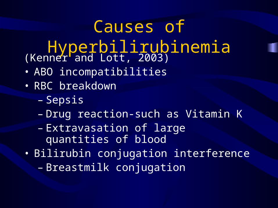
Causes of Hyperbilirubinemia(Kenner and Lott, 2003)• ABO incompatibilities• RBC breakdown
– Sepsis– Drug reaction-such as Vitamin K– Extravasation of large quantities of blood
• Bilirubin conjugation interference– Breastmilk conjugation

Causes of Hyperbilirubinemia
(Kenner and Lott, 2003)• Transplacental and neonatal drug interactions• Hypothyroidism-affects up to 3 to 4 weeks• Acidosis and hypoxia• Decreased bowel motility or hepatocellular
damage can cause abnormal bilirubin excretion• Congestive heart failure can cause abnormal
bilirubin excretion

What will you see in a jaundiced infant?
• Yellow discoloration that starts on the face and spreads caudally
• Yellow discoloration of the sclera and mucus membranes
• Signs and symptoms of kenicterus

Hyperbilirubinemia
• Kenicterus is the yellow staining of brain tissue and leads to brain damage, such as cerebral palsy and deafness.
• Kenicterus occurs in 50% of infants with bili levels of 30 of greater and in 10% with bili levels 20 to 25

Hyperbilirubinemia
• Risk factors (Kenner and Lott, 2003)– Inadequate intake/dehydration– Acidosis and hypoxia– Certain drugs and substances may compete with
available bilirubin binding sites such as sufisoxazole, salicylates and sodium benzoate
– Hypoproteinemia (lower albumin levels)-places preterm infants at higher risks

Hyperbilirubinemia• Clinical manifestations of
kenicterus– usually evident in the first 5 days of
life– In a sick or preterm neonate,
kenicterus can occur at levels as low as 5 mg/dl (Nathan & Oski, 1998)

Hyperbilirubinemia
• Generally, the neonate is stable and otherwise healthy
• Because of early discharge, physiological hyperbilirubinemia is often diagnosed in the home care setting
• Key to diagnosis is parent education
• Home phototherapy can be used
Hyperbilirubinemia
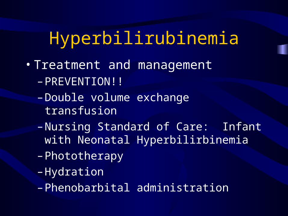
• Treatment and management– PREVENTION!!
– Double volume exchange transfusion
– Nursing Standard of Care: Infant with Neonatal Hyperbilirbinemia
– Phototherapy
– Hydration
– Phenobarbital administration

Phototherapy
• Phototherapy lights causes the bilirubin to become more water-soluble; this bilirubin can be excreted in bile/stool and urine
• May use bank lights and/or bili blanket

Phototherapy
• Use bank lights and/or bili blankets– Use bili mask to protect
eyes.– Monitor temperature
closely by using continuous skin temperature monitoring; take temperature Q 2-4 hours
– Bili levels at least every 8-12 hours, turning off phototherapy lights with lab draws only

Phototherapy
• Important to expose as much skin as possible, covering only genitals and eyes for protection
• Turn infant frequently to allow for maximum exposure
• Avoid use of ointments and lotions

Phototherapy
• Important that the phototherapy light levels are within acceptable range– Bilimeter readings q
shift with approximate goal of 25 to 45 (combined readings of biliblanket and bili lights)

Phototherapy• Side effects-(Kenner and Lott, 2003)
– Parental teaching-signs and symptoms of hyperbilirubinemia and dehydration
– Dermal Rash– Lethargy– Abdominal distention– Possible eye drainage– Dehydration– Thrombocytopenia– Hypocalcemia– Temporary lactose intolerance– “Bronze” baby syndrome

Nursing Standard of Care
• Patient will not experience neurological injury– Assess and document skin color from head,
sclera, and trunk in daylight or under fluorescent light at least q shift
– Obtain bili levels as ordered– Observe for and report abnormal neurological
findings– Check bilimeter readings

Nursing Standard of Care
• Patient will not experience injury from phototherapy– Cover eyes– Inspect eyes q 2 hours and prn with phototherapy lights
off, notify physician of any drainage– Change eye shields prn– Avoid tight head band on eye shield to reduce the risk
of increased ICP, especially in preterm infants– Maximize exposed skin surface, protecting genitalia– Avoid exposure of skin temperature probe to
phototherapy lights
Nursing Standard of Care
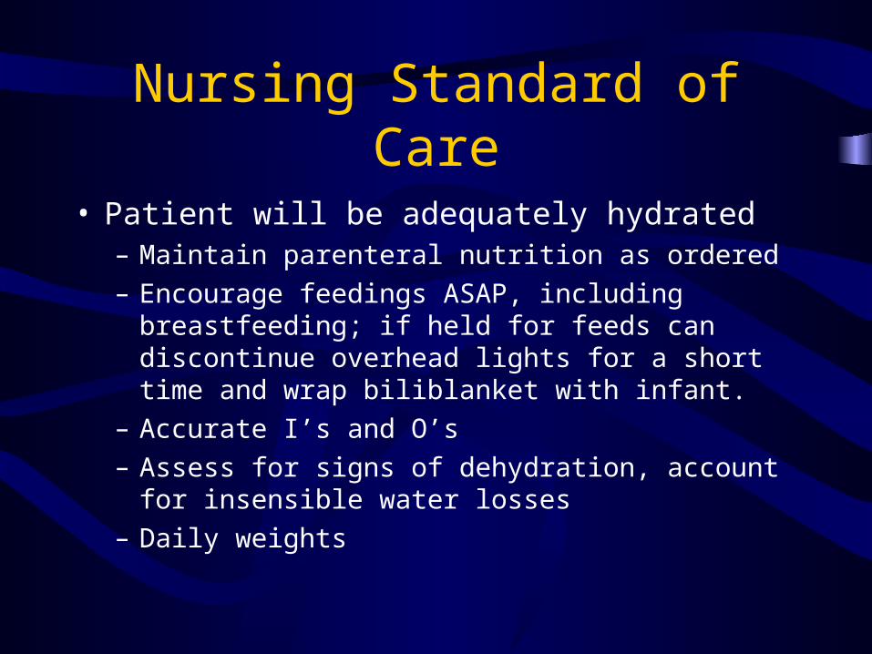
• Patient will be adequately hydrated– Maintain parenteral nutrition as ordered
– Encourage feedings ASAP, including breastfeeding; if held for feeds can discontinue overhead lights for a short time and wrap biliblanket with infant.
– Accurate I’s and O’s
– Assess for signs of dehydration, account for insensible water losses
– Daily weights

Nursing Standard of Care
• Patient will maintain thermoregulation– Provide neutral thermal environment
• Maintain axillary temperature of at least 36.5 degrees Celsius
• Avoid cold stress
• Q 4 hour vitals and prn
• Properly secure temperature probe to abdomen or back

Nursing Standard of Care
• The parents/caregivers will participate in the child’s care and health goals/outcomes– Provide information and explanations of
hyperbilirubinemia and phototherapy to the parents/care givers
– Explore with parents/caregivers their willingness to provide care

Phototherapy
• Follow-up Care– Maintain adequate hydration,
including enteral feeds if possible
– Check bili levels at least 4-8 hours after phototherapy is D/C’ed; there is a chance for rebound

Exchange Transfusion
• Early exchange transfusion is indicated with the presence of hemolytic disease
• Carefully monitor fluid and electrolyte status
• Provide adequate hydration

Exchange Transfusion
• Double volume Exchange Transfusion– Purpose-to remove the infant’s bilirubin
and antibody-coated red blood cells from circulation by removing the infant’s blood volume and replacing the volume with blood or another volume expander

Phenobarbital Administration
• Increases the uptake and conjugation of bilirubin by the liver and increasing its excretion by increasing bile flow

Care of the Neonate with Anemia

Anemia
• Definition– “low hemoglobin concentration and/or
decreased number of red blood cells diminishes the oxygen-carrying capacity of the blood and the level of oxygen available to tissues” (Blanchette & Zipursky, 1994; Hume, 1997; Miller, 1995; Oski, Brugnara, & Nathan, 1998)

Anemia
• Physiology– removal or loss of 10% or more of total blood volume
over 24 to 48 hours can lead to anemia
– In general, there are 100 ml of blood volume per kg in a preterm infant and 82-85 ml of blood volume per kg in a term infant (Kenner and Lott, 2003)
– Hgb and HCT levels determine the type and degree of anemia; in general, Hct less than 40% is considered anemia
– Hgb may not accurately the extent of acute blood loss
– low HCT or Hgb may be acceptable if retic count is NL
Anemia
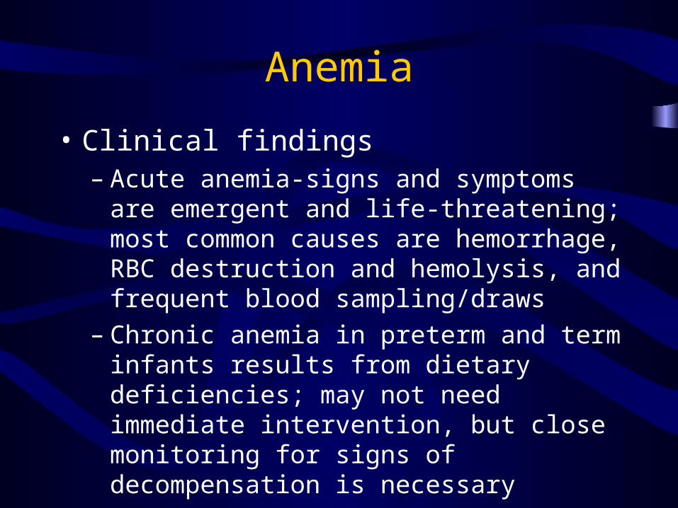
• Clinical findings– Acute anemia-signs and symptoms are emergent
and life-threatening; most common causes are hemorrhage, RBC destruction and hemolysis, and frequent blood sampling/draws
– Chronic anemia in preterm and term infants results from dietary deficiencies; may not need immediate intervention, but close monitoring for signs of decompensation is necessary

Anemia
• Risk factors and indications– Frequent lab draws– Family history of anemia or jaundice– History of bleeding, splenectomy, consanguinity, and/or blood
group incompatibilities– Some ethnic groups and natives of specific geographical origins,
such as African-American population and sickle cell anemia– Maternal history– Presence of cephalohematoma– Abnormal distention of mass-damage or ruptured liver, spleen , adrenal,
kidney – Cardiovascular abnormalities-tachycardia, murmur, gallop rhythm
– Hydropic changes

Anemia
• Normal values for the neonate (0 to 30 days)– RBC-4.1 to 6.1– Hemoglobin (Hgb)-16 to 21– Hematocrit (Hct)-44 to 60– Reticulocyte count (Retic)-2 to 6– Values vary depending on infant’s gestational
age and actual age

Anemia
• Acute Anemia– Symptoms are more emergent and life-
threatening– Most common causes include hemorrhage,
RBC destruction and hemolysis, and frequent blood sampling

Acute Anemia
• Signs and Symptoms of Acute Anemia– Pallor– Tachycardia – Shallow, rapid, irregular respirations– Low or absent blood pressure, low venous
pressures– Weak or absent peripheral pulses– Poor perfusion
Acute Anemia
• Signs and Symptoms of Acute Anemia– Capillary refill time greater than 4 seconds– Mottling– Lethargy– Low HCT– Hgb may be initially NL, with decline over the
next 6-12 hours

ANEMIA
• Chronic Anemia in preterm and term infants is a result in dietary deficiencies and may not need immediate intervention
• Close monitoring for signs of decompensation is necessary

Anemia
• Signs and symptoms of chronic anemia– Pallor without signs of acute distress– Increased incidence of apneic and/or
bradycardic episodes or increased severity of apneic and/or bradycardic episodes
– Hepatosplenomegaly – Signs of congestive heart failure– Tachycardia

Anemia• Signs and symptoms of chronic anemia
– Increased oxygen requirement – Increased respiratory effort (dyspnea) or
tachypnea
– Lethargy, decreased activity/energy level-infant just does not “seem like oneself”
– Poor feeding– Poor weight gain – Low RBC’s, HCT, and Hgb levels; Retic counts
may be low, normal, or high

Anemia
• Management of acute anemia– Remember your ABC’s of resuscitation!!– Hemodynamic support– CBC with differential, type and cross, Coombs
testing– Blood transfusion
• acute-CMV safe, HgbS negative, and O-negative PRBC’s
• in infants less than 1 kg or immuno-suppressed use irradiated

Anemia
• Management of acute anemia– Provide warmth, monitoring of vital signs, and
continuous and accurate assessment of I’s and O’s
– Lab tests and physical exams are necessary in order to determine the cause of acute anemia
– Modifications in care that eliminate recurrence of precipitous events and prevent blood loss
Anemia
• Chronic anemia
– Goal of treatment-the control or eradication of the cause or symptoms

ANEMIA• Management of chronic anemia
– Nutritional management/replacement therapy• Iron-ferrous sulfate (Ferinsol), iron-fortified
formulas• Folic acid• Vitamin E (Aquasol E)
– Erythropoeiten
– Transfusion therapy

Transfusion Therapy
• Blood products for neonates are CMV safe, leukofiltered, and Hgb-S negative; irradiated for immunosuppressed infants such as micropreemies (less than 1 kg) and septic infants
• Use MDX (minimal donor exposure) protocol for infants less than 1 kg
• The decision to transfuse is dependent on many factors

Transfusion Therapy
• Assess for S/S of transfusion reactions-usually occurs during the first 15 minutes of the transfusion:– Shivering/chills -Vomiting– Dyspnea -Presence of blood in urine– Hyperthermia -Tachycardia– Hypertension -Rash– Irritability

Transfusion Therapy
• Vitals– TPR and B/P before(within 5
minutes of the start of the transfusion) and after administration (within 5 minutes of the end of the transfusion), at 15 minutes of the start of the transfusion, and every hour until transfusion is complete
Transfusion Therapy
• If signs of transfusion reaction appear– Stop transfusion immediately– Immediately send transfused blood
and administration set to blood bank.– Follow instructions on “report of
transfusion reaction” form

Anemia
• Long-term follow-up and prognosis– Continue to assess for S/S of anemia– Maintain adequate nutritional support– Improved oxygenation and control or
eradication of symptoms are indicative of a positive prognosis
– Long-term prognosis is determined by the underlying causes & degree of anemia and the infant’s response to interventions

Care of the Neonate with Polycythemia

Polycythemia
• Definition– the condition in which an excess mass of
RBC’s is in circulation, resulting in increased blood viscosity
– Venous HCT greater that 65% and venous Hgb is greater than 22 g/dl
• Occurs in the first week of life

Polycythemia
• Incidence (Kenner and Lott, 2003)– 4-5% of all infants
– 2-4% of AGA infants
– 10-15% of SGA and LGA infants
– Not seen in infants less than 34 weeks gestation

Polycythemia-Risk factors (Kenner and Lott, 2003)
• PIH, pre-eclampsia/eclampsia
• Increased maternal age • Maternal renal or heart
disease• Severe maternal diabetes• Oligohydramnios• Maternal smoking• Placental infarction
• Placental previa• Viral infections, especially
TORCH infections• Postmaturity• Placental dysfunction
leading to SGA• Cyanotic cardiac
abnormalities• Trisomies 13, 18, 21• Beckwith-Wiedemann
Syndrome

Polycythemia
• Physiology– Active-results as a response to tissue hypoxia,
usually in utero– Passive-results from increased blood volume
secondary to maternal-fetal or twin-twin transfusion.
• Clinical findings – infants may be asymptotic

Polycythemia- Clinical findings
• Respiratory distress• Pleural effusions• Pulmonary
congestion and edema
• Central cyanosis• Plethora (extreme
ruddiness)
• Cardiomegaly• Arrhythmias,
dysrhythmias, ECG changes
• Tachycardia• Lethargy• Elevated
reticulocyte count

Polycythemia- Clinical Findings
• Seizures• Apnea• Vomiting • Poor suck• Exaggerated startle• Tremors• Hypotonia
• Jitteriness• Hypocalcemia• Hypoglycemia• hyperbilirubinemia• Hepatospleno-
megaly• Thrombocytopenia

Polycythemia
• Management– Basic resuscitation and stabilization– Adequate hydration– Assessment of symptoms determines treatment– Venous Hgb and Hct, CBC with differential,
blood cultures– Thorough H & P– Partial volume exchange transfusion

Partial Volume Exchange Transfusion
• Also known as Single Volume Exchange
• Generally similar to double-volume exchange transfusion, except for the use of 5% Albumin or crystalloid instead of RBC’s for blood replacement and the partial removal of blood

Partial Volume Exchange Transfusion
• Goals– Relieves congestive failure and helps
improve CNS function
– Corrects hypoglycemia
– Reduces cyanosis
– Improve renal function
– Desired decrease in HCT less than 60%
Polycythemia
• Long-term follow-up/prognosis– Early treatment and management of symptoms
can prevent persistent problems and adverse effects
– Problems are related to:• The underlying causes or disease processes• The extent of the CNS complications-gross
and fine-motor delays may occur; speech delays may be evident around the age of 2; and learning deficits may be seen in school age children
Care of the Neonate with Thrombocytopenia

Thrombocytopenia
• Definition-the disease process in which the platelet count is less than 100,000– infants may be asymptotic– most common bleeding disorder of the neonate
• Physiology– Normal-150,000 to 450,000– Abnormal-less than 150,000; watch for active
S/S

Thrombocytopenia
• Decisions for platelet transfusion are dependent on the infant’s specific condition, underlying disease process, severity of symptoms, and the ability of the infant for hemostasis

Thrombocytopenia- Neonatal Risk Factors
• Birth asphyxia• Giant hemangiomas• Presence of
thrombosis• Side effect of
exchange transfusion
• Cold stress• polycythemia
• Sepsis/infection• Some congenital
diseases or syndromes
• Apgars less than 7• DIC• Meconium
aspiration syndrome

Thrombocytopenia- Neonatal Risk Factors
• NEC• PPHN• SGA• Isoimmune (Rh
incompatibility• Cephalohematoma• Absence of Vitamin
K administration• Renal failure
• Hepatic disease• Cardiopulmonary
bypass• Congenital
Anomalies such as Trisomies 13,18, & 21, Fanconi Anemia, Congenital Leukemia
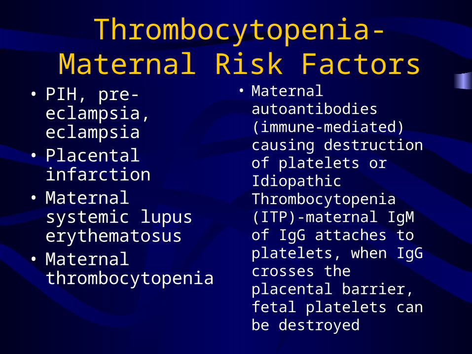
Thrombocytopenia-Maternal Risk Factors
• PIH, pre-eclampsia, eclampsia
• Placental infarction• Maternal systemic
lupus erythematosus• Maternal
thrombocytopenia
• Maternal autoantibodies (immune-mediated) causing destruction of platelets or Idiopathic Thrombocytopenia (ITP)-maternal IgM of IgG attaches to platelets, when IgG crosses the placental barrier, fetal platelets can be destroyed

Thrombocytopenia-Risks from Drug Side Effects
• Maternal or Neonatal– Indomethacin– Demerol– Phenergan– Aspirin– Sulfonamides
– Quinide– Quinine – Nitric Oxide-
prevents adhesion of platelets to endothilial cells

Thrombocytopenia• Clinical Findings
– Presence of petichiae, purpura, ecchymosis– Bleeding (GU, GI, umbilical, wound, puncture
sites, integumentary)– Hepatosplenamegaly– Jaundice– Septic shock in severe cases– Anemia– Low platelet count– PT/PTT and other coagulation factors are
normal– Elevated forms of immature platelets

Thrombocytopenia
• Management– Basic resuscitation and stabilization– Control of bleeding and fluid resuscitation– CBC with diff, PT, PTT, clotting factors,
fibrinogen, FDP, blood cultures– Administer blood products as necessary– Thorough H & P for risk factors and causes

Thrombocytopenia
• Management (continued)– Antenatal treatment with
corticosteroids– Postnatal steroid therapy– Strict I’s and O’s– Guiac stools, gastrocult gastric
secretions,dipstick urine

Thrombocytopenia
• Management (continued)– Control and prevention of S/S-
• only necessary heelsticks• constant assessment all PIV sites,
umbilical line sites, puncture sites, drain sites, foley site, and wound sites
• minimal tape use

Thrombocytopenia
–Management (continued)– Control and prevention of S/S-
• treatment of anemia• assess of S/S of intracranial
hemorrhage,NEC, GI bleeding, hyperbilirubinemia
• administration of Vitamin K

Thrombocytopenia• Management (continued)
– Treatment of underlying pathophysiology– Treatment of anemia– Exchange transfusion using blood less than 2
days old– Platelet transfusion-using single donor platelets
when possible– Administration of clotting factors
• FFP• Specific clotting factors• Cryoprecipitate

Thrombocytopenia
• Long-term follow-up/prognosis– Assess for recurrence of S/S– Follow-up platelet counts and clotting factor
levels– Prognosis is dependent on the degree of
thrombocytopenia, underlying disease, and existing syndromes

Care of the Neonate with DIC

Disseminated Intravascular Coagulation
• Definition-”an acquired hemorrhagic disorder with an underlying disease manifested as an uncontrollable activation of coagulation and fibrinolysis. Consumption of clotting factors is thought to be initiated by the release of thromboplastic material from damaged or diseased tissue into circulation. Fibrinogen converts to fibrin to form microthrombi” (Andrew, 1997: Beardsley and Nathan, 1998: Fuse et al, 1996; Hilgartner and Corrigan, 1995; Kuehl, 1997; Pugh, 1997 ). DIC presents with depletion of platelets, PT, fibrinogen, and Factors V, VII, and VIII. PT and PTT are prolonged.

DIC-Risk Factors
• PIH, pre-eclampsia, eclampsia
• Placental abruption
• Placental abnormalities
• Infection/sepsis
• Fetal distress
• Hypoxia and acidosis
• Obstetrical complications, traumatic delivery
• Dead fetal twin

DIC-Risk Factors
• Severe Rh incompatibility
• Thrombocytopenia
• Respiratory distress
• Hypotension
• Persistent pulmonary hypertension

DIC• Physiology
– results from a pre-existing disorder and does not develop independently; the underlying problem must be identified and treated
• Lab values-– PT/PTT are prolonged
– Fibrinogen is low
– FDP is high
– Platelet count is low
– D-dimer is greater than 1.0
– Abnormal red blood cell shape, cell fragmentation, and decreased number of platelets on peripheral smears

DIC
• Clinical Findings– S/S depend on the underlying disease– Continued/prolonged bleeding or oozing from
puncture sites, wound sites, and/or umbilicus– Presence of petechiae, purpura, and ecchymosis– Hemorrhage-often from every orifice– Thrombosis of peripheral vessels resulting in
localized necrosis and gangrene

DIC
• Clinical Findings (continued)– Generalized multiple site bleeding– Organ and tissue ischemia secondary to
microvascular occlusion by thrombi– Septic shock– Presence of anemia– “Blueberry muffin” spots
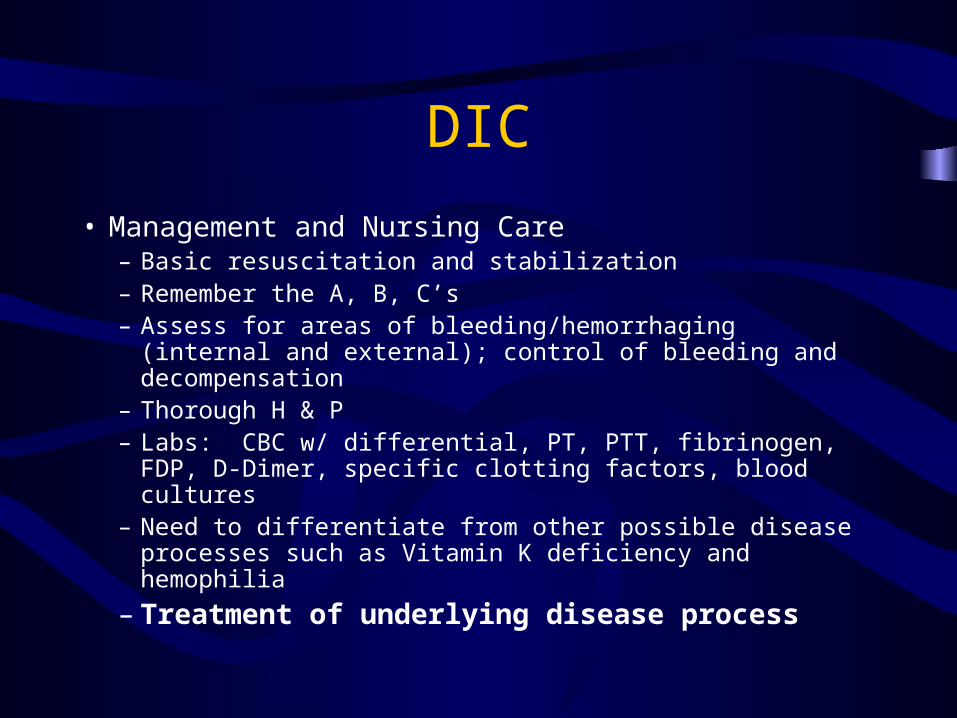
DIC
• Management and Nursing Care– Basic resuscitation and stabilization– Remember the A, B, C’s– Assess for areas of bleeding/hemorrhaging (internal and
external); control of bleeding and decompensation– Thorough H & P– Labs: CBC w/ differential, PT, PTT, fibrinogen, FDP, D-
Dimer, specific clotting factors, blood cultures– Need to differentiate from other possible disease
processes such as Vitamin K deficiency and hemophilia
– Treatment of underlying disease process

DIC
• Management & Nursing Care (continued)– Treatment of clinical symptoms– Administration of applicable blood products– Hemodynamic stabilization– Strict I’s and O’s– Assess for signs and symptoms of anaphylactic blood
products– Maintain fluid and electrolyte balance, adequate
hydration– Keep infant warm– Minimal tape use

DIC• Long-term follow-up/prognosis
– Prognosis is related to the expected outcome and successful management of the disease process, severity of DIC, and severity of complications
– Need to assess for:• Complications• S/S of organ failure• GI bleeding• Intraventricular, parenchymal hemorrhage• Severe depletion, hypovolemic shock• Continue to assess for S/S of DIC

Care of the Neonate with ABO and Rh Incompatibilities

ABO and Rh INCOMPATILITIES
• Definition– ABO-RBC destruction or adverse clustering of
RBC’s as a result of exposure of antibodies or agglutinins of one blood type to another
– Rh-more severe; occurs when an Rh negative mother’s antibodies to Rh positive factor are exposed to the antigens of an Rh positive infant leading to the destruction of the infant’s RBC’s

ABO Incompatibilities
• Risk factors-Exposure of mixing of fetal & maternal circulation usually occurs via hemorrhage during labor, delivery, amniocentesis, abortion, and ectopic pregnancy
ABO
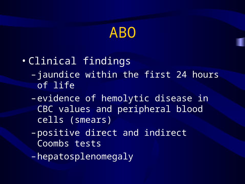
• Clinical findings– jaundice within the first 24 hours of life
– evidence of hemolytic disease in CBC values and peripheral blood cells (smears)
– positive direct and indirect Coombs tests
– hepatosplenomegaly

Rh
• Clinical findings– Jaundice/hyperbilirubinemia– Hepatosplenomegaly– Hydrops fetalis-
• Anemia• Hypoxia• Congestive heart failure• Hypoalbuminemia
– Erythroblastosis Fetalis

ABO and Rh
• Management-(NICU)– Basic resuscitation and stabilization– Blood products– RhoGam to Rh negative mothers after delivery– Hydration, fluid and electrolyte balance– Maternal and obstetrical H & P– Type & Cross, Coombs testing, CBC with diff,
Bili levels
ABO and Rh
• Management (continued)-(NICU)– Phototherapy– Exchange transfusion– Parancentisis or thorancentesis– Hemodynamic monitoring and support– Strict I’s and O’s– Double volume exchange transfusions