Rare Malignant Tumors of the Parotid Glands: Oncocytic Neoplasms
Rare Parotid Myxofibrosarcoma:
Discussion of Radiologic-Pathological Correlation
Sara Boyd, D.O. PGY-IVJulia Cameron-Morrison, D.O. PGY-IV
Amber Berry, D.O., Department of Otolaryngology Head and Neck Surgery
Timothy McKnight, D.O., Department of Radiology
Michigan State University Consortium-Beaumont Farmington Hills Hospital
Farmington Hills, Michigan1
Disclosure StatementNo disclosures or conflicts of interest for
any contributing author.
This case report was formally acknowledged by the Beaumont
Farmington Hills Resident Research Committee on March 14, 2017.
Institutional Review Board approval is not required according to the Resident
Research Committee.
2
Learning Objectives• Describe and acknowledge a reported case of
rare malignant myxofibrosarcoma arising in the parotid gland.
• Discuss etiology, histopathology, prognosis, and treatment of malignant myxofibrosarcoma in the head and neck.
• Review of myxofibrosarcoma imaging features in radiology literature.
• Discuss and briefly summarize the differential diagnoses for myxofibrosarcoma.
3
IntroductionMalignant myxofibrosarcoma is a soft tissue sarcoma that
typically presents in adults over 60 years of age and most commonly involves the subcutaneous layers of the lower extremities1. Rarely, these tumors can arise in the head and neck, reportedly in 2-15% of all cases2, most commonly arising from the nasal cavity and paranasal sinuses3. Following resection, the risk of local recurrent disease can reach 50%, with worsening tumor grade and prognosis with each recurrence4. Less than twenty case reports have been published describing myxofibrosarcomas of the head and neck5, the majority of which have been published in oncology and otolaryngology journals. Information regarding these tumors is scant in radiologic literature. The goal of this presentation is to highlight the non-specific radiologic features of head and neck myxofibrosarcoma, demonstrating the role of imaging as a part of a comprehensive clinical work-up which must include pathological confirmation.
4
Case PresentationAn eighty five year old African American female
presented to a local otolaryngologist with left sided facial swelling and total left facial paralysis (House Brackmann VI) that began in March 2016. A contrast enhanced CT of the neck was performed in May 2016 which demonstrated marked diffuse enlargement of the left parotid gland with extension into the parapharyngeal space with slightly heterogeneous enhancement but no discrete soft tissue mass. Histology from an ultrasound guided biopsy demonstrated mostly stroma, suggesting a mixed tumor-pleomorphic adenoma. Clinical correlation was recommended.
5
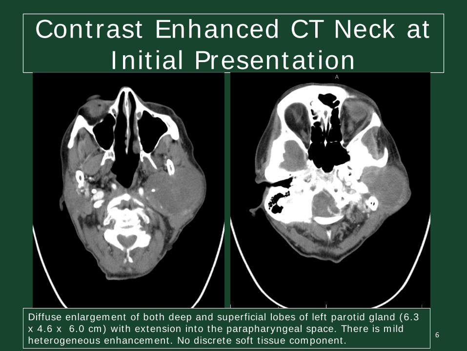
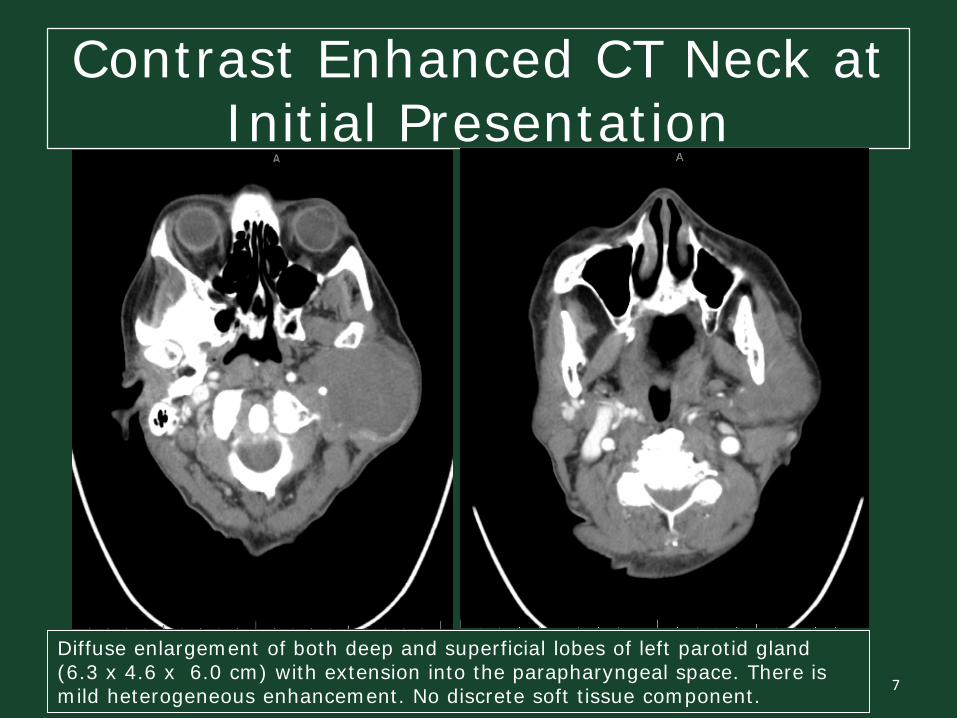
Contrast Enhanced CT Neck at Initial Presentation
6
Diffuse enlargement of both deep and superficial lobes of left parotid gland (6.3 x 4.6 x 6.0 cm) with extension into the parapharyngeal space. There is mild heterogeneous enhancement. No discrete soft tissue component.
Contrast Enhanced CT Neck at Initial Presentation
7
Diffuse enlargement of both deep and superficial lobes of left parotid gland (6.3 x 4.6 x 6.0 cm) with extension into the parapharyngeal space. There is mild heterogeneous enhancement. No discrete soft tissue component.
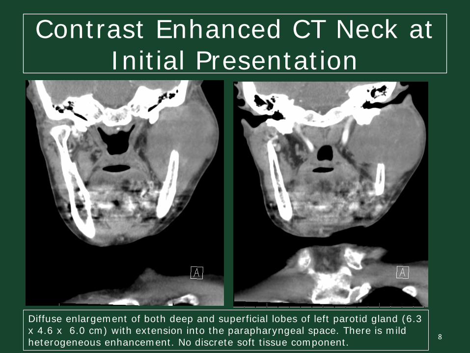
Contrast Enhanced CT Neck at Initial Presentation
8
Diffuse enlargement of both deep and superficial lobes of left parotid gland (6.3 x 4.6 x 6.0 cm) with extension into the parapharyngeal space. There is mild heterogeneous enhancement. No discrete soft tissue component.
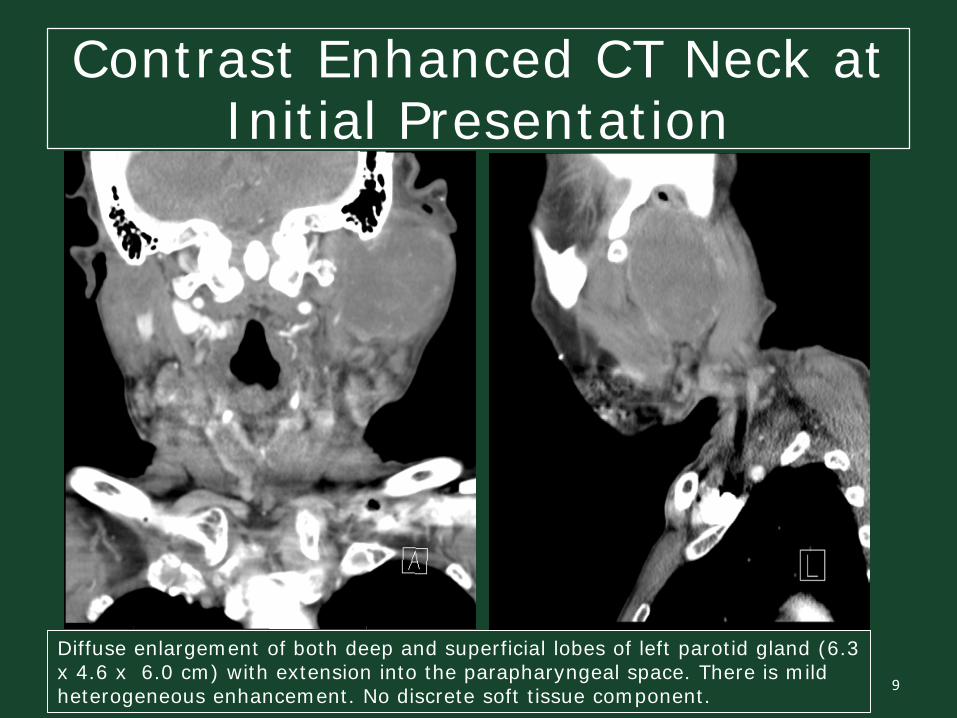
Contrast Enhanced CT Neck at Initial Presentation
9
Diffuse enlargement of both deep and superficial lobes of left parotid gland (6.3 x 4.6 x 6.0 cm) with extension into the parapharyngeal space. There is mild heterogeneous enhancement. No discrete soft tissue component.
Contrast Enhanced CT Neck at Initial Presentation
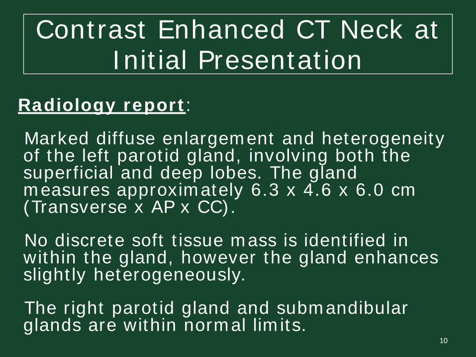
Radiology report:
Marked diffuse enlargement and heterogeneity of the left parotid gland, involving both the superficial and deep lobes. The gland measures approximately 6.3 x 4.6 x 6.0 cm (Transverse x AP x CC).
No discrete soft tissue mass is identified in within the gland, however the gland enhances slightly heterogeneously.
The right parotid gland and submandibularglands are within normal limits.
10
Case Presentation
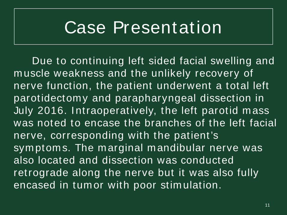
Due to continuing left sided facial swelling and muscle weakness and the unlikely recovery of nerve function, the patient underwent a total left parotidectomy and parapharyngeal dissection in July 2016. Intraoperatively, the left parotid mass was noted to encase the branches of the left facial nerve, corresponding with the patient’s symptoms. The marginal mandibular nerve wasalso located and dissection was conducted retrograde along the nerve but it was also fully encased in tumor with poor stimulation.
11
Intraoperative and Gross Specimen Images
12
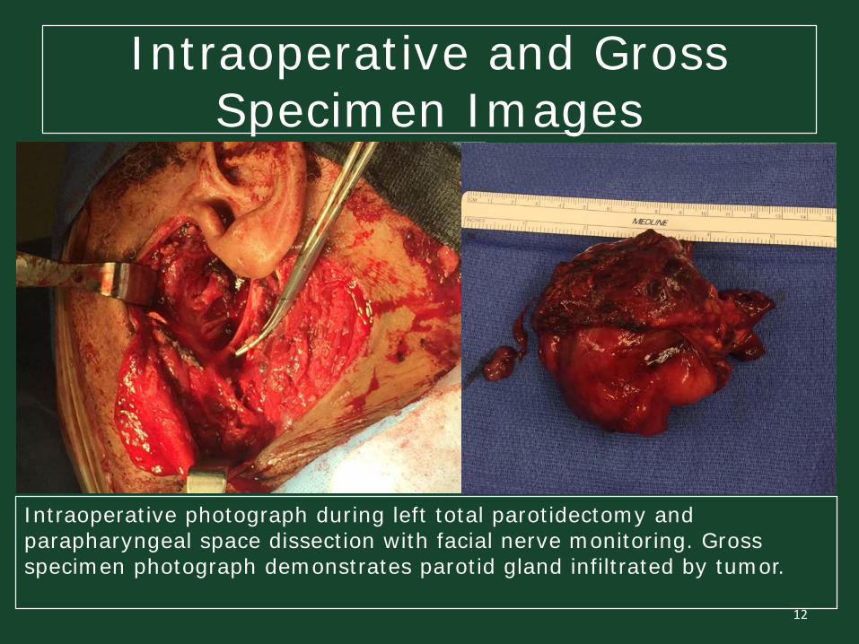
Intraoperative photograph during left total parotidectomy and parapharyngeal space dissection with facial nerve monitoring. Gross specimen photograph demonstrates parotid gland infiltrated by tumor.
Case Presentation
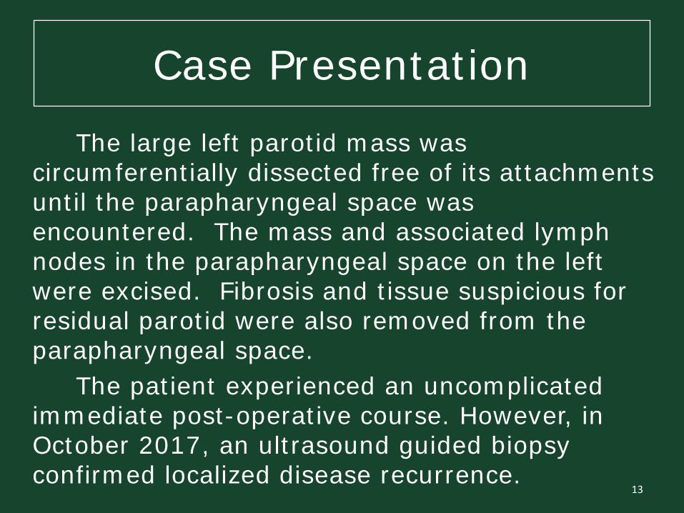
The large left parotid mass was circumferentially dissected free of its attachments until the parapharyngeal space was encountered. The mass and associated lymph nodes in the parapharyngeal space on the left were excised. Fibrosis and tissue suspicious for residual parotid were also removed from the parapharyngeal space.
The patient experienced an uncomplicated immediate post-operative course. However, in October 2017, an ultrasound guided biopsy confirmed localized disease recurrence.
13
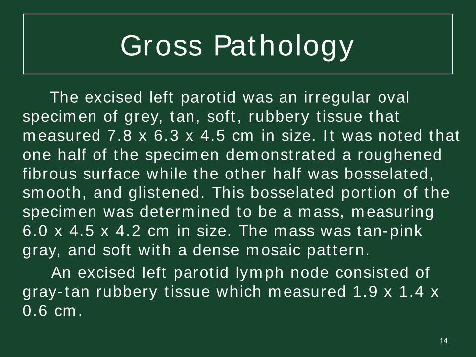
Gross PathologyThe excised left parotid was an irregular oval
specimen of grey, tan, soft, rubbery tissue that measured 7.8 x 6.3 x 4.5 cm in size. It was noted that one half of the specimen demonstrated a roughened fibrous surface while the other half was bosselated, smooth, and glistened. This bosselated portion of the specimen was determined to be a mass, measuring 6.0 x 4.5 x 4.2 cm in size. The mass was tan-pink gray, and soft with a dense mosaic pattern.
An excised left parotid lymph node consisted of gray-tan rubbery tissue which measured 1.9 x 1.4 x0.6 cm.
14
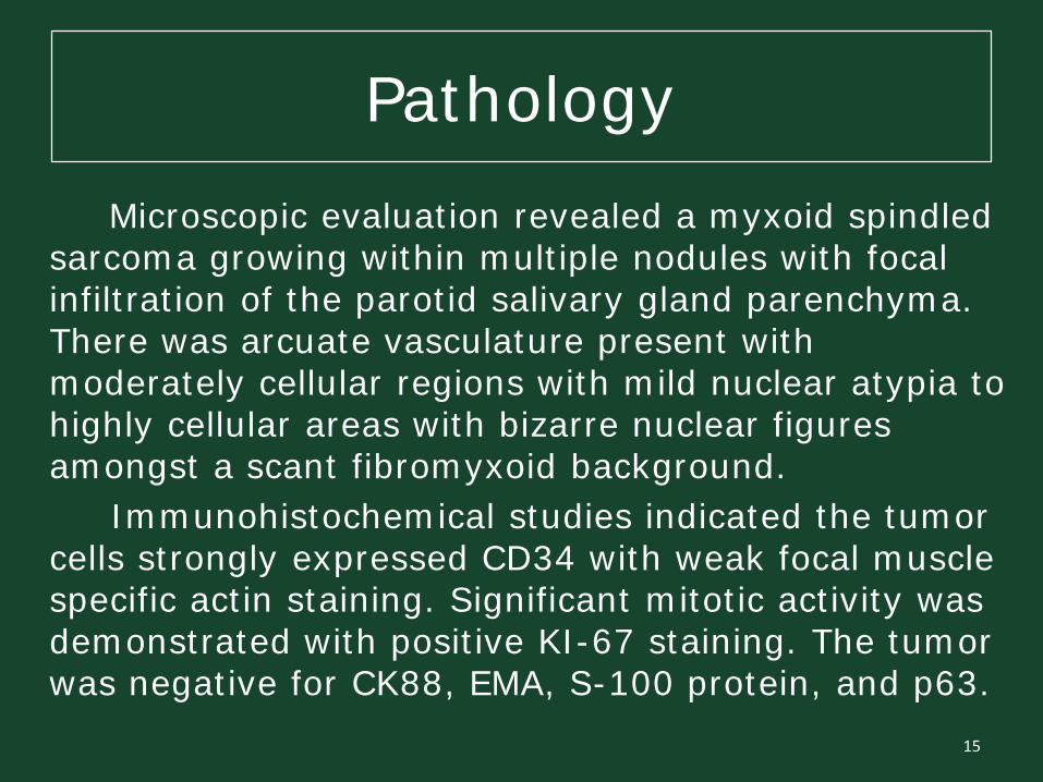
PathologyMicroscopic evaluation revealed a myxoid spindled
sarcoma growing within multiple nodules with focal infiltration of the parotid salivary gland parenchyma. There was arcuate vasculature present with moderately cellular regions with mild nuclear atypia to highly cellular areas with bizarre nuclear figures amongst a scant fibromyxoid background.
Immunohistochemical studies indicated the tumor cells strongly expressed CD34 with weak focal muscle specific actin staining. Significant mitotic activity was demonstrated with positive KI-67 staining. The tumor was negative for CK88, EMA, S-100 protein, and p63.
15
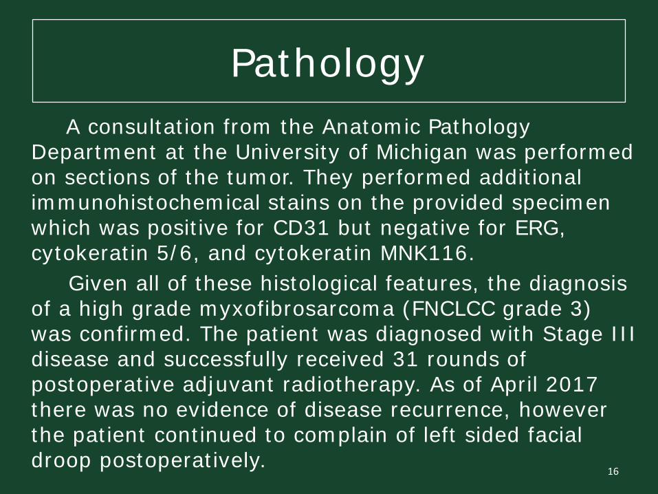
PathologyA consultation from the Anatomic Pathology
Department at the University of Michigan was performed on sections of the tumor. They performed additional immunohistochemical stains on the provided specimen which was positive for CD31 but negative for ERG, cytokeratin 5/6, and cytokeratin MNK116.
Given all of these histological features, the diagnosis of a high grade myxofibrosarcoma (FNCLCC grade 3) was confirmed. The patient was diagnosed with Stage III disease and successfully received 31 rounds of postoperative adjuvant radiotherapy. As of April 2017 there was no evidence of disease recurrence, however the patient continued to complain of left sided facial droop postoperatively.
16
Pathology Slides
17
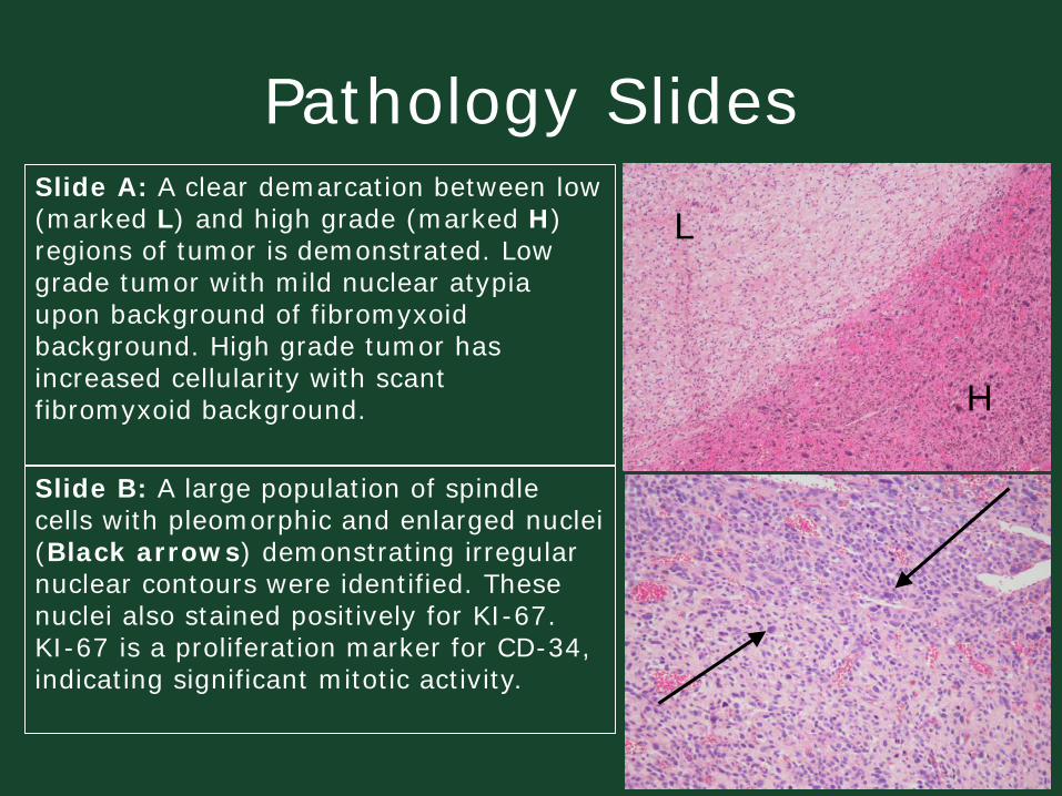
Slide B: A large population of spindle cells with pleomorphic and enlarged nuclei (Black arrows) demonstrating irregular nuclear contours were identified. These nuclei also stained positively for KI-67. KI-67 is a proliferation marker for CD-34, indicating significant mitotic activity.
L
H
Slide A: A clear demarcation between low (marked L) and high grade (marked H) regions of tumor is demonstrated. Low grade tumor with mild nuclear atypia upon background of fibromyxoid background. High grade tumor has increased cellularity with scant fibromyxoid background.
Pathology Slides
18
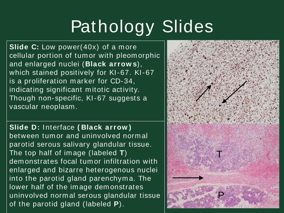
Slide C: Low power(40x) of a more cellular portion of tumor with pleomorphic and enlarged nuclei (Black arrows), which stained positively for KI-67. KI-67 is a proliferation marker for CD-34, indicating significant mitotic activity. Though non-specific, KI-67 suggests a vascular neoplasm.
Slide D: Interface (Black arrow) between tumor and uninvolved normal parotid serous salivary glandular tissue. The top half of image (labeled T) demonstrates focal tumor infiltration with enlarged and bizarre heterogenous nuclei into the parotid gland parenchyma. The lower half of the image demonstrates uninvolved normal serous glandular tissue of the parotid gland (labeled P).
T
P
Pathology Slides
19
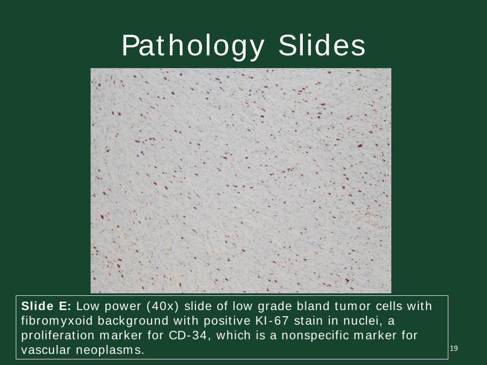
Slide E: Low power (40x) slide of low grade bland tumor cells with fibromyxoid background with positive KI-67 stain in nuclei, a proliferation marker for CD-34, which is a nonspecific marker for vascular neoplasms.
Pathology Slides
20
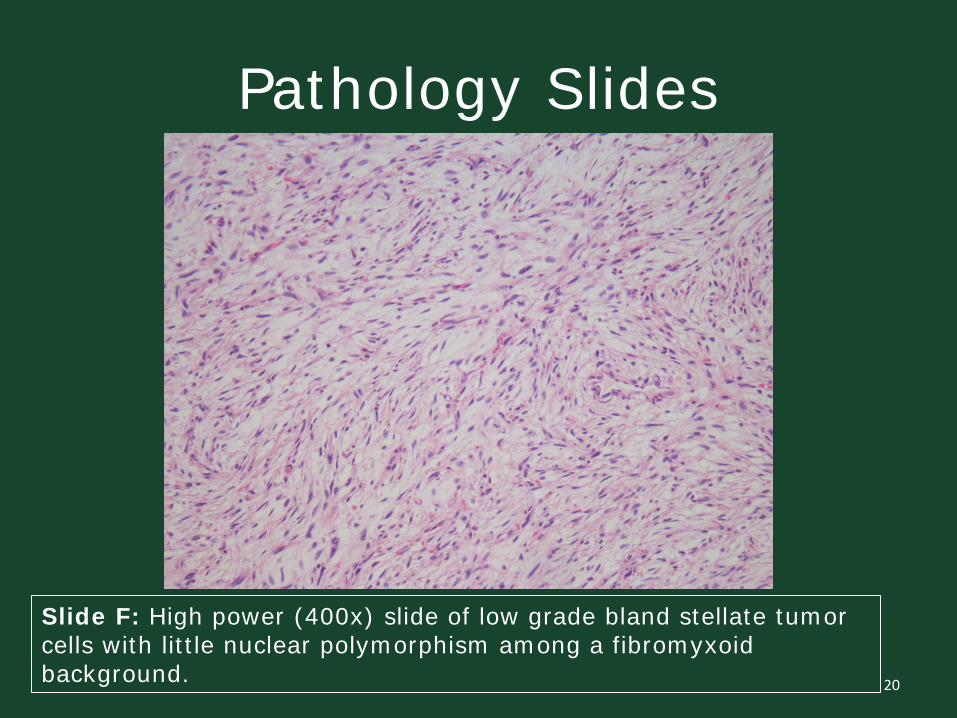
Slide F: High power (400x) slide of low grade bland stellate tumor cells with little nuclear polymorphism among a fibromyxoidbackground.
DiscussionAs the most common malignant connective tissue
sarcoma in older adults, myxofibrosarcoma was initially classified as a myxoid variant of malignant fibrous histiocytoma1,2, which was later declassified by the World Health Organization and determined to be its own diagnostic entity in 20025. These mesodermal derived neoplasms most often arise from the subcutaneous layer of the lower extremities in patients in their seventh decade of life. They incite a wide spectrum of clinical presentations from slowly enlarging painless lesions3 that enlarge over weeks to months10 with minimal symptoms to destructive and aggressive tumors with earlier onset of symptoms and a higher risk of metastatic spread7. Advanced disseminated cases of myxofibrosarcomas have been known to metastasize to the lung, bone, and liver3. 21
DiscussionThese tumors have also been found to arise primarily
in the chest in 12% and the retroperitoneum or mediastinum in 8% of reported cases5. Academic literature appears to be in disagreement about gender predilection, as some articles suggest a male predominance, while many others indicate no significant difference in incidence between male and female patients diagnosed with myxofibrosarcoma10.
22
DiscussionThough the majority of myxofibrosarcomas develop
primarily within lower extremities, they can rarely be found in the head and neck region. Few published studies and articles in both Otorhinolaryngology and Radiology literature have determined that the incidence of myxofibrosarcomas arising in the head and neck region ranges from 3-10%3,8 and typically portend a poorer prognosis compared to those arising in the extremity, chest, or mediastinum. Amongst the anatomic head and neck regions, myxofibrosarcomas have been reported to involve the nasal cavity and paranasal sinuses, maxillary sinus, sphenoid sinus, mandible, orbit, larynx, parotid gland, oral cavity, ear, eyelid, and infratemporal space2,3.
23
DiscussionThe most common head and neck location is the nasal
cavity and paranasal sinuses, in up to 30% of cases3. Primary myxofibrosarcoma of the parotid gland is rare, with approximately 30 cases reported worldwide8. Patients with tumor invasion into the aerodigestive tract tend to present earlier due to onset of symptoms. Similarly, in our case the patient presented with left sided facial swelling and weakness secondary to involvement of the facial nerve.
24
DiscussionWith identifiable pleomorphism of the nuclei and highly
variable mitotic activity, these soft tissue tumors have been divided into five histopathological subtypes, including storiform-pleomorphic (most common)3, myxoid, giant cell, inflammatory, and angiomatoid2. These lesions demonstrate a spectrum in the amounts of contained background myxoid matrix and developed cellularity- from less cellular tumors containing minimal atypia to more densely cellular lesions with significant cell atypia.
25
DiscussionA three or four-tiered grading system is currently in
clinical use based on histology, which is based on the cellular degree, nuclear pleomorphism, and the mitotic activity rate1. Low-grade myxofibrosacomas demonstrate a relatively small to moderately cellular structure with a prominent background of myxoid matrix2 with very few mitoses visualized. Low-grade neoplastic cell shapes can be fusiform, round, or stellate and their nuclei can be hyperchromatic with irregular margins. It has also been reported that low-grade tumor cells are commonly found in the periphery of a blood vessel.
26
DiscussionIntermediate-grade myxofibrosarcomas are more
hypercellular than low-grade lesions but typically do not demonstrate intratumoral necrosis. Immunohistocheimcalstaining of low-grade neoplasms can show positive staining for CD-34, vimentin, and Ki-672. High-grade myxofibrosarcomas are much more densely packed with atypical cells that demonstrate scant interspersed myxoidmaterial and tend to be more solid than low-grade tumors. Their cellular pleomorphism is severe and the cells exhibit significantly raised mitotic rates6.
27
DiscussionLike many other high-grade neoplasms, necrosis and
resultant intratumoral hemorrhage are commonly found in higher grade myxofibrosarcomas. The rates of metastatic spread also increase with increasing grade. There are rare reports of low-grade myxofibrosarcomashaving distant metastasis. The recurrence rate of disease however, does not correlate with histologic grade, and is therefore independent, as there is up to a 50-60% local recurrence rate reported in both low and high grade tumors2. With each local recurrence of disease following surgical resection, the patient’s prognosis worsens with an additional relative increase in the risk of metastasis4.
28
DiscussionBecause of this unique malignant feature, it is
absolutely crucial for complete surgical excision of myxofibrosarcoma with clean margins to be performed to achieve curative therapy6 with many oncologists opting to add subsequent adjuvant radiotherapy5. The patient discussed in this case report underwent surgical resection with parotidectomy and parapharyngeal dissection as well as completed 31 rounds of adjuvant radiotherapy, in conjunction with the generally accepted mainstay of treatment.
29
DiscussionCross sectional imaging with magnetic resonance
imaging (MRI) and computed tomography (CT) of myxofibrosarcomas augments the evaluation of the presence or absence of surrounding adjacent structural invasion2 and the degree of invasion if present. MRI is the preferred modality and should be performed before biopsy4. The extent of soft tissue involvement and osseous destruction are crucial features that must be identified and commented on by radiologists. Ultrasound has not been shown to contribute any significant radiologic information and is thus not recommended4, as it tends to delay diagnosis and possibly appropriate treatment.
30
DiscussionAmong the scarce literature published on head and
neck myxofibrosarcomas, there are even fewer articles published that evaluate or discuss the radiologic features of this rare malignancy. In a study by Park et. al, their investigation into the CT and MR features determined that these tumors are largely nonspecific on imaging studies. On CT, they most often appeared as a lobulatedsoft tissue mass, similar to muscle in attenuation, with a centralized hypoattenuation which may related to underlying necrosis, hemorrhage or be secondary to the myxoid makeup of the tumor3. In up to 20% of the patients in their study, ossification or calcification was detected3.
31
DiscussionOn MRI, myxofibrosarcomas often had heterogeneous
signal intensity on all pulse sequences, which is likely a result of the intricate and complex histologic features3. Fibrous tissue tended to be low in signal on MRI, in opposition to the hyperintense myxoid stroma. An aggressive myxofibrosarcoma can contain intratumoralhemorrhage, presenting as fluid-fluid levels of differing signal intensities3. Nodular solid components, when present, will heterogeneously enhance following the administration of contrast.
32
DiscussionDue to their nonspecific imaging features,
myxofibrosarcomas arising in the head and neck region is often confused with several other soft tissue and fibrous abnormalities and neoplasms. This reiterates how crucial it is for a percutaneous biopsy to be performed as a part of a comprehensive evaluation of a head and neck mass. Additional diagnostic considerations that may appear similarly on imaging include fibrosarcoma, pleomorphicrhabdomyosarcoma, nodular fasciitis, fibromatosis, and malignant mesenchymal tumors3 to name just a few.
33
DiscussionFibrosarcomas are malignant neoplasms that are
associated with prior irradiation of the head and neck region and demonstrate a characteristic herringbone-like structure9. These tumors can arise in the sinonasalcavities, larynx, or neck and demonstrate nonspecific heterogeneity on MRI sequences. Rhabdomyosarcoma, the most common pediatric soft tissue sarcomatousneoplasm, can also be seen in adult patients, and can arise in the head and neck 40% of the time9. On imaging they commonly show lytic destruction of osseous structures with similar nonspecific features on CT and MRI to many other etiologies.
34
DiscussionNodular fasciitis occurs in the head and neck region in
approximately 15-20% of patients and presents as proliferation of fibrous tissue, often felt as a palpable abnormality in affected patients9. CT imaging of nodular fasciitis demonstrates a fluid attenuating mass and on MRI, these masses are heterogeneous with increased signal on both T1 and T2 weighted sequences9.
35
ConclusionAny patient presenting with swelling in the parotid
gland area should always raise a red flag for clinicians due to the possibility of an underlying neoplasm1. In the evaluation of parotid gland tumors, CT and MRI can be invaluable in suggesting pathologic diagnosis based on imaging characteristics established in radiology literature, as well as providing preoperative staging and guidance for surgical planning3. This case report illustrates how radiologic imaging of myxofibrosarcomas arising in the parotid gland, as well as other locations in the head and neck, are non-specific3 on cross sectional imaging and must be performed in conjunction with histologicsampling to confirm the diagnosis of this rare malignancy.
36
ConclusionIt is critical for any focal invasion of
myxofibrosarcomas into surrounding structures be identified. This neoplasm requires complete surgical excision with clear margins for curative therapy6 , often with subsequent adjuvant radiotherapy. These patients should remain in a long-term clinical follow-up regimen given the change in prognosis with each recurrence2. Therefore, although rare, myxofibrosarcoma must be considered in the differential diagnosis of parotid gland tumors.
37
References1Li X, Chen X, Shi ZH, et al. Primary myxofibrosarcoma of the parotid: case
report. BMC Cancer. 2010;10:246.2Qiubei Z, Cheng L, Yaping X, Shunzhang L, Jingping F. Myxofibrosarcoma of
the sinus piriformis: case report and literature review. World J Surg Oncol. 2012;10(1):245.
3Park SW, Kim HJ, Lee JH, et al. Malignant fibrous histiocytoma of the head and neck: CT and MR imaging findings. AJNR ASm J Neuroradiol. 2009;30:71-76.
4Darouassi Y, Attifi H, Zalagh M, et al. Mxyofibrosarcoma of the thyroid gland. European Annals of Otorhinolaryngology, Head and Neck Diseases. 2014;131:385–387.
5Dell'aversana Orabona G, Iaconetta G, Abbate V, et al. Head and neck myxofibrosarcoma: a case report and review of the literature. J Med Case Rep. 2014;8:468.
6Udaka T, Yamamoto H, Shiomori T, Fujimura T, Suzuki H. Myxofibrosarcoma of the neck. J Laryngol Otol. 2006;120(10):872-4.
7Gugatschka M, Beham A, Stammberger H, Schmid C, Friedrich G. First case of a myxofibrosarcoma of the vocal folds: case report and review of the literature. J Voice. 2010;24(3):374-6.
38
References8Mačák J, Šmardová J, Zavřelová I, Vránová V, Kuglík P. Malignant fibrous
histiocytoma of the parotid gland. Česko-slovenská patologie. 2007; 4:148-152.
9Razek AA, Huang BY. Soft tissue tumors of the head and neck: imaging-based review of the WHO classification. Radiographics. 2011;31(7):1923-54.
10Srinivasan B, Ethunandan M, Hussain K, Ilankovan V. Epitheloidmyxofibrosarcoma of the parotid gland. Case Rep Pathol. 2011;2011:641621.
39

Contact Information
Questions or Comments?
Feel free to contact us:
[email protected]@beaumont.org
40