A rare case of parotid duct sialolithiasis with sialo-oral fistula
4
A A rare case e of parot sialo- tid duct s -oral fistu ialolithia ula sis with
-
Upload
apollo-hospitals -
Category
Health & Medicine
-
view
108 -
download
2
Transcript of A rare case of parotid duct sialolithiasis with sialo-oral fistula

A
A rare case
e of parotsialo-
tid duct s-oral fistu
ialolithiaula
sis with
ww.sciencedirect.com
a p o l l o m e d i c i n e x x x ( 2 0 1 4 ) 1e2
Available online at w
ScienceDirect
journal homepage: www.elsevier .com/locate/apme
Case Report
A rare case of parotid duct sialolithiasis withsialo-oral fistula
Surya Kanta Pradhan*
Department of ENT e Head Neck Surgery, Apollo Hospitals, 251, Sainik School Road, Unit-15, Bhubaneswar 751005,
Orissa, India
a r t i c l e i n f o
Article history:
Received 8 July 2014
Accepted 18 July 2014
Available online xxx
Keywords:
Parotid gland
Sialolithiasis
Sialo-oral fistula
* Tel.: þ918093060163 (mobile).E-mail address: [email protected]
Please cite this article in press as: Pradha(2014), http://dx.doi.org/10.1016/j.apme.2
http://dx.doi.org/10.1016/j.apme.2014.07.0030976-0016/Copyright © 2014, Indraprastha M
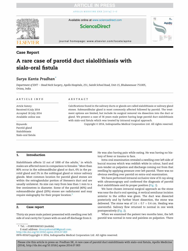
a b s t r a c t
Calcifications found in the salivary ducts or glands are called sialolithiasis or salivary gland
stones. Submandibular gland is most commonly affected followed by parotid. The treat-
ment options are limited, but include its surgical removal via dissection into the duct or
gland. We present a case of 36 years male patient having large parotid duct sialolithiasis
with sialo-oral fistula which was treated by intraoral surgical approach.
Copyright © 2014, Indraprastha Medical Corporation Ltd. All rights reserved.
1. Introduction
Sialolithiasis affects 12 out of 1000 of the adults,1 in which
males are affectedmore in comparison to females.2 More than
80% occur in the submandibular gland or duct, 6% in the pa-
rotid gland and 2% in the sublingual gland or minor salivary
glands. Most common location for parotid gland stones are
within the extraglandular portion of Stensen's duct and are
usually unilateral. Its size can vary from less than 1 mm to a
few centimetres in diameter. Some of the parotid (40%) and
submandibular gland (20%) stones are radiolucent and may
require sialography for their proper location.2
2. Case report
Thirty six yearsmale patient presented with swelling over left
side of oral cavity for 3 years with on and off discharge from it.
m.
n SK, A rare case of paro014.07.003
edical Corporation Ltd. A
He was also having pain while eating. He was having no his-
tory of fever or trauma to face.
Intra-oral examination revealed a swelling over left side of
buccal mucosa which was reddish white in colour, hard and
non-tender on palpation and discharge coming out from that
swelling by applying pressure over left parotid. There was no
obvious swelling over parotid on extra-oral examination.
We have performed intraoral occlusive view of X-ray along
with ultrasonograpy and confirmed the diagnosis of parotid
duct sialolithiasis and its proper position (Fig. 1).
We have chosen intraoral surgical approach as the stone
was near the duct's oral opening. A vertical semilunar incision
anterior to the orifice was given. The duct was dissected
posteriorly and by further blunt dissection, the stone was
delivered. The stone was of 1.6 � 0.7 � 0.4 cm. Healing was
uneventful. Salivary flow returned to normal in one week
postoperatively (Fig. 2).
When we examined the patient two months later, the left
parotid was normal in tone and painless on palpation. There
tid duct sialolithiasis with sialo-oral fistula, Apollo Medicine
ll rights reserved.
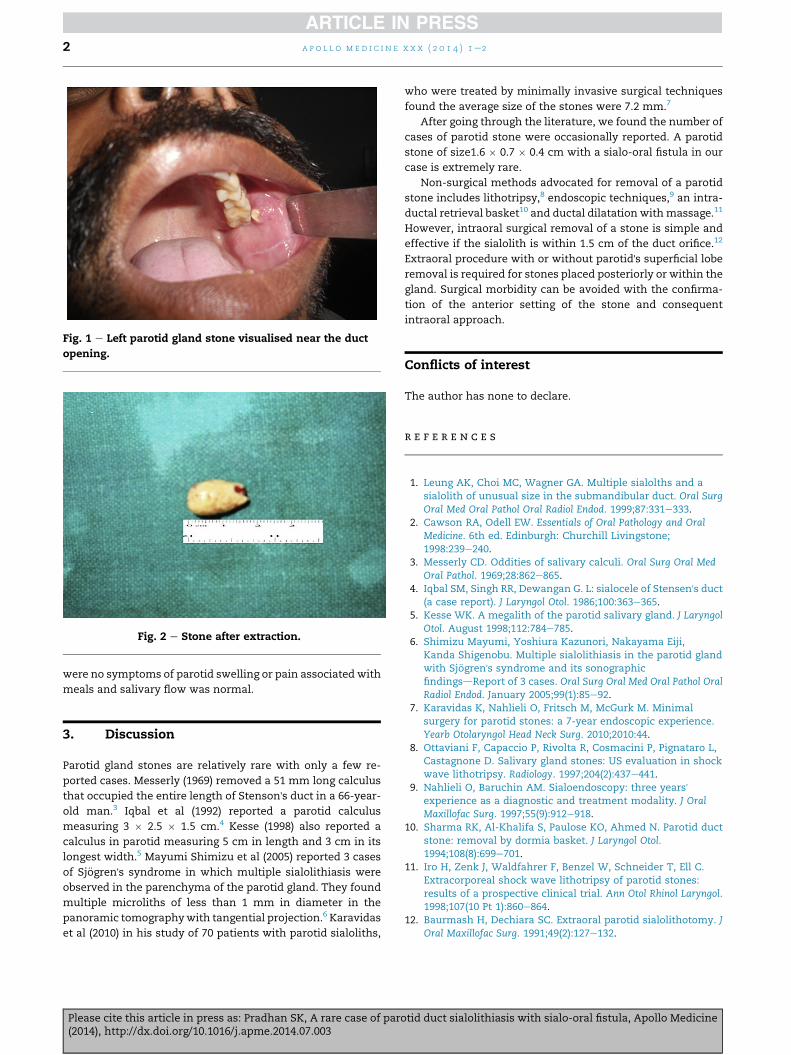
Fig. 1 e Left parotid gland stone visualised near the duct
opening.
Fig. 2 e Stone after extraction.
a p o l l o m e d i c i n e x x x ( 2 0 1 4 ) 1e22
were no symptoms of parotid swelling or pain associated with
meals and salivary flow was normal.
3. Discussion
Parotid gland stones are relatively rare with only a few re-
ported cases. Messerly (1969) removed a 51 mm long calculus
that occupied the entire length of Stenson's duct in a 66-year-
old man.3 Iqbal et al (1992) reported a parotid calculus
measuring 3 � 2.5 � 1.5 cm.4 Kesse (1998) also reported a
calculus in parotid measuring 5 cm in length and 3 cm in its
longest width.5 Mayumi Shimizu et al (2005) reported 3 cases
of Sj€ogren's syndrome in which multiple sialolithiasis were
observed in the parenchyma of the parotid gland. They found
multiple microliths of less than 1 mm in diameter in the
panoramic tomographywith tangential projection.6 Karavidas
et al (2010) in his study of 70 patients with parotid sialoliths,
Please cite this article in press as: Pradhan SK, A rare case of par(2014), http://dx.doi.org/10.1016/j.apme.2014.07.003
who were treated by minimally invasive surgical techniques
found the average size of the stones were 7.2 mm.7
After going through the literature, we found the number of
cases of parotid stone were occasionally reported. A parotid
stone of size1.6 � 0.7 � 0.4 cm with a sialo-oral fistula in our
case is extremely rare.
Non-surgical methods advocated for removal of a parotid
stone includes lithotripsy,8 endoscopic techniques,9 an intra-
ductal retrieval basket10 and ductal dilatation withmassage.11
However, intraoral surgical removal of a stone is simple and
effective if the sialolith is within 1.5 cm of the duct orifice.12
Extraoral procedure with or without parotid's superficial lobe
removal is required for stones placed posteriorly or within the
gland. Surgical morbidity can be avoided with the confirma-
tion of the anterior setting of the stone and consequent
intraoral approach.
Conflicts of interest
The author has none to declare.
r e f e r e n c e s
1. Leung AK, Choi MC, Wagner GA. Multiple sialolths and asialolith of unusual size in the submandibular duct. Oral SurgOral Med Oral Pathol Oral Radiol Endod. 1999;87:331e333.
2. Cawson RA, Odell EW. Essentials of Oral Pathology and OralMedicine. 6th ed. Edinburgh: Churchill Livingstone;1998:239e240.
3. Messerly CD. Oddities of salivary calculi. Oral Surg Oral MedOral Pathol. 1969;28:862e865.
4. Iqbal SM, Singh RR, Dewangan G. L: sialocele of Stensen's duct(a case report). J Laryngol Otol. 1986;100:363e365.
5. Kesse WK. A megalith of the parotid salivary gland. J LaryngolOtol. August 1998;112:784e785.
6. Shimizu Mayumi, Yoshiura Kazunori, Nakayama Eiji,Kanda Shigenobu. Multiple sialolithiasis in the parotid glandwith Sj€ogren's syndrome and its sonographicfindingsdReport of 3 cases. Oral Surg Oral Med Oral Pathol OralRadiol Endod. January 2005;99(1):85e92.
7. Karavidas K, Nahlieli O, Fritsch M, McGurk M. Minimalsurgery for parotid stones: a 7-year endoscopic experience.Yearb Otolaryngol Head Neck Surg. 2010;2010:44.
8. Ottaviani F, Capaccio P, Rivolta R, Cosmacini P, Pignataro L,Castagnone D. Salivary gland stones: US evaluation in shockwave lithotripsy. Radiology. 1997;204(2):437e441.
9. Nahlieli O, Baruchin AM. Sialoendoscopy: three years'experience as a diagnostic and treatment modality. J OralMaxillofac Surg. 1997;55(9):912e918.
10. Sharma RK, Al-Khalifa S, Paulose KO, Ahmed N. Parotid ductstone: removal by dormia basket. J Laryngol Otol.1994;108(8):699e701.
11. Iro H, Zenk J, Waldfahrer F, Benzel W, Schneider T, Ell C.Extracorporeal shock wave lithotripsy of parotid stones:results of a prospective clinical trial. Ann Otol Rhinol Laryngol.1998;107(10 Pt 1):860e864.
12. Baurmash H, Dechiara SC. Extraoral parotid sialolithotomy. JOral Maxillofac Surg. 1991;49(2):127e132.
otid duct sialolithiasis with sialo-oral fistula, Apollo Medicine

Apollo hospitals: http://www.apollohospitals.com/Twitter: https://twitter.com/HospitalsApolloYoutube: http://www.youtube.com/apollohospitalsindiaFacebook: http://www.facebook.com/TheApolloHospitalsSlideshare: http://www.slideshare.net/Apollo_HospitalsLinkedin: http://www.linkedin.com/company/apollo-hospitalsBlog:Blog: http://www.letstalkhealth.in/