Primary Malignant Parotid Melanoma: Searching for a Rare ... · origin and localized in the parotid...
6
International Journal of Otolaryngology and Head & Neck Surgery, 2014, 3, 228-232 Published Online September 2014 in SciRes. http://www.scirp.org/journal/ijohns http://dx.doi.org/10.4236/ijohns.2014.35042 How to cite this paper: Villarreal, I.M., Arellano, B., Sáenz, J., Martin, L., Castello, J.R. and García-Berrocal, J.R. (2014) Pri- mary Malignant Parotid Melanoma: Searching for a Rare Entity. International Journal of Otolaryngology and Head & Neck Surgery, 3, 228-232. http://dx.doi.org/10.4236/ijohns.2014.35042 Primary Malignant Parotid Melanoma: Searching for a Rare Entity Ithzel Maria Villarreal 1 , Beatriz Arellano 1 , Jeanette Sáenz 1 , Luz Martin 2 , José Ramón Castello 3 , José Ramón García-Berrocal 1* 1 Department of ENT, “Puerta de Hierro-Majadahonda” University Hospital, Madrid, Spain 2 Department of Pathology, “Puerta de Hierro-Majadahonda” University Hospital, Madrid, Spain 3 Department of Plastic Surgery, “Puerta de Hierro-Majadahonda” University Hospital, Madrid, Spain Email: * [email protected] Received 7 July 2014; revised 5 August 2014; accepted 1 September 2014 Copyright © 2014 by authors and Scientific Research Publishing Inc. This work is licensed under the Creative Commons Attribution International License (CC BY). http://creativecommons.org/licenses/by/4.0/ Abstract Objectives: The aim of this paper is to establish a correct approach to the diagnosis of a primary malignant melanoma of the parotid gland. Methods: We present a case of a patient with a painless mass situated in the left parotid region after an initial cytological and radiological study suggest- ing a Warthin tumor whose treatment involved a total parotidectomy with a facial nerve recon- struction. Results: The histopathological result showed a malignant melanoma that after various studies and evaluations showed no other primary origin different from the parotid gland itself. Conclusions: Salivary gland melanomas are relatively rare entities that when being of a primary origin and localized in the parotid gland become even more uncommon. Upon a melanoma found in the parotid gland, the first suspicion should be a metastatic origin, being the most common sites the oral cavity and the skin from the head and neck region. Keywords Primary Parotid Melanoma, PET-Scan, S100 Protein, Facial Nerve Reconstruction 1. Introduction Malignant melanoma is one of the most aggressive malignant tumors accounting for 15% of all cancers [1] and represents approximately 0.7% of all malignant neoplasms within the parotid gland [2]. They are characterized by a difficult late diagnosis and poor prognosis. Most of them are considered to be metastasis although some- times the primary origin cannot be established [1]. Primary melanoma of the parotid gland is considered ex- tremely rare [2] [3]. * Corresponding author.
Transcript of Primary Malignant Parotid Melanoma: Searching for a Rare ... · origin and localized in the parotid...

International Journal of Otolaryngology and Head & Neck Surgery, 2014, 3, 228-232 Published Online September 2014 in SciRes. http://www.scirp.org/journal/ijohns http://dx.doi.org/10.4236/ijohns.2014.35042
How to cite this paper: Villarreal, I.M., Arellano, B., Sáenz, J., Martin, L., Castello, J.R. and García-Berrocal, J.R. (2014) Pri-mary Malignant Parotid Melanoma: Searching for a Rare Entity. International Journal of Otolaryngology and Head & Neck Surgery, 3, 228-232. http://dx.doi.org/10.4236/ijohns.2014.35042
Primary Malignant Parotid Melanoma: Searching for a Rare Entity Ithzel Maria Villarreal1, Beatriz Arellano1, Jeanette Sáenz1, Luz Martin2, José Ramón Castello3, José Ramón García-Berrocal1* 1Department of ENT, “Puerta de Hierro-Majadahonda” University Hospital, Madrid, Spain 2Department of Pathology, “Puerta de Hierro-Majadahonda” University Hospital, Madrid, Spain 3Department of Plastic Surgery, “Puerta de Hierro-Majadahonda” University Hospital, Madrid, Spain Email: *[email protected]
Received 7 July 2014; revised 5 August 2014; accepted 1 September 2014
Copyright © 2014 by authors and Scientific Research Publishing Inc. This work is licensed under the Creative Commons Attribution International License (CC BY). http://creativecommons.org/licenses/by/4.0/
Abstract Objectives: The aim of this paper is to establish a correct approach to the diagnosis of a primary malignant melanoma of the parotid gland. Methods: We present a case of a patient with a painless mass situated in the left parotid region after an initial cytological and radiological study suggest-ing a Warthin tumor whose treatment involved a total parotidectomy with a facial nerve recon-struction. Results: The histopathological result showed a malignant melanoma that after various studies and evaluations showed no other primary origin different from the parotid gland itself. Conclusions: Salivary gland melanomas are relatively rare entities that when being of a primary origin and localized in the parotid gland become even more uncommon. Upon a melanoma found in the parotid gland, the first suspicion should be a metastatic origin, being the most common sites the oral cavity and the skin from the head and neck region.
Keywords Primary Parotid Melanoma, PET-Scan, S100 Protein, Facial Nerve Reconstruction
1. Introduction Malignant melanoma is one of the most aggressive malignant tumors accounting for 15% of all cancers [1] and represents approximately 0.7% of all malignant neoplasms within the parotid gland [2]. They are characterized by a difficult late diagnosis and poor prognosis. Most of them are considered to be metastasis although some-times the primary origin cannot be established [1]. Primary melanoma of the parotid gland is considered ex-tremely rare [2] [3].
*Corresponding author.

I. M. Villarreal et al.
229
2. Case Report A 50-year-old male presented at our institution with a 3 months history of a painless mass in the left mandibular angle which had rapidly increased in size. No other symptoms were present and his past medical history was unremarkable.
Physical examination showed a round, painless approximately 1 cm in size mass located in the left parotid compartment. No other clinical features were perceived.
Complementary studies were done, beginning with an ultrasound that revealed both parotid glands enlarged finding also in the left side a solid mass within the superficial lobe. It was well defined, slightly lobulated, ap-proximately 3.8 × 2 cm large, hypoechogenic and heterogeneous with very little vascularization, with contrast dye the mass enhanced except for small filiform vessels found interiorly and a small enhancing found peripheri-cally that suggested a Warthin tumor as a first possible diagnosis. Ultrasonography guided fine needle aspiration cytology was informed as inconclusive. Consequently an MRI (Magnetic Resonance Imaging) was also per-formed, in which a small intraparotid nodule with heterogeneous signal also suggesting a possible Warthin tu-mor as well as the ultrasound was described but since it did not have the typical enhancement pattern of a Warthin tumor, a pleomorphic adenoma had to be ruled out too (Figure 1). A PET-scan performed before treat- ment showed no other sites affected.
With a suspicion of a possible pleomorphic adenoma a left superficial lobe parotidectomy was scheduled. During the intervention, we observed some dark areas in the tumor making necessary an intraoperative biopsy that confirmed a melanoma (T3N0M0). The intervention had to be transformed into a total parotidectomy and facial nerve reconstruction with a sural nerve graft (Figure 2).
Other complementary studies were done searching for a possible primary tumor, considering that a primary malignant melanoma of the salivary glands is so uncommon.
Digestive tests such as colonoscopy and an upper endoscopy reported the following results: in the colono-scopy the caecum, the ileocaecal valve and the appendicular orifice were identified and from there down to the rectum, 2 small polyps in the hepatic angle and rectum were the only pathological findings which were his-tologically analyzed being identified as hyperplastic polyps, and the upper endoscopy showed a small hiatal her-nia and a Schatzki ring with no other pathological findings
A cranial/brain MRI was performed and informed as normal and a thoracic-abdominal Computerized Tomo-graphy (CT scan) showed no signs of a possible primary tumor in this area.
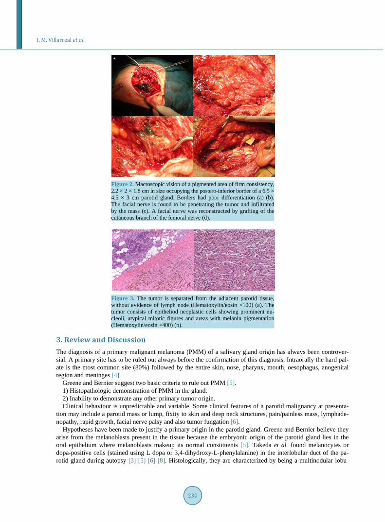
He also was evaluated by a Dermatologist, Neurologist and Ophthalmologist not finding any suggestion in the physical examination or imaging testing that lead to a suspicion of a possible primary tumor in their areas of expertise. The histopathological final result confirmed an intraglandular melanoma whose cells were S100 posi-tive, melan A positive and HMB-45 positive. No lymphoid tissue was identified on the periphery of the tumor (Figure 3). He was referred then to the department of Oncology for evaluation whose decision was to make a close follow-up. A 6 months follow-up has shown no surgical complications. Diagnostic imaging performed re-cently (PET-scan) showed postsurgical changes and was free of highly metabolic activity in the parotid region and in other areas that could suggest extraglandular primary tumor localization.
(a) (b)
Figure 1. Axial and coronal T1-weighted (a) and T2 (b) respec-tively MRI, demonstrates a high-signal-intensity in T1, heteroge-neous mass in the left parotid gland (arrow).
I. M. Villarreal et al.
230
Figure 2. Macroscopic vision of a pigmented area of firm consistency, 2.2 × 2 × 1.8 cm in size occupying the postero-inferior border of a 6.5 × 4.5 × 3 cm parotid gland. Borders had poor differentiation (a) (b). The facial nerve is found to be penetrating the tumor and infiltrated by the mass (c). A facial nerve was reconstructed by grafting of the cutaneous branch of the femoral nerve (d).
Figure 3. The tumor is separated from the adjacent parotid tissue, without evidence of lymph node (Hematoxylin/eosin ×100) (a). The tumor consists of epitheliod neoplastic cells showing prominent nu-cleoli, atypical mitotic figures and areas with melanin pigmentation (Hematoxylin/eosin ×400) (b).
3. Review and Discussion The diagnosis of a primary malignant melanoma (PMM) of a salivary gland origin has always been controver-sial. A primary site has to be ruled out always before the confirmation of this diagnosis. Intraorally the hard pal-ate is the most common site (80%) followed by the entire skin, nose, pharynx, mouth, oesophagus, anogenital region and meninges [4].
Greene and Bernier suggest two basic criteria to rule out PMM [5]. 1) Histopathologic demonstration of PMM in the gland. 2) Inability to demonstrate any other primary tumor origin. Clinical behaviour is unpredictable and variable. Some clinical features of a parotid malignancy at presenta-
tion may include a parotid mass or lump, fixity to skin and deep neck structures, pain/painless mass, lymphade-nopathy, rapid growth, facial nerve palsy and also tumor fungation [6].
Hypotheses have been made to justify a primary origin in the parotid gland. Greene and Bernier believe they arise from the melanoblasts present in the tissue because the embryonic origin of the parotid gland lies in the oral epithelium where melanoblasts makeup its normal constituents [5]. Takeda et al. found melanocytes or dopa-positive cells (stained using L dopa or 3,4-dihydroxy-L-phenylalanine) in the interlobular duct of the pa-rotid gland during autopsy [3] [5] [6] [8]. Histologically, they are characterized by being a multinodular lobu-

I. M. Villarreal et al.
231
lated tumor, with cells that show large nuclei, prominent nucleoli and plentiful eosinophilic cytoplasm. Within the tumor cells, brown granular pigment, melanosomes and compound melanosomes are found [1].
The presence of neuromelanin within a tumor or characteristic cytologic features such as intranuclear invagi-nation of the eosinophilic cytoplasm or prominent nucleolation can be helpful diagnostic clues when differenti-ating melanomas from carcinoma [7].
Immunohistochemistry in these neoplasms is characterized by S100 protein, melan A and HMB-45 positivity [1] [6] [8].
Woodward et al. proposed that four conditions should be met before making a diagnosis of primary parotid gland melanoma [3] [4] [7] [8]: • The predominant tumor mass or bulk should be intraparotid. • There should be no identifiable lymph node tissue present in the mass. • There should be no evidence of malignant melanoma elsewhere after diligent search of eyes, skin, nose,
pharynx, mouth, oesophagus, anogenital region or meninges. • There should be no evidence of previous excision of a malignant melanoma or progressing pigmented lesion.
In our case, all of these conditions were fulfilled. A thorough study should include a careful analysis of the integument, ocular examination, ultrasonography/
CT scan of the abdomen, panendoscopy, PET-scan, and CT scan of thorax and brain [9]. Lopez-Cedrun et al. suggest that even with normal results there is a doubt about the primary origin suggesting the location of the primary malignant melanoma in a hardly explorable internal organ [2].
According to a study by Shah et al., patients submitted to neck dissection for a primitive melanoma of the face, ear or anterior region of the scalp should be considered for parotidectomy [2] [10].
Lymph node involvement seems to be one of the more useful predictive parameters [4]. Metastases involving the intraparotid lymph nodes tend to have well-demarcated interfaces between the capsule of the node and the adjacent salivary gland tissue, in contrast to supposed primary melanoma that tend to infiltrate with poor de-marcation [3].
Neck dissection should always be performed because of the high incidence of hidden metastases in these nodal groups [2] [3] [10]. The presurgical PET scan was negative for a possible primary origin which made us decide not to perform a cervical lymphadenectomy.
Radiologically, MRI is shown to be a better option than CT scan because it provides more detailed anatomical information and may better define the position of the parotid mass in relation to the facial nerve [6].
Standard treatment in parotid gland cancers is surgical and a total parotidectomy should be performed [2]. Radiotherapy and chemotherapy are considered to be of low value [3] [11]. In this type of tumors, adjuvant ra-diotherapy is not part of a regular treatment protocol but it is recommended for high risk patients, based on grade, stage and margin of excision to improve local control.
High incidence of local recurrences is a fact even after apparently adequate surgery also in early and localized lesions. Facial nerve palsy is the most common complication of the surgical management of the parotid malig-nancies. In our case the facial nerve infiltration leads to its sacrifice and reconstruction.
4. Conclusion Parotid cancer accounts for a histologically and biologically diverse group of neoplasms. Malignant melanoma represents approximately 0.7% of all malignant neoplasms within the parotid gland [2] [3] [9]. The biological behaviour of primary parotid melanomas is variable and unexpected. Prognosis is influenced by several factors, particularly stage and tumor grade. The diagnostic protocol suggests that a histological diagnosis of a melanoma should always be followed by a thorough search for primary or metastatic disease elsewhere because a primary lesion is a candidate for a proper surgical treatment whereas a metastatic lesion is not [4]. The average survival rate hardly exceeds 2 years [2].
References [1] Huguet, P., Gabaldon, M., Sansano, I., et al. (2010) Melanoma maligno amelanótico en un ganglio de glándula
parótida. Estudio clinicopatólogico, inmunohistoquímico y molecular de un caso. Revista Española de Patología, 43, 144-147.
[2] Barbieri, M., Gentile, R., Cordone, M.P., et al. (2002) Primitive Malignant Melanoma of the Parotid Gland. ORL, 64,

I. M. Villarreal et al.
232
297-299. http://dx.doi.org/10.1159/000064131 [3] Tsutsumida, A., Yamamoto, Y., Sekido, M., et al. (2008) Suspected Case of Primary Malignant Melanoma of the Pa-
rotid Gland. Scandinavian Journal of Plastic and Reconstructive Surgery and Hand Surgery, 42, 105-107. http://dx.doi.org/10.1080/02844310601004707
[4] Woodwards, R.T., Shepherd, N.A. and Hensher, R. (1993) Malignant Melanoma of the Parotid Gland: A Case Report and Literature Review. British Journal of Oral and Maxillofacial Surgery, 31, 313-315. http://dx.doi.org/10.1016/0266-4356(93)90068-8
[5] Bahar, M., Anavi, Y., Abraham, A., et al. (1990) Primary Malignant Melanoma in the Parotid Gland. Oral Surgery, Oral Medicine, Oral Pathology, Oral Radiology, 70, 627-630. http://dx.doi.org/10.1016/0030-4220(90)90412-L
[6] Malata, C.M., Camilleri, I.G., McLean, N.R., et al. (1997) Malignant Tumours of the Parotid Gland: A 12-Year Re-view. British Journal of Plastic Surgery, 50, 600-608. http://dx.doi.org/10.1016/S0007-1226(97)90505-1
[7] Prayson, R.A. and Sebek, B.A. (2000) Parotid Gland Malignant Melanomas. Archives of Pathology Laboratory Medi-cine, 124, 1780-1784.
[8] Gao, N., Li, L.J., Li, Y., et al. (2008) Primary Amelanotic Malignant Melanoma of the Parotid Gland: A Case Report. Journal of International Medical Research, 36, 1435-1439. http://dx.doi.org/10.1177/147323000803600633
[9] Bussi, M., Cardarelli, L., Riontino, E., et al. (1999) Primary Malignant Melanoma Arising in the Parotid Gland: Case Report and Literature Review. Tumori, 85, 523-525.
[10] Renaut, A.J. (1996) Melanoma Arising within the Parotid Salivary Gland a Case Report and Review of Management. European Journal of Surgical Oncology, 22, 201-202. http://dx.doi.org/10.1016/S0748-7983(96)90983-X
[11] Bron, L.P., Traynor, S.J., McNeil, E.B., et al. (2003) Primary and Metastatic Cancer of the Parotid: Comparison of Clinical Behavior in 232 Cases. Laryngoscope, 113, 1070-1075. http://dx.doi.org/10.1097/00005537-200306000-00029


![Parotid Lesions in Children Undergoing Parotidectomy. The … · 2018. 8. 8. · of salivary gland masses occur within the parotid gland [1-4]. Parotid gland lesions are infrequent](https://static.fdocuments.us/doc/165x107/60d3cf2c7c14947d7f31fea4/parotid-lesions-in-children-undergoing-parotidectomy-the-2018-8-8-of-salivary.jpg)