Rapid hemostasis of arterial puncture sites with collagen...
12
Clin. Cardiol. 20, 981-992 (1997) Rapid Hemostasis of Arterial Puncture Sites with Collagen in Patients Undergoing Diagnostic and Interventional Cardiac Catheterization SIGMIJND SII.BER, M.D., FACC Dr. Muller Hospital, Munich, Germany Summary: Despite the continuous reduction of sheath sizes in diagnostic and interventional cardiac catheterizations and the discontinuation of coumadin use after coronary stent implan- tation, a challenging role remains for hemostatic devices in the sealing femoral puncture sites. Since the introduction of the vascular hemostatic device (VHD) in 199 1 and the hemostatic puncture closing device (HPCD) in 1992, numerous studies investigating these devices have been published. The deploy- ment success rates reported in 2,292 patients for VHD is 97%, ranging from 88 to 100%. For HPCD, the mean deployment success rate resulting from 622 published patients leads to an identical result of 97%, ranging between 91 and 100%. For time to hemostasis, data have been analyzed according to the four different clinical situations, depending on level of antico- agulation (none or full) and the time of sheath removal (imme- diate or delayed). In randomized studies, when compared with the manual control groups, both devices revealed a statistical- ly significantreduction in time to hemostasis: 12 to 16 minutes less for diagnostic catheterization and 14 to 30 minutes less for PTCA. As for minor local complications, no clinically rele- vant diflerences seem to exist. None of these devices has been proven to reduce major local complications. Prospective trials addressing early mobilization after percutaneous transluminal coronary angioplasty and the cost effectiveness of arterial clo- sure devices in defined subgroups are warranted. Key words: collagen, hemostasis, complication, cardiac cath- eterization, percutaneous transluminal coronary angioplasty Introduction Despite the continuous reduction of sheath sizes in diag- nostic and interventional cardiac catheterizations and the dis- Address for reprints: Sigmund Silber, M.D. Associate Professor of Medicine Dr. Muller Hospital Am Isarkanal 36 8 1379 Munich, Germany Received: May I, 1997 Accepted with revision: August 21, 1997 continuation of coumadin use after coronary stelit implanta- tion, a challenging role for hemostatic devices in sealing femoral puncture sites remains: patients undergoing diagnos- tic coronary angiography may be ambulated almost iinmedi- ately and discharged many hours earlier than currently prac- ticed in most centers utilizing a supine restriction period of 6 h after diagnostic catheterization.' On the other hand, patients undergoing percutaneous transluminal coronary angioplasty (PTCA) by the femoral approach (which is the access used far more frequently than the brachial or radial approach') are usually immobilized overnight, which may result in signifi- cant discomfort with increased back pain and need for anal- gesics? In patients with low-risk procedures, when prolonged vascular access does not seem to be needed, sheath pulling immediately after PTCA increases patients' comfort (return- ing to their rooms without a sheath), decreases burden for the medical staff, and may reduce hospital costs by shortening the length of stay. Hemostatic devices may allow patients to walk 2 to 3 h after the end of the procedure and hence further in- crease their comfort. In addition, even with the current stent protocols using aspirin and ticlopidine, major local bleeding complications may still occur in 1% [STRESS 111 (Stent Restenosis Study), ISAR (Intracoronary Stenting Anti- thrombotic Regimen)], up to 2.2% [STARS (Stent Antico- agulation Regimen Study)], and 2.4% [FANTASTIC (Full Anticoagulation versus Ticlopidine plus Aspirin after Stent Implantati~n)].~~ Furthermore, glycoprotein IIb/IIla inhib- itors are increasingly used in both high- and low-risk patients: although the increased rate of bleeding complications in the EPIC study (Evaluation of c7E3 for the Prevention of Ischemic Complications) could be significantly reduced by decreasing the concomitant heparin dosage,8 the EPILOG (Evaluation of PTCA to Improve Long-Term Outcome by c7E3 GP IIb/IIIa Receptor Blocker) and RESTORE (Ran- domized Efficacy Study of Tirofiban for Outcomes and Restenosis) trials still revealed a rate of major bleeding of I .8 and 2.5%, respectively, in the patients treated with glycopro- tein IIb/IIIa inhibitors, and even of 2.3 to 3.1 %. in the placebo groups.8%9 The use of low molecular weight heparin in pa- tients at high risk for stent thrombosis may also be associated with a higher bleeding risk.I0 Since there has been no published overview summarizing and analyzing the results for collagen devices. it is the pur- pose of this paper to review the data and to provide a differ- entiated analysis of success and local complication rates for
Transcript of Rapid hemostasis of arterial puncture sites with collagen...

Clin. Cardiol. 20, 981-992 (1997)
Rapid Hemostasis of Arterial Puncture Sites with Collagen in Patients Undergoing Diagnostic and Interventional Cardiac Catheterization
SIGMIJND SII.BER, M.D., FACC
Dr. Muller Hospital, Munich, Germany
Summary: Despite the continuous reduction of sheath sizes in diagnostic and interventional cardiac catheterizations and the discontinuation of coumadin use after coronary stent implan- tation, a challenging role remains for hemostatic devices in the sealing femoral puncture sites. Since the introduction of the vascular hemostatic device (VHD) in 199 1 and the hemostatic puncture closing device (HPCD) in 1992, numerous studies investigating these devices have been published. The deploy- ment success rates reported in 2,292 patients for VHD is 97%, ranging from 88 to 100%. For HPCD, the mean deployment success rate resulting from 622 published patients leads to an identical result of 97%, ranging between 91 and 100%. For time to hemostasis, data have been analyzed according to the four different clinical situations, depending on level of antico- agulation (none or full) and the time of sheath removal (imme- diate or delayed). In randomized studies, when compared with the manual control groups, both devices revealed a statistical- ly significant reduction in time to hemostasis: 12 to 16 minutes less for diagnostic catheterization and 14 to 30 minutes less for PTCA. As for minor local complications, no clinically rele- vant diflerences seem to exist. None of these devices has been proven to reduce major local complications. Prospective trials addressing early mobilization after percutaneous transluminal coronary angioplasty and the cost effectiveness of arterial clo- sure devices in defined subgroups are warranted.
Key words: collagen, hemostasis, complication, cardiac cath- eterization, percutaneous transluminal coronary angioplasty
Introduction
Despite the continuous reduction of sheath sizes in diag- nostic and interventional cardiac catheterizations and the dis-
Address for reprints:
Sigmund Silber, M.D. Associate Professor of Medicine Dr. Muller Hospital Am Isarkanal 36 8 1379 Munich, Germany
Received: May I , 1997 Accepted with revision: August 21, 1997
continuation of coumadin use after coronary stelit implanta- tion, a challenging role for hemostatic devices in sealing femoral puncture sites remains: patients undergoing diagnos- tic coronary angiography may be ambulated almost iinmedi- ately and discharged many hours earlier than currently prac- ticed in most centers utilizing a supine restriction period o f 6 h after diagnostic catheterization.' On the other hand, patients undergoing percutaneous transluminal coronary angioplasty (PTCA) by the femoral approach (which is the access used far more frequently than the brachial or radial approach') are usually immobilized overnight, which may result in signifi- cant discomfort with increased back pain and need for anal- gesics? In patients with low-risk procedures, when prolonged vascular access does not seem to be needed, sheath pulling immediately after PTCA increases patients' comfort (return- ing to their rooms without a sheath), decreases burden for the medical staff, and may reduce hospital costs by shortening the length of stay. Hemostatic devices may allow patients to walk 2 to 3 h after the end of the procedure and hence further in- crease their comfort. In addition, even with the current stent protocols using aspirin and ticlopidine, major local bleeding complications may still occur in 1% [STRESS 111 (Stent Restenosis Study), ISAR (Intracoronary Stenting Anti- thrombotic Regimen)], up to 2.2% [STARS (Stent Antico- agulation Regimen Study)], and 2.4% [FANTASTIC (Full Anticoagulation versus Ticlopidine plus Aspirin after Stent Implan ta t i~n ) ] .~~ Furthermore, glycoprotein IIb/IIla inhib- itors are increasingly used in both high- and low-risk patients: although the increased rate of bleeding complications in the EPIC study (Evaluation of c7E3 for the Prevention of Ischemic Complications) could be significantly reduced by decreasing the concomitant heparin dosage,8 the EPILOG (Evaluation of PTCA to Improve Long-Term Outcome by c7E3 GP IIb/IIIa Receptor Blocker) and RESTORE (Ran- domized Efficacy Study of Tirofiban for Outcomes and Restenosis) trials still revealed a rate of major bleeding of I .8 and 2.5%, respectively, in the patients treated with glycopro- tein IIb/IIIa inhibitors, and even of 2.3 to 3.1 %. in the placebo groups.8%9 The use of low molecular weight heparin in pa- tients at high risk for stent thrombosis may also be associated with a higher bleeding risk.I0
Since there has been no published overview summarizing and analyzing the results for collagen devices. it is the pur- pose of this paper to review the data and to provide a differ- entiated analysis of success and local complication rates for

982 Clin. Cardiol. Vol. 20, December I997
patients undergoing diagnostic or interventional cardiac catheterizations.
Characterization of Protocols and Patients Enrolled
To analyze the studies, it is important to differentiate be- tween the protocols investigated. With regard to hemostasis, four different clinical situations may be encountered: (1) im- mediate sheath pulling after diagnostic catheterization is usu- ally related to smaller sheath sizes and to no or a low level of anticoagulation; (2) after PTCA (or other coronary interven- tions), which are usually performed using larger catheters, sheaths may have been pulled with delay and without contin- ued anticoagulation at a time when little or no anticoagula- tion is effective; ( 3 ) on the other hand, delayed sheath-pulling in patients under continued full anticoagulation (prolonged heparin administration or on coumadin according to previous stent protocols) must be strictly discerned; and (4) sheath pulling immediately after PTCA is, of course, always per- formed under full anticoagulation. Unfortunately, many of the published studies did not differentiate between these clin- ical settings and thus reported a mixture of overall results.
The exclusion criteria used in most of the studies were quite homogeneous: inadvertent penetration of the dorsal ar- terial wall with the puncture needle, previous application of collagen sealing of the femoral access site, known allergy to collagen, clinical signs of or known peripheral artery disease, acute myocardial infarction, status post thrombolytic therapy,
known coagulation defects or known platelet dysfunction. se- vere and uncontrolled arterial hypertension (systolic > 220 mmHg or diastolic > 120 mmHg), preexisting hematoma or hematoma developed during the procedure, or patients with a venous femoral sheath.
Definition of Hemostasis
Basically, two parameters of measuring the success of hemostasis have been reported: the mean value of the times until complete hemostasis occurred in each individual patient (time to hemostasis) and the percentage of patients showing complete hemostasis after a specified time interval (hemosta- sis success rate). Although theoretically both parameters can be measured simultaneously, most ofthe studies reported only either one parameter or incomplete data.
Time to Hemostasis
Time to hemostasis is defined as the time elapsed from ini- tial compression at removal of the sheath until the comple- tion of compression. The first time at which no bleeding oc- curs is taken as the time to hemostasis in a particular patient. However, the measurement of time to hemostasis is not stan- dardized: results for time to hemostasis intrinsically depend on the time interval to the first and between the subsequent checks for bleeding; using a minimum time resolution of, for example, 15 min, one cannot expect to find a time to hemo- stasis of < 15 min. Table I summarizes the time intervals be-
TABLF 1 Published time intervals between sheath removal and checking for hemostatsis in collagen studies for determination of time to hemo\ta- sis. Some studies did not clearly define all time intervals ("?")
First author (reference) Patientdgroups First check interval (min) Subsequent intervals (min)
VHD Sanbom ( I 1 ) Collagen ( diagn + PTCA) 1 -2 Oozing: 2-5
Brisk: 5-- I0 Control-diagn 10 7
Control-PTCA 15 ?
Ernst ( 12) Collagen (diagn + PTCA) 2-3 > Schrider ( 13) Collagen (diagn + PTCA) 3-5 7
Control (diagn + PTCA) 15 I0 Foran ( 14) Collagen (diagn+ PTCA) 3 '? Rartorelli ( 15) Collagen (PTCA) 1 1 von Hoch ( 16) Collagen (PTCA) 2
Control (PTCA) <? ,? Silber ( I 7) Collagen (PTCA) 2 5
Control (PTCA) 2 5
Kussmaul ( 18) Collagen (diagn + PTCA) ,? '? Control (diagn + PTCA) 15 10
de Swart (19) Collagen (diagn + PTCA) 0.5 1.2.10 Murrary (20) Collagen (diagn + PTCA) 5 I0
HPCD
Abhrevicitions: diagn =diagnostic, PTCA = percutaneous transluminal coronary angioplasty. VHD = vascular hemostatic device. HPCD = hemostatic puncture closing device.

S. Silber: Rapid hemostasis with collagen 9x3
\ Mechanic
n dn- -- Technique Plug Sandwich Gel Gel Heat
Material Collagen Collagen + Thrombin/ Thrombin/ Radio- Suture anchor collagen fibrin frequency
Prostar"' (VHD) (HPCD) (GVSD) Techstar'
Company Datascope Shewood/ Vascular Global Scimed Perclose Quinton Solutions Therapeutics
Name VasoSeal" Angio-Seal'" Duet'" BioSeal ?
FIG. I = Gcnhony vascular hemostatic device.
Devices used for sealing arterial puncture sites. VHD=vascular hemostatic device, HPCD = hemostatic puncture closing device. GVSD
tween sheath removal and the first check as well as the subse- quent time intervals used in studies determining time to he- mostasis with collagen devices. As one can see, time inter- vals between 30 s and IS min have been used for the first check interval, and between 1 min and 10 min for the subse- quent intervals. Furthermore, not all studies clearly defined the time intervals for all the groups investigated; in some studies, even the time intervals within the same study varied. The choice of the time interval between the bleeding checks is an ambiguous decision: too short intervals may not give sufficient time for thrombus formation and may artificially increase-particularly in the manual control groups-the time to hemostasis.
In some studies, after deployment of a hemostatic device, all patients automatically received a vascular C-clamp2[ or an air cushion device.?' Therefore, the determination of time to hemostasis was not possible in these studies.
Hemostasis Success Rate
This parameter reveals the percentage of patients showing complete hemostasis at a specified point of time. The shorter the time interval defined, the lower the success rate. Thus, in addition to sheath size and level of anticoagulation, when comparing the results for hemostasis success rates one must consider possible differences of the time points at which the success rate was measured. The time intervals for determina- tion of hemostasis success rates range from 0 s (immediate
to 1 h." hemostasis~Y,".'"02to~ min!2. 13. 17. 18.22.25.2handevenup
Vascular Complications
In this analysis, the definitions of major and minor vascular complications were used according to the US. multicenter tri- al. I I The following complications were classified as major: thrombosis or loss of distal pulses, large pseudoaneurysm or atrioventricular (AV) fistula, and bleeding with need for trans- fusion or vascular surgery. Bleeding from puncture site that did not need transfusion and/or vascular surgery, as well as a small pseudoaneurysm treated medically, were classified as a minor complications.
Hemostatic Devices
Sandbags do not reduce vascular complications and even increase patient di~comfort.?~ Mechanical devices such as C-clamps, stasis buttons, or air cushions were used :IS ;I
replacement for manual compression, but upon physical ex- amination did not clearly show a reduction i n hemutoma for- mation.28, 29 Recent data suggest a reduction in ultrasound- detected AV fistulae and pseudoaneurysms with the C- clamp."' Mechanical devices cannot. of course. reduce the time to hemostasis and therefore cannot decrease the mini- mum time required for bed rest.
Hemostatic devices for rapid closure of arterial puncture sites may be classified according to their mechanisms. a s il- lustrated in Figure l . The vast majority of clinical experience has been gained using bovine collagen devices: the prototype was the vascular hemostatic device (VHD) (Fig. 2. VasoSeal' , Datascope Corp., Montvale, N.J.). a pure collagen plug tlc- vice, followed by the hemostatic puncture closing device (HPCD) (Fig. 3, Angio-Seal"', originally developed by the
F a . 2 The VHD (vascular hemostatic device. VrisoSeal" ) coml"-i.;- es a blunt-tipped 1 I-F dilator (center) which is inseitzd u4ng the short guide wire, a short 1 1.5-F sheath (right), and two 90 mg colla- gen cartridges (between the above).

9x4 Clin. Cardiol. Vol. 20, December I997
FIG. 3 The HPCD (hemostatic puncture closing device, Angio- Seal"') consists ofa short guide wire, a dilator with 2 luinina, an 8-F sheath (mounted together, center), the "can-ier" device (containing thc anchor. the collagen, and the suture. above), and a tamper (below).
Kensey Nash Corporation, Exton, Pa.; it is now a trademark of Quinton Instrument Company, Bothell, Wash., within the USA, and of Sherwood Davis & Geck, St. Louis, Mo., out- side the USA). In addition to the collagen, the HPCD applies an intra-arterial anchor. Both devices are discussed in detail below.
Another approach is the installation of a fibrin sealant via the arterial sheath. Fibrin sealant is a well-known tissue adhe- sive which combines fibrinogen (from human plasma) and (bovine) thrombin to form fibrin. The first encouraging results were obtained in animals and in 20 patients.31s32 The wide- spread application of fibrin may, however, be limited by the need for use of human plasma products. Therefore, the Ger- shony Vascular Sealing Device (GVSD) (Duet"', Vascular So- lutions. Inc., Minneapolis, Minn.), using a mixture of bovine thrombin and collagen, may be more promising.33 A different way of inducing hemostasis is the application of low-energy radiofrequency (30-35 W) via a guide wire through subcuta- neous tissue to the periarterial First clinical results in 55 patients have been reported; the success rate seems to be related to the level of anticoagulation.34
The clinical findings using suture devices (Perclose, Inc.) are controversial. The Prostar"' device uses four needles (two sutures); the Techstar"' device uses two needles (one suture). After predilation of the subcutaneous tissue (requiring a 2 I -F tissue track), the suture-containing device is advanced into the artery, the needles are retracted, and knots are tied against the arterial wall, facilitated by a knot-pushing tool.35 Whereas preliminary results with the Prostar device were encouraging, the relatively high rate of local vascular complications in the U.S. multicenter trial make further analysis necessary.35. 36
Modifications of the Prostar Plus and Techstar devices seem to provide more reliable result^;^^,^* the use ofbothdevices in a single center with high volume experience was. however, associated with a need for vascular repair in 2.1% of the pa-
tients undergoing PTCA.jY Furthermore, the deployment suc- cess rate of 89.6% (786/877 patients) even wiih the 6F- Techstar device appears relatively I o w . ~ "
Comparing the Vascular Hemostatic and Hemostatic Puncture Closing Devices
The basis of VHD and HPCD is collagen. Putified hovine collagen has been used in vascular, abdominal untl dental sur- gical procedures since late in 1960 as an adjunct to hcmostasis when control of bleeding by ligature or other conventional methods was The biodegradablc collagen plug induces platelet activation and aggregation, releasing co- agulation factors and resulting i n the formation of fibrin arid the subsequent generation of a thrombus!' I t is assumed that anticoagulation with heparin or even antiaggregation with as- pirin do not affect hemostasis induced by collagen.", Ji Col- lagen is ultimately degraded and resorbed by granulocytes and niacrophages. These cells, releasing their colltigenase en- zymes, invade the plug and selectively digest the collagen a s II function of the physical properties ofthe different collugens.4" The immunogenicity ofcollagen has been a subject of debate. focusing in particular on injectable collagen. which is uscd t o correct dermal defects such as acne or wrinkles. The reports 01' apossible link of collagen to autoimmune disease haw impli- cated only injectable collagen, which is quite different i n structure and the degree of cross-linking from the collagen sponge used in VHD and HPCD. Antigenicity of purilied col- lagen is considerably reduced and. although allergies to colla- gen have been de~cribed?~ allergic reactions to VHD h a w not been aclinical
Device Description and Deployment
The VHD consists of a purified collagen plug that induces the formation of a hemostatic cap directly over the arterial puncture site when inserted adjacent to the arterial wall. The methodofitsdeployment isdescribed intletailelscwhere.". In brief, VHD deploys acollagen plug onto the external arte- rial wall after dilation of the skin and subcutmeoiis tissue to 1 1.5 F. It comprises four parts: a blunt-tipped I I -F dilator. one of seven differently sized I 1 3-F sheaths selected by length using a preprocedure needle depth measurement techniqie. and two 90 mg collagen cartridges. When the sheath is to he pulled, ashortguide wire is inserted and the existing sheath is removed while complete hemostasis is maintained with trim
ual compression. Then the blunt-tipped I I -F dilator is insert- ed over the guide wire just down to the site of the arterinl puncture. Guidance is obtained by feeling the resistance of' the dilator against the outer surface ofthe artery a s well as by the marker on the dilator. The 1 1 .S-F sheath is then advanced over the dilator down to the arterial sut-tice. While still holding pressure, the dilator and the guide wire itre removed and the collagen cartridge deployed with a "push and pull" move- ment. We saw in a previous study that one collagen plu, 0 IS ' as effective as two, but is better tolerated.25.JX
The HPCD provides a mechanical block of the arterial puncture site with an anchor from inside the artery. guiding

S. Silber: Rapid hemostasis with collagen 985
and holding the collagen in the tract. It consists of four com- ponents within a single delivery device (“carrier”) requiring an 8-F sheath: anchor, collagen plug, connecting suture, and a tamper. All three components deployed into the patient (an- chor, suture, and collagen) are completely resorbable; the an- chor and the suture are made from polyglycolic and polylactic acids. The small plug contains only about 14 mg collagen. The technique of its deployment has been described in detail elsewhere. 18, l9 In brief, a short guide wire is inserted and the existing sheath is removed while hemostasis is maintained with manual compression. First, the location of the end of the 8-F sheath within the artery is determined by inserting a mod- ified dilator with two lumina: one (distal) for the guide wire and one (proximal) at the end of the 8-F sheath. The location of the end of the 8-F sheath is determined by the presence of blood flow through the modified dilator. Our preferred loca- tion of the sheath is ca. l cm further down the puncture site in- side the artery lumen. The dilator is then removed and the car- rier device is introduced into the 8-F sheath. The anchor is secured against the intraluminal arterial wall (we check three times at different angles) and the collagen plug is deployed on the outer arterial wall. A tamper is pushed downward to com- press the collagen against the outer arterial wall (“sandwich technique”). Finally, a spring is attached between the tamper and a metal tag fixed to the positioning suture, thus applying continuous pressure on the tamper.24 Although the deploy- ment technique may sound complicated, it usually takes < 60 s to deploy the device.
The material used for the intra-arterial anchor of the HPCD is a 50:50 D,L polylactic-coglycolic acid copolymer and well established in medical use. It has a safe history, for it is widely used in sutures, bioresorbable meshes, and sustained release drug delivery systems. Some concern about the concept ofin- serting a foreign body (although resorbable) into the lumen of the artery has been expressed. After initial experience in ani- m a l ~ ? ~ ultrasound studies in patients have shown that HPCD caused no more changes in flow pattern than those observed in the control group with manual compression. 5o Therefore, the described changes of flow pattern after intra-arten, ‘1 I an- choring are related to the puncture procedure itself rather than to the hemostatic d e v i ~ e . ’ ~ , ~ ~ In the majority of patients. the anchor is absorbed within 4 weeks;s0 after 2 months, complete absorption of the device was documented by ultrasound in all patientsz3 U.S. and European single and multiceriter trials have established the safety of the concept of intra-arterial an- choring.l8.19,51
Published Data Regarding Vascular Hemostatic and Hemostatic Puncture Closing Devices
Deployment Success Rates
Table I1 lists the results reported for a successful deploy- ment of either VHD or HPCD: In 2292 patients reported. the deployment success rate for VHD is 97%, ranging from 88
TABLE I1 Deployment success rates of collagen devices First author No. of patients No. of No. of Deployment (reference) receiving device diagnostic patients PTCA patients success nrte ((5) VHD Sanborn(l1) 246 90 156 98 Ernst ( 12) 252 105 140 98 Schrader (13) 50 30 20 I00 Slaughter (26) 51 - 51 98 Foran ( 14) 63 46 17 91 Bartorelli ( I 5 ) 100 100 100 Gibbs (52) 10 10 I 00 v Hoch (16) 154 - 154 88 Webb (21) 32 - 32 100
98 Kuhn (53) 600 600 - 98 Silber (25) 660 660 -
Silber ( 17) 74 - 74 100 Total 2292 91
Kussmaul(23) 68 - 68 93 deSwart(l9) 20 4 16 95 Aker(S1) 30 26 4 91 Kussmaul(18) 218 168 46 96 Chevalier (54) 52 ? ? 98 Blengino (5.5) 29 - 29 93 Silber (56) 65 65 - I00 Silber (24) 140 - 140 I (x) Total 622 97
-
-
HPCD
Abbreviations as in Table I.

986 Clin. Cardiol. Vol. 20, December 1997
to 100%. For HPCD, the mean deployment success rate re- sulting from the 622 published patients leads to an identical result of97%, ranging between 91 and 100%.
Time to Hemostasis
The published data on time to hemostasis for VHD and HPCD in patients undergoing diagnostic cardiac catheter- ization with no systemic or little administration of heparin is listed in Table 111. The mean values for time to hemostasis vary between 1.7 and 4.3 min. In randomized studies, both devices revealed statistically significant reductions of time to hemostasis of about 12 to 16 min less than in the manual con- trol groups.
In patients undergoing FTCA, the level of anticoagulation at the time of sheath removal was reported by most but not all groups (Table 111). For VHD, a statistically significant reduc- tion in time to hemostasis has been shown for immediate and for delayed sheath removal without prolonged anticoagulation (Table 111). The one study using VHD in patients with delayed sheath removal and prolonged anticoagulation had no control group. For HPCD, a statistically significant reduction in time
to hemostasis has been shown for inmediate and for delayed sheath removal with prolonged anticoagulation (Table 111). For both devices, the average gain in time to hemostasis was approximately 14 to 30 min and therefore somewhat more than the gain in diagnostic patients. A gain in time to hemosta- sis for immediate sheath removal cannot be calculated. since there is no real control group for immediate sheath removal and manual compression.
The results of reports comprising a mixture of diagnostic and therapeutic interventions also showed an overall statisti- cally significant reduction in time to hemostasis for VHD and HPCD (Table 111). Not all authors, however, revealed data on the level of anticoagulation at the time of sheath removal (Table 111).
Time to Ambulation
One of the primary goals using hemostatic devices is en- abling patients to walk earlier. In most studies, however. early ambulation was not an end point, therefore the data of time to ambulation cannot be used for this purpose.". To analyie the reported time to ambulation, it is important to know the
TABLE 111 Time to hemostasis using collagen devices. Differences can be attributed to different study designs regarding time of sheath reinovul and levels of anticoagulation
ACT(s)/F"IT(s)/INR Time to hemostasis (min) First author No. of patients at sheath removal and Collagen Control (reference) receiving device collagen application group group
Diagnostic patients Sanborn ( I I ) 90 (VHD) p7T=35.6+ 13.8 Ernst ( 12) I05 (VHD) - Kussiiiaul(18) 168 (HPCD) ACT= 166k58 Condon (57) 31 (HPCD) -
Sanbom ( 1 1) 7 1 (VHD) €TI = 36.2 f 16.9 Silber (17) 74 (VHD) €TI = 49.4 f 31 .O
Rartorelli (15) 100 VHD lNR=3.2+2 Kussmaul(23) 68 (HPCD) ACT = 220 f 94 Kussmaul(18) 46 (HPCD) ACT = 213 k 89
Sanborn ( 1 I ) 85 (VHD) PTT = 52.9 f 50.9 Slaughter (26) 5 I (VHD) ACT=381 f 152 von Hoch ( 16) 117 (VHD) - Emst ( 1 2) 140 (VHD) -
Blengino (55) 29 (HPCD) ACT = 274 k 61
PTCA-delayed sheath removal without prolonged anticoagulation
PTCA-delayed sheath removal and prolonged anticoagulation
PTCA-immediate sheath removal
Mixed patient groups Schdder ( 13) 50 (VHD) - deSwart(l9) 20 (HECD) - Chevalier (54) 52 (HPCD) -
de Swart (58) 55 (HPCD) ACT= lS9+- 129 Murray (20) 95 (HPCD) ACT= 141 a57
4.1 k2.8" 4.3 f 2.8 (0.5-20) 2.3i- 16.7"
I .7 "
4.3 f 3.7 l J
3.0 rt 3.0 (2- 1 5 )
2.2 k 2.1 (1-8) 4.4 k 8.9 3.5 * 8.5
7.6 k 1 1.6" 5 (3-1 5) 5" (4-6) 5.3 k 7.6 (20-32)
2+6"
4.3 a 3.0" (2-15) 1.2 k 2.1 2.3 f 6.7 1.2f 1.6" 1.9 +- 5
17.6+ 9.2
13.65 1 1 . 0 18.4
-
33.6 k 24.2 17.4f7 (5-75)
-
19.6r 126
33.6a 24.2 27 ( I 8 4 0 ) 27 (20-33
16+5 -
42.3 k 18.9 (20-120)
29.3 rt 23.2 12.9 i- 5.6 20.0 9
-
~ ~
" p < 0.05. Ahhrevicition.c: ACT = activated clotting time, I" = partial thromboplastin time, INR = International Normalized Ratio. Other abbreviations as in Table I.

S. Silber: Rapid hemostasis with collagen 9x7
time of sheath removal (Table IV). Therefore, with delayed sheath removal after PTCA, a time to ambulation of 2 to 3 days is not surprising.'s- s2 Unfortunately, not all publications revealed the time of sheath removal (Table IV).
For diagnostic cardiac catheterization, two studies ad- dressed the primary end point of early ambulation: with im- mediate sheath removal, patients were successfully ambu- lated 30 min after the deployment of VHDZs and even 20 min after HPCD.56 For patients undergoing PTCA, no study specifically investigating early ambulation has been pub- lished. In PTCA studies, the shortest time intervals to ambu- lation were in the range of 6 to 7 h for VHD,26, 53 and 8 h for HPCD.18 In mixed patient populations, after diagnostic or therapeutic cardiac catheterization, attempts were made to ambulate patients within 2 h after the deployment of VHDI4 or starting at 4 h after HPCD.'9-s1
Local Complications
Coniplications after manual compression: Vascular com- plications in patients undergoing diagnostic cardiac cath-
eterizations occur in the order of 0.5"/0.6° The incidence of major vascular complications (requiring blood transfusion o r vascular surgery) in patients undergoing diagnostic cardiac catheterization has been reported to range from 0.35 to S%.6'45 While in earlier decades these incidences often in- cluded thrombosis and distal embolism, later studies reported lower vascular complication rates of < 0.59. retlecting im- proved equipment and extensive operator experience.('". h('
With the use of SF-catheters for outpatient coronary angiog- raphy, major and minor complications were less frequent than reported earlier.@ In recent studies, major vascular compl ica- tions (as defined above) did not occur in patients undergoing diagnostic cardiac catheterization. I . 19. Small hematomus are only scarcely reported; many centers do not report them and/or consider them unimportant.', 6o Kern o r trl. reported 8% (23/287) minor hematomas in outpatients undergoing SF- catheterizations.66
Complications after collagen devices: The complication rates reported for collagen plugging are somewhat confusing. because several studies did not differentiate between diagnos-
TABLE 1V Time to ambulation using collagen devices First author No. of patients Time of (reference) receiving device sheath removal Time to ambulation
Diagnostic patients
_ _ _
VHD Sanbom ( I I ) 90 '? I 3 .3k 12.1 h Silber (25) 660 Immediately 30 m i n Silber (56) 65 Immediately 70 ntin
Kussmaul( 18 j 168 56+ 171 min 4 1 2 h Silber(S6j 65 Immediately 20 niin
HPCD
PTCA patients VHD Bartorelli (15) 100 Next day >3 days Gibbs (52) 10 Immediately >'days Sanbom ( I I ) 85 (on heparin) Immediately 16.1 * 1 1 . 1 I1
Camenzind (22) 62 Immediately >I211
Sanbom ( I 1 j 7 1 (off heparin) Delayed 73.OklI.I h Slaughter (26) 51 Immediately X.S(7-17jh
Nagtegaal(59) 80 Immediately 9 k S h Kuhn (53) 600 Immediately & l 2 h
HPCD Blengino (55) 29 '? 1 s.x k 3.3 11 Kussinaul( 18) 46 465 k 523 min 8 I1
Mixed patient groups VHD Emst(l2j Schrader (1 3) Foran (14)
Aker(51) Chevalier (54) 52 deSwart(l9)
105 Diagn + 140 PTCA 30 Diagn + 20 PTCA 46 Diagn + 17 PTCA
26 Diagn + 4 PTCA
4 Diagn + 16 PTCA
HPCD
Immediately Immediately
Immed./delayed
? ?
Immediately
8.3 (I-24)h 6.4k2.2(&12) h '-within 2 hor 1 h '
16.5 (4-57) I1
10.8 * 7 h 6.7 k 3.5 (4 I X ) h
Abbreviations as in Table I.
988 Clin. Cardiol. Vol. 20, December 1997
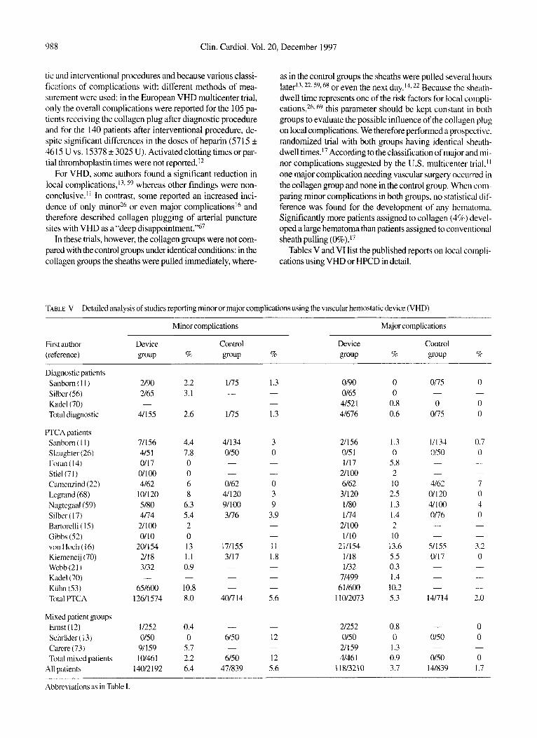
tic and interventional procedures and because various classi- fications of complications with different methods of mea- surement were used: in the European VHD multicenter trial, only the overall complications were reported for the 105 pa- tients receiving the collagen plug after diagnostic procedure and for the 140 patients after interventional procedure, de- spite significant differences in the doses of heparin (5715 k 46 IS U vs. 15378 k 3025 U). Activated clotting times or par- tial thromboplastin times were not reported.I2
For VHD, some authors found a significant reduction in local complications,13- 59 whereas other findings were non- conclusive.” In contrast, some reported an increased inci- dence of only minor26 or even major complications16 and therefore described collagen plugging of arterial puncture sites with VHD as a “deep di~appointment.”~’
In these trials, however, the collagen groups were not com- pared with the control groups under identical conditions: in the collagen groups the sheaths were pulled immediately, where-
as in the control groups the sheaths were pulled several hours later13-22,59,68 or even the next day.14.2’ Because the sheath- dwell time represents one of the risk factors for local compli- cations,26, 69 this parameter should be kept constant in both groups to evaluate the possible influence of the collagen plug on local complications. We therefore performed a prospective. randomized trial with both groups having identical sheath- dwell times.]’ According to the classification of major and mi- nor complications suggested by the U.S. multicenter trial,11 one major complication needing vascular surgery occurred in the collagen group and none in the control group. When com- paring minor complications in both groups. no statistical dif- ference was found for the development of any hematoma. Significantly more patients assigned to collagen (4%) devel- oped a large hematoma than patients assigned to conventional sheath pulling (0%).l7
Tables V and VI list the published reports on local compli- cations using VHD or HPCD in detail.
TABLE V Detailed analysis of studies reporting minor or major complications using the vascular hemostatic device (VHD)
Minor complications Major complications
Fir\t author Device Control Device Control (reference) group % group % group % P O U P o/c
Diagnostic patients Sanbom ( 1 I ) Silber(S6) Kadel(70) Total diagnostic
PTCA patients Sanbom ( I I ) Slaughter (26) Foran ( 14) Stiel(7 1 ) Camenzind (22) Legrand (68) Nagtegaal(59) Silber ( 17) Bartorelli ( 15) Gibbs (52) von Hoch ( 16) Kiemeneij (70) Webb(21) Kadel(70) Kuhn (53) Total PTCA
Mixed patient groups Emst (12) Schriider ( 13) Carere (73) Total mixed patients
All patients
2190 2165
41155 -
71156 415 I 0117 01100 4162
101120 5/80 4174 21100 0110
201 1 54 2/18 3/32
651600 12611 574
-
11252 Of50 91 1 59 1 0146 1
14012 I 92
2.2 3.1
2.6 -
4.4 7.8 0 0 6 8
6.3 5.4 2 0 13 1.1 0.9
10.8 8.0
-
0.4 0
5.7 2.2 6.4
1175 - -
1 I75
41134 0150 - -
0162 41120 91100 3116 -
-
171155 3/17 -
- -
4017 14
- 6/50
6150 471839
-
1.3 - -
1.3
3 0 -
- 0 3 9
3.9 - - I 1 1.8 - - -
5.6
- 12
12 5.6
-
0190 0165 41521 41676
21156 Of5 1 1/17
211 00 6162 31 120 1 I80 1 174
Ul00 1110
2111.54 1118 1/32
71499 611600
1 1012073
21252 0150 21159 4/46 1
I18/32lO
0 0
0.8 0.6
1.3 0
5.8 2 10 2.5 I .3 1.4 2 10
13.6 5.5 0.3 1.4 10.2 5.3
0.8 0 1.3 0.9 3.7
Of75
0 0175
-
11133 Of50 -
- 4162
01 1 20 41100 Of76 - -
51155 0117 - __ -
141714
- Of50
0150 141839
-
0
0 0
-
0.7 0 - -
7 0 4 0 -
- 3.2 0
__ -
- 2.0
0 0
0 1.7
-
- Abbreviations as in Table 1.

S. Silber: Rapid hemostasis with collagen ‘189
TABLE VI Detailed analysi5 of studies reporting minor or major complications using the hemostatic puncture closing device (HPCD)
Minor complications Major comphcation\ Fint author Device Control Device Control (reference) group % group % ErouP % group [k
Diagnostic patients Kussmaul ( 1 8) Condon (57) Silber (56) Total diagnostic
PTCA patients Kussmaul (23) Kussmaul ( I 8) Silber (24) Total PTCA patients
Mixed patient groups Aker(51) Chevalier (54) de Swart (58) Murray (20) Total mixed patients
All patients
91168 113 1 0165
101264
9/68 6146 01 140 151254
2/29 4/52 415 5 6195
16/23 1 4 1 1749
5.3 3.2 0
3.8
13 I3 0
5.9
7 7
7.3 6.3 6.9 5.5
171152 1/18
181170 -
- 16/63
16/63 -
- 1 1148 3/54 2192
161194 501421
11.2 5.6
11 -
- 25
25 -
- 23 5.5 2,1 8.2 11.7
611 68 013 1 0165
61264
0168 1 I46
01 I 40 11254
0129 0152 0.155 1 195 11231 81749
3.6 0 0
2.3
0 2 0
0.4
0 0 0 1
0.4 1 . 1
41152 0118
41170 -
- 1/63
I 163 __
-
0148 0154 1/92 11194 61327
3.6 0
2.3 -
.-
I .6
1 6
-
~-
0 0 I
0.5 1.4
Abbreviations as in Table I
Although both devices carry the inherent risk of inadvertent intra-arterial collagen insertion, published reports on this de- vice-related complication exist only for VHD. (Table VII).
Cost Effectiveness
Hemostatic closure devices are expensive and have to prove their cost effectiveness.
Effectiveness can be defined as hemostatic success. So far, no study has proven one hemostatic device to be clearly better than others. Therefore, differences between these devices may be related to different costs.
TABLE VII Reports on intra-arterial complications (insertions or protruions) wing the vascular hemostatic device (VHD)
First author (reference) Number %
PTCA patient5 Camenzind (22) Kuhn (53) Sanbom ( I 1 ) Stiel(7 1 ) von Hoch ( I 6)
Carere (73) Form ( I 4) Kadel(70)
Mixed patient groups
Total
1 162 21600 1/71
21100 21154
21 159 1 I63
41 1020 1512229
1.6 0.3 1.4 2.0 1.3
1.3 1.6 0.4 0.7
Costs may be divided into procedural costs, hospital costs, and follow-up costs. Procedural costs reflect the price of the device plus the time for deployment (cathlab time, doctors’ time spent for deployment). In our facilities, VHD has the lowest price, HPCD is in the middle range, and suture devices are the most expensive. The time for deployment of either VHD or HPCD is similar (< 1 min), whereas the suture de- vices usually take us 4 to 5 min. Hos@d c~)st.s: i n Germany. hospitals are paid by the day (length of stay). In diagnostic pa- tients undergoing catheterization in the afternoon, all henio- static devices may make an overnight stay unnecessary and therefore save the costs for 1 day. There is, however, a conflict of interest between health insurers and hospitals: saving overnight stay is important to the health insurers, but leads to a loss of income for the hospitals. In patients undergoing PTCA, usually 1 or 2 days may be saved, with the same con- flicting interests a$ described for diagnostic procedures. In o u r study, the decision to discharge was left to the ward physi- cians. Nevertheless, patients with collagen sealing were dis- charged 1 day earlier than the control group.17 This is in gocd agreement with a U.S. study that reported a significant dr- crease from 2.4 f 0.98 days (control group, 56 patients) to 1.53 2 0.8 days (collagen group, 47 patient^).'^ Fo/hv-~tp costs are predominantly defined by local vascular complica- tions; these complications may lead to substantial costs, even higher than that of the catheterization itself.’ Hemostatic de- vices should at least not increase the rate of local vascular complications or, even better, lead to a decrease of complica- tions. There is, however, no clear evidence that hemostatic de- vices decrease the rate of local complications; some devices even show a tendency to increase local complications.

990 Clin. Cardiol. Vol. 20, December 1997
Future Aspects
The data suggest that mechanical forces play a more impor- tant role in sealing arterial puncture sites with collagen than previously anticipated.I2 The collagen itself may not be that important for devices using intra-arterial anchorings6 and could perhaps be replaced by nonbiological, resorbable mate- rials. Besides device changes (like 6F-devices for 6F-PTCAs or 4F-devices for 4F-diagnostics), it is important to fill the gaps of missing studies that have early ambulation as primary end point after PTCA. Newer antiplatelet protocols with ticlo- pidine pretreatment andor the administration of IIb/IIIa in- hibitors also need to be investigated. Furthermore, prospective trials addressing the cost effectiveness of arterial closure de- vices in defined subgroups of patients are warranted.
References
I.
2.
3.
4.
5.
6.
7.
8.
9.
10
I I
Krause PB, Klein LW Utility of apercutaneous collagen hemosta- sis device: To plug or not to plug? J Am Coll Curdiol 1993;22: 1280-1282 Krone RJ, Johnson L, Noto T, and the Registry Committee of the Society for Cardiac Angiography and Interventions: Five year trends in cardiac catheterization: A report from the registry of the Society for Cardiac Angiography and Interventions. Cuthet Curdi- O V N S ~ Diugn 1996;39:31-35 Waksman R, Scott NA, Ghazzal ZMB, Mays R, Frerichs FA, Petersen JY, King SB 111: Randomized comparison of flexible ver- sus nonflexible femoral sheaths on patient comfort after angioplas- ty. A m Heart J 1996; 13 1 : 1076-1 078 Stress 111 Investigators: Early outcome after coronary stent place- ment with high pressure inflation and antiplatelet therapy: Interim results of the STRESS 111 trial. Circularion 1996;94:1-684 Schomig A, Neumann FJ, Kastrati A, Schiihlen H, Blasini R, Hadamitzky M, Walter H, Zitzmann-Roth E-M, Richardt G, Alt E, Schmitt C, Ulm K A randomized comparison of antiplatelet and anticoagulant therapy after the placement of coronary artery stents. NEnglJMed 1996;334: 1084-1089 Leon M, Baim DS, Giambartolomei A, Williams DO, Diver DJ, Senerchia C, Fitzpatrick M, Popma JJ, Kuntz RE: Clinical and an- giographic results from the Stent Anticoagulation Regimen Study (STARS). Circulation 1996;94:I-685 Bertrand M, Legrand V, Boland J, Fleck E, Bonnier J, Emmanuel- son H, Vrolix M, Missault L, Chierchia S, Casaccia M, Niccoli L, Oto A: Full anticoagulation versus ticlopidine plus aspirin after stent implantation: A randomized multicenter European study: The FANTASTIC trial. Circulation 1996;94:1-685 1-incoff AM, Tcheng JE, Miller DP, Booth JE, Montague EA, Topol El: Marked enhancement of clinical efficacy of platelet GP IIb/lIIa blockade with c7E3Fab (abciximab) linked to reduction in bleed- ing complications: Outcome in the EPILOG and EPIC trials. Circirlution 1996;94:1-375 Anderson HV, Bertrand M, Whitworth HB, Sax FL, Willerson JT for the RESTORE Investigators: Bleeding risk with platelet inhi- bition using tirofihan: The RESTORE Trial. Circulation 1996;94:
Goods CM, Liu MW, Jain SP, Mathur A, Yadav JS, Al-Shalbi KF, Dean LS, lyer SS, Parks JM, Roubin GS: Low molecular weight heparin versus standard heparin in patients at high risk for stent thrombosis: Clinical outcomes. Circulutian 1996;94:1-684 Sanbom TA, Gibbs HH, Brinker JA, Knopf WA, Kosinski EJ, Roubin GS: A multicenter randomized trial comparing a percuta- news collagen hemostasis device with conventional manual com- pression after diagnostic angiography and angioplasty. J Am Coll Curio1 1993;22: 1273-1 279
1-553
12.
13.
14.
15.
16.
17.
18.
19.
20.
21.
22.
23.
24.
25.
26.
27.
28.
Emst SMPG, Tjonjoegin RM, Schrader R, Kaltenbach M, Sigwart U, Sanborn TA, Plokker HWT Immediate sealing of arterial punc- ture sites after cardiac catheterization and coronary angioplasty us- ing a biodegradable collagen plug: Results of an intern;itional rcg- istry. JArn CollCarLlioll993;21:85 1-855 Schrader R, Steinbacher S, Burger W, Kadel C. Vallbracht C. Kaltenbach M: Collagen application for sealing of arterial puncture sites in comparison to pressure dressing: A randomi& trinl. Crrt/w/ Curdiovasc Diagn 1992;27:29&302 Foran JPM, Patel D, Brookes J, Wainwright RJ: Early mohiiisation after percutaneous cardiac catheterisation using collagen plug (VasoSeal) haemostasis. Br HenrrJ 1993;69:42442Y Bartorelli AL, Sganzerla P, Fabbiocchi F, Montorsi P, De Cesare N, Child M, Tavasci E, Passaretti B, Loaldi A: Prompt and safe fem- oral hemostasis with a collagen device after intracoronary iniplan- tation of Palmaz-Schatz stents. Am H e m J 1995; 130:26-32 von Hoch F, Neumann F-J, Theiss W , Kastrati A, Schiiniig A: Efficacy and safety of collagen implants for haemostasis of the vas- cular access site after coronary balloon angioplasty and coronary stent implantation. Eur Heart J 1995; I6:64(M6 Silber S, Bjorvik A, Rosch A, Muhling H: Advantages of sealing arterial puncture sites after FTCA with a single collagen plug: A randomized, prospective trial. JAm Coll Curdiol 1995;23:262A Kussmaul WG 111, Buchbinder M, Whitlow PL, Aker UT. Heuser RR, King SB, Kent KM, Leon MB, Kolansky DM, Sandza JG Jr: Rapid arterial hemostasis and decreased access site complications after cardiac catheterization and angioplasty: Results ofa random- ized trial of a novel hemostatic device. J A m Cull Curtiiol I995;25:
de Swart H, Dijkman L. Hofstra L. Biir FW, Van Onimen V, Tordoir J, Wellens HJJ: A new hemostatic puncture closure device for the immediate sealing of arterial puncture sites. A m J Ctrrtliol lY93; 72 :44549 Murray CR, Mortazavi A, Warth DC, Buchbinder M: A randon- ized controlled prospective study of the Angio-Seal device versus manual pressure for rapid arterial puncture closure. C'ircdrrriorr
Webb JG, Carere RA, Dodek AA: Collagen plug hemostatic clo- sure of femoral arterial puncture sites following implantation ofin- tracoronary stents. Curher Curdiowsc Diugn I993;30:3 14-3 I6 Camenzind E, Grossholz M, Urban P, Dorm7 PA. Didier D. Meicr B: Collagen application versus manual compression: A prospectivc randomized trial for arterial puncture site closure after coronary ail-
gioplasty. JArn Coll Curdiol 1994;24:655-662 Kussmaul WG, Buchbinder M, Whitlow PL, Aker UT. Heuser RR. King SB, Kent KM, Leon MB, Kolansky DM, SandLa JG: Femoral artery hemostasis using an implantable device (AngioSeal"') after coronary angioplasty. Cuthet Curdiovusc Dirr~n 1 Y96;37:362-365 Silber S, Dorr R, Miihling H, Konig U: Sheath pulling immediate- ly after PTCA: Comparison of two different deployment tech- niques for the hemostatic puncture closure device. A prospectivc, randomized study. Cuthet Curdi-diovusc Diugn 1997:4 I 378-383 Silber S, Haentsch C, Seidel N, Miihling H: Early ambulation (30 minutes) after cardiac catheterization using the collagen plug. Efficacy and follow-up comparing two different plug dosages in 660cases. J Am Coll Curdiol I Y93;2 1 : I S0A Slaughter PM, Cherty R, Flintoft VF, Lewis S, Sykora K. Beatlie DM, Schwartz L A single center randomized trial assessing use of a vascular hemostasis device vs. conventional manual compression following PTCA: What are the potential resource savings'? Cutliet CurdiovuscDiagn 1995;34:2 16214 Christensen B, Lacarella C, Manion R. Bruhn-Ding B. Meyer S. Wilson R: Sandbags do not prevent complications after catheteriza- tion. Circularion 1994;901-205 Bogart DB, Bogart MA, Miller JT, Farrar MW, Barr WK, Montgomery MA: Femoral artery catheterization complications: A study of 503 consecutive patients. Cnflief Curtliovusc D i q n 1995; 34:8-13
1685-1 692
1996;94:1-376

29.
30.
31.
32.
33.
34.
35.
36.
37.
38.
3Y.
40.
41.
42.
43.
44.
45.
46.
47.
48.
S. Silber: Rapid hen
Semler HJ: Transfemoral catheterization: Mechanical versus man- ual control of bleeding. Radiology 1985; 154234-235 Pracyk JB, Wall, TC, Longabaugh JP, Tice FD, Hochrein J, Cox GC. Lee KL, Tcheng JE: Femoral vascular complications follow- ing coronary intervention: Is mechanical clamp hemostasis better than hand pressure? Circulation 1996;94:I-377 Ismail S , Combs MJ, Goodman NC, Teotia SS, Teates CD, Abbott RD, Fechner RE, Nolan SP, Powers ER, Spotnitz WD: Reduction of femoral arterial bleeding post catheterization using percutaneous application of fibrin sealant. Cathet Curdiovasc Diugn 199.534: 88-95 Kipshizde N, Ferguson JJ, Macris MP, Clubb F, Cloy M, Horn J, Cummins F, Keane S, Nikolaychik V, Baker JE: Percutaneous de- livery of a biosealant to achieve peripheral artery hemostasis: Experimental and clinical studies. Circulution 1995;921-410 Gershony G, Kasprzyk DJ, Hussain HM, Powell J, Horzewski J: A novel vascular sealing device for closure of arterial puncture sites. Circithtion 1995;92:1-409 Goy JJ, Debbas N, Depairon M, Mische H, Bottum P, Desombre L: Preliminary results with a new arterial sealing device. Circulution
Carere RG, Webb JG, Ahmed T, Dodek AA: Initial experience us- ing Prostar”’: A new device for percutaneous suture-mediated clo- sure of arterial puncture sites. Curhet Curdiovasc Diagn I996;37:
Vetter J, Ribeiro E, Hinohara T, Carrozza J Jr, Simpson J: Suture mediated percutaneous closure of femoral artery access sites in ful- ly anticoagulated patients following coronary interventions. Cirmlatiori I994;90:1-62 I Cattelaens N, Cerckens U, Muller R, Staberock M, Grzan 0, Lampe EG, Simpson J, Grube E: The Prostar plus percutaneous closure device versus manual compression following coronary in- terventions. Circulation 1996;94:1-484 Gerckens U, Cattelaens N, Muller R, Staberock M, Grzan 0, Lampe EG, Simpson J , Grube E: Early ambulation following elec- tive diagnostic coronary angiography using a percutaneous arterial closure device (Techstar): A randomized trial versus manual com- pression. Circulation 1 996;94:I-484 Loubeyre CH, Fajadet J, Karam C, Jordan CH, Cassagneau B, Laurent J-P, Marco J: Prospective use of a percutaneous vascular closure device in patients undergoing PTCA and stenting. Circu-
Cerckens U, Cattelaens N, Muller R, Lampe EG, Grube E: Perku- taner Nahtverschluss von Femoralarterienzugangen nach diagnos- tischer Herxkatheteruntersuchung oder Koronarintervention. Dtsch rned Wdir 1996;121:1487-1491 Abbott WM, Austen WG: The effectiveness and mechanism of collagen-induced topical hemostasis. Surgery 1975;78:723-729 Peacock E. Siegler H, Biggers P: Use of tannedcollagen sponges in the treatment of liver injuries. AnnSurg 1965;161:238-243 Silverstein ME, Chvapil M: Experimental and clinical experiences with collagen fleece as a hemostatic agent. J Trauma 1981;21: 388-393 Chvapil M, Holubec H: Effect of hemostatic fleece on 14C-sero- tonin release by human platelets. Jpn Phamacol Ther 1990;18: 57(2913)-62(2918) Chvapil M. Chvapil TA: Hemostatic effectiveness of hemostatic collagen fleece (Novacol) in heparinized and apirin treated rabbits. Jpn Phamucol Ther 1990; 18:43(2899)-48(2904) Chvapil M: Tissue reaction and biodegradation of implanted he- mostatic collagen fleece in rats. Jpn Pharniucol Ther 1990;18:
Kitamura K, Yasuoka R, Ohara M, Shimotsuma M, Hagiwara A, Yamane T, Yamaguchi T, Takahashi T How safe are the xenogene- ic hemostats? Report of a case of severe systemic allergic reaction. Surg Elday 1995;25:433435 Silber S. Dorr R, Rosch A, Muhling H: Complications after colla- gen plugging for hemostasis following femoral puncture: Exper- ience in over 3.000 patients. Circulation 1995;92:1-56
1995;92:1-56
367-372
klfioil 1996;94:I-376
179 (3927)-192(3940)
nostasis with collagen 99 I
49. Kensey KR, Evans DE, McGill LD, Nash JC: In vivo feasibility testing of a bioresorbable hemostatic puncture closure device. J Am Coll Cardiol I99 1 ; 17:263A
SO. de Swart H, Dijkman L. van Ommen V, B2r F, Wellens H: The hemostatic puncture closure device causes similar changes in femoral artery flow as a conventional pressure bandage: Results of a randomized study. JAm Coll Curdiol1994:22:286A
51. Aker UT, Kensey KR, Heuser RR, Sandza JG, Kussinaul WG 111: Immediate arterial hemostasis after cardiac catheterization: Initial experience with a new puncture closure device. Crirhet Cirrr/iowi.sc
52. Gibbs JSR, Slade AKB, Blake J, Nordrehaug JE, Rickards AF. Buller NP, Sigwart U: Femoral arterial hemostasis using a collagen plug after coronary artery stent implantation. J hrtervenr (iirtliol
53. Kuhn C, Sumpelmann D, Geiger B, Siems R, Kunze KP. Geiger M, Mathey D, Schofer J: Friihzeitige Blutstillung nach koronathera- peutischen Eingriffen durch Anwendung von Kolhgenplugh. Z
54. Chevalier B, Lancelin B, Berthaux X: Hemostatic puncture closure device versus regular compression: A randomized study. J A m CoII Cardiol I995;23:93A
55. Blengino S, Hann B, MaielloL, NakamuraS, Hall P. Biagi G, Finci L, Colombo A: A randomized study of the 8 French hemostatic puncture closure device vs. manual compression after coronary in- terventions. JAin Coll Cardiol1995;25:262A
56. Silber S, Luckas C, Dorr R, Muhling H. Zindler C: Intra-arterial an- choring is superior to plain collagen plugging for sealing arterial puncture sites and ambulating after 20 minutes. Circiilution 199.5;
57. Condon JV, Elsner GB, Nootens MT. Gowan SA. Coverdale J. Linnemeier TJ, Rothbaum DA: Comparison of hemostasis time and vascular complications between the AngioSeal hemostatic puncture closure device and manual pressure. Circdufiotz 1905;
58. de Swart H, Dijkman L, van Ommen V, Blr F. Wellens H: The hemostatic puncture closure device shortens time to hem ambulation after arterial catheterization: Results of a ra study. / A m CoIl Curdiol1994;22:355A
59. Nagtegaal EM, Schalij MJ, Buis B: Routine use of collagen to seal the femoral artery puncture site after percutaneous transluminal coronary angioplasty in fully anticoagulated patients: A clinical evaluation. J A m ColZ Cardiol 1993;2 1 :23 I A
60. Johnson LW, Krone R Cardiac Catheterization 1991: A report of the registry of the Society for Cardiac Angiography and Inter- ventions. Cathet Curdiowsc Diugii I 993;28:2 19-220
a MG, Noble J: Complication rate of coronary angiogra- phy: A review of 5,250 cases studied by a percutaneous femoral technique. Circiilation 197653: 106- I 14
62. Davis K, Kennedy JW, Kenip HG, Judkins MP, Gosselin AJ, Killip T Complications of coronary arteriography from the Collaborative Study of Coronary Artery Surgery (CASS). Circrrlution IY79;59: I I 05-1 1 1 2
63. Gersh BJ, Kronmal RA, Frye RL, Schaff HV, Ryan TJ, Gosselin AJ, Kaiser GC, Killip T 111: Coronary arteriography and coronary artery bypass surgery: Morbidity and mortality in patients aged 65 years or older. Circulation 1983;67:48349 I
64. Green GS, McKinnon CM, Rosch J, Judkins M P Complications of selective percutaneous transfemoral coronary arteriography and their prevention: A review of 445 consecutive examinations. Circulation 1972;45:552-557
65. Wyman RM, Safian RP, Portway V, Skillman JJ, McKay RG, Baim DS: Current complications of diagnostic and therapeutic cardiac Catheterization. / A m Coll Cardiol 1988; 12: 1400-1406
66. Kern MJ, Cohen M, Talley JD, Litvack F. Serota H. Aguirre F, Deligonul U, Bashore TM: Early ambulation after 5 French diag- nostic cardiac catheterization: Results of a multicenter trial. Am J Curdioll990; IS: 1475-1483
Diugn 1994;3 1 :228-232
1992;5:85-88
KardiOl 1993;82:5 15-520
9211-225
92~1-599

992 Clin. Cardiol. Vol. 20, December 1997
67.
68.
69.
70.
Geschwind HJ: Percutaneous arterial approach revisited. Eur HeurtJ 1995;16:579-580
Legrand V, Doneux P, Tilman Chu S: Comparison of puncture site closure with collagen plug (Vasoseal) or by early manual compres- sion following FTCA. Circularion 1993;88:1-72
Muller DWM, Shamir KJ, Ellis SG, Topol EJ: Peripheral vascular complications after conventional and complex percutaneous coro- nary interventional procedures. Am J Curdiol 1992;69:63-68
Kadel C, Burger W, Skupin M, Kaltenbach M, Schrader R: Seal- ing of femoral artery puncture site with percutaneously applied col- lagen in 1,000 patients: Complications requiring surgical repair. Circulation 1988;88:1-25 1
71. Stiel GM, Beythien C, Kalkowski H. Hamper K, Kohwer HD. Nienaber CA, Hamm CW: Periphere Embolie von Hdmostasekol- lagen (VasoSeal). ZKurdiol1992;8 1 :543-545
72. Kiemeneij F, Laarman GJ: Improved anticoagulation m;inagcment after Palmaz-Schatz coronary stent implantation by sealing the x - terial puncture site with a vascular hemostasis dcvi<c. Cdic. / Cardiovusc Diagn 1993;30:3 17-322
73. Carere R, Webb J, Dodek A: Collagen plug closure offemoral a l e - rial punctures. Are complications excessive'! Circuh7im 19%:
74. Spokojny AM, Fahey F, Gibbs HH, Molloy T. Shaftel PA. B w a L, Sanbom TA: Use of collagen plug immediately post angioplasty i n heparinized patients allows earlier discharge. Ci/W/t/ti~Jil 1993:
90:1-62 I
88:l-252