Hemostasis: Dr.Faten Hemostasis: Hemo/Stasis Hemo=BleedigStasis=Stop.
29
-
Upload
marcia-lane -
Category
Documents
-
view
248 -
download
2
Transcript of Hemostasis: Dr.Faten Hemostasis: Hemo/Stasis Hemo=BleedigStasis=Stop.

Hemostasis:
Dr.Faten
Hemostasis:Hemo/Stasis
Hemo=Bleedig Stasis=Stop
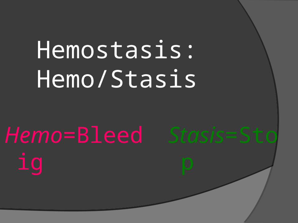
Nomenclature Plasma – the liquid portion of the blood.
Plasma can clot. Serum – the liquid left over after blood has
clotted. Serum cannot clot. Hemostasis – the normal, protective process
whereby blood loss is limited following vascular injury [i.e. It is the stoppage of bleeding in case of small vascular injury.].
Thrombosis – a pathologic process in which blood clots form inside blood vessels.
Hemostasis
It is the stoppage of bleeding in case of small vascular injury.
It depends on the vascular wall, platelets and the clotting factors.
Mechanism:(1) Extravascular:-Physical effects of skin and elastic tissue inclosing the
blood vessels.-Biochemical effects of released substances from tissue
in activation of clotting.(2) Vascular phase = vascular constriction (3) Platelets plug (4) Blood coagulation
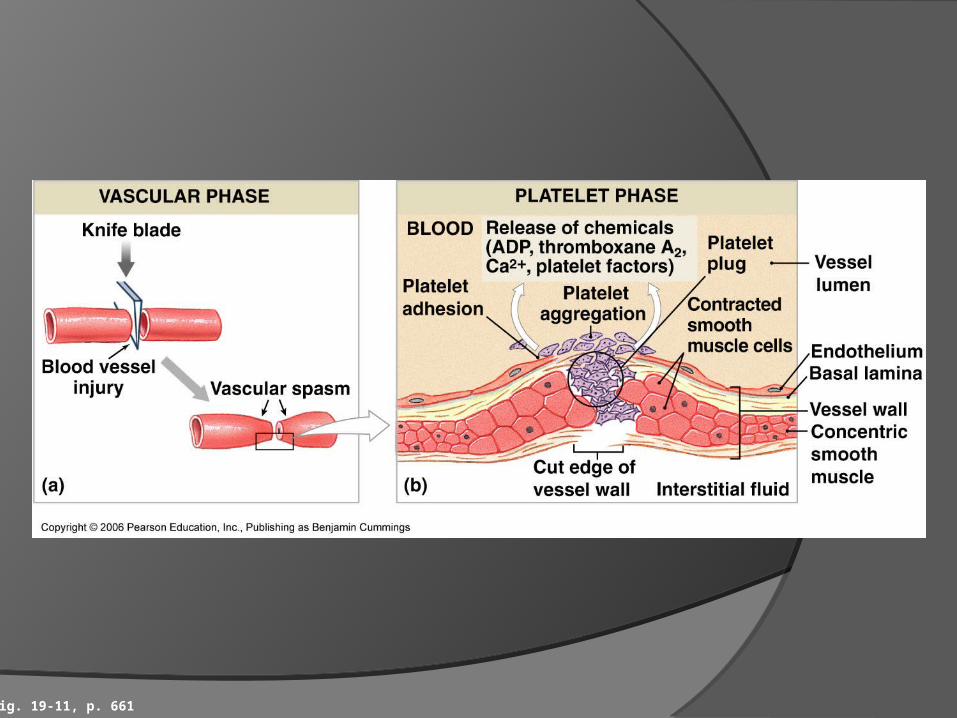
Fig. 19-11, p. 661
The Blood - Constituents ThrombocytesFunction: – haemostasis – form a plug to block damaged vessels.- Help initiate clotting process
Structure:-- cytoplasmic fragments of cells- roughly disc shaped - anucleated
Lifespan – 5 – 9 daysNumber – 250 – 500000/mm3
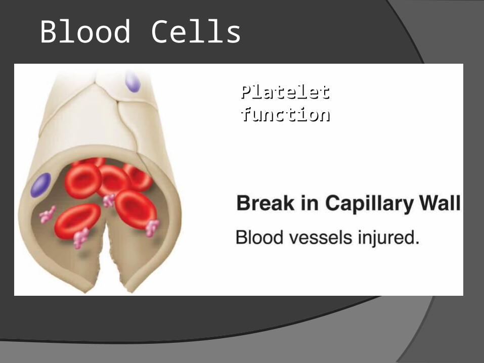
Formation of platelet plug (Platelet function)
The main function of platelets is the formation of mechanical plug to close the vascular injury in the following steps:
1)Platelet adhesion: Following blood vessel injury, platelets
adhere to the exposed subendothelial collagen and myofibrils.
.2) Platelets release occurs as the following: a-The platelets swell and becomes irregular with numerous
processes which facilitate the interaction between adjacent platelets.
b- The contractile proteins of platelets contract causing the release of the contents of platelets such as ADP, serotonin, VWF, fibrinogen and platelet factor .
c- Platelets enzymes form thromboxane A2 which released in the blood. Thromboxane A2 platelet cAMP release of platelets contents required for aggregation and severe V.C.
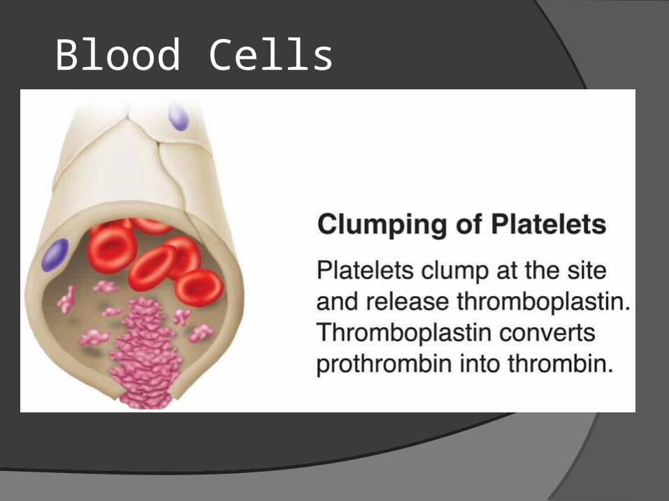
3)Platelet aggregation:-Released ADP and thromboxaneA2 cause the platelet to
swell and adhere to each other (aggregation) leading to further release and more aggregation and so on.
-The result is the formation of platelet plug which closes the injured area.
4)Platelet plug:
At first, the plug is loose then becomes firm and tight by contraction of platelets contractile proteins and by fibrin threads which attached to the injured vessel wall forming tight plug.
The normal endothelial cells around the injury release prostacyclin which causes limitation of the plug to site of the injury by inhibition of platelet aggregation and causes vasodilatation to limit the vasoconstrictive effect of thromboxaneA2.
.5) Platelet fusion: by fibrinogen, ADP and enzymes.
6) Platelet release thrombasthenin & ATP clot retraction.
7) Platelet derived growth factor stimulate growth of the vascular endothelial cells, smooth muscles and fibroblasts which help the repair of the damaged vessels.
Blood Cells
Platelet functionPlatelet function
Blood Cells
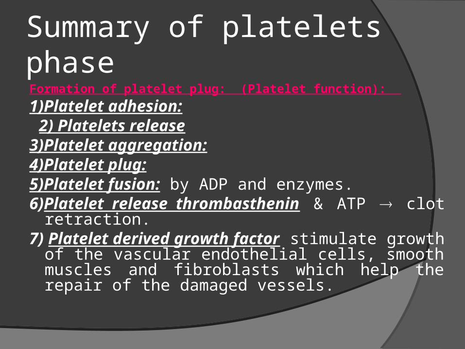
Summary of platelets phaseFormation of platelet plug: (Platelet function):
1)Platelet adhesion: 2) Platelets release 3)Platelet aggregation:4)Platelet plug:5)Platelet fusion: by ADP and enzymes.6)Platelet release thrombasthenin & ATP clot
retraction.7) Platelet derived growth factor stimulate growth
of the vascular endothelial cells, smooth muscles and fibroblasts which help the repair of the damaged vessels.
Blood Cells
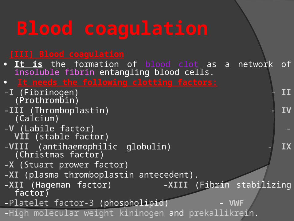
Blood coagulation [III] Blood coagulation It is the formation of blood clot as a network of insoluble fibrin
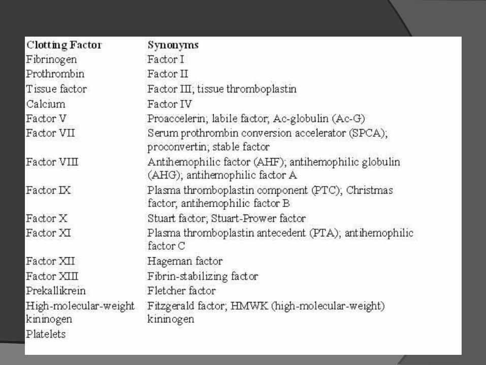
entangling blood cells. It needs the following clotting factors:-I (Fibrinogen) - II (Prothrombin)-III (Thromboplastin) - IV (Calcium)-V (Labile factor) - VII (stable factor)-VIII (antihaemophilic globulin) - IX (Christmas factor)-X (Stuart prower factor)-XI (plasma thromboplastin antecedent).-XII (Hageman factor) -XIII (Fibrin stabilizing factor)-Platelet factor-3 (phospholipid) - VWF -High molecular weight kininogen and prekallikrein.
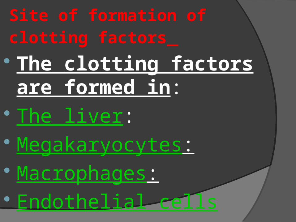
Site of formation of clotting factors The clotting factors are
formed in: The liver: Megakaryocytes: Macrophages: Endothelial cells
Steps of blood clotting
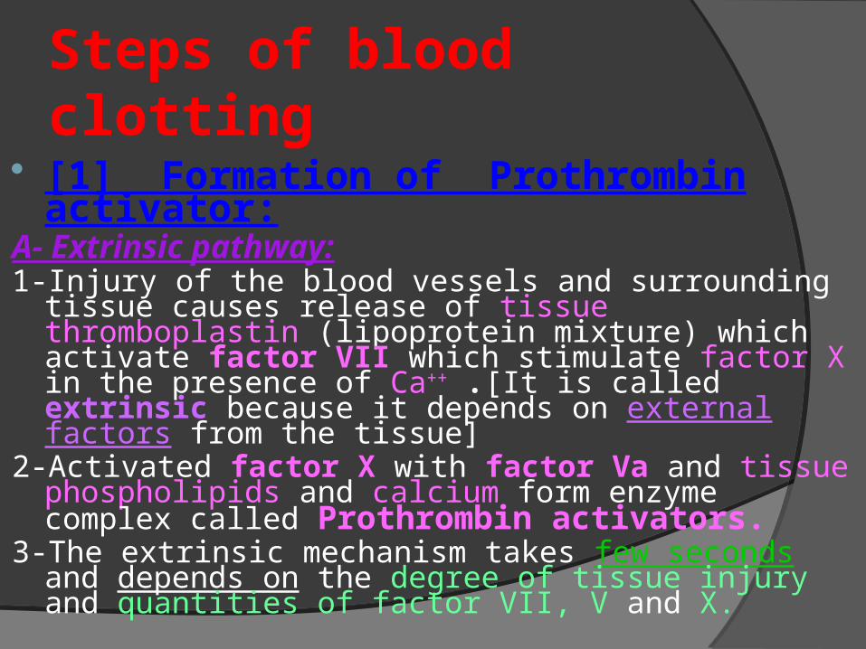
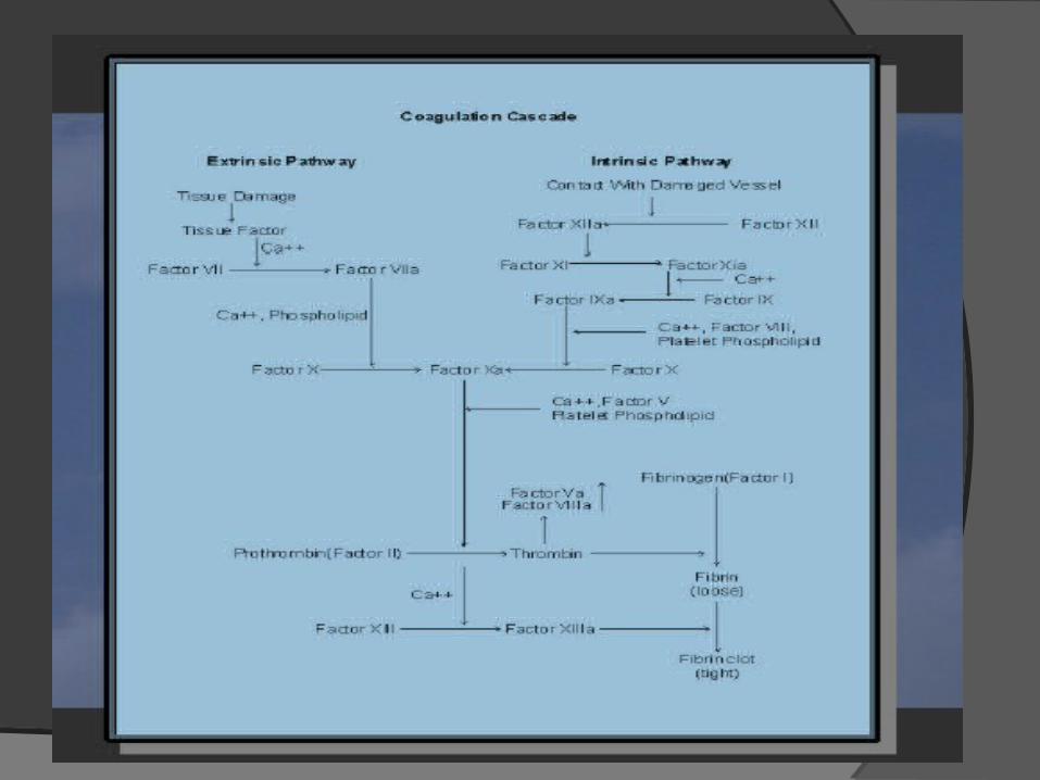
[1] Formation of Prothrombin activator:
A- Extrinsic pathway:1-Injury of the blood vessels and surrounding tissue
causes release of tissue thromboplastin (lipoprotein mixture) which activate factor VII which stimulate factor X in the presence of Ca++ .[It is called extrinsic because it depends on external factors from the tissue]
2-Activated factor X with factor Va and tissue phospholipids and calcium form enzyme complex called Prothrombin activators.
3-The extrinsic mechanism takes few seconds and depends on the degree of tissue injury and quantities of factor VII, V and X.
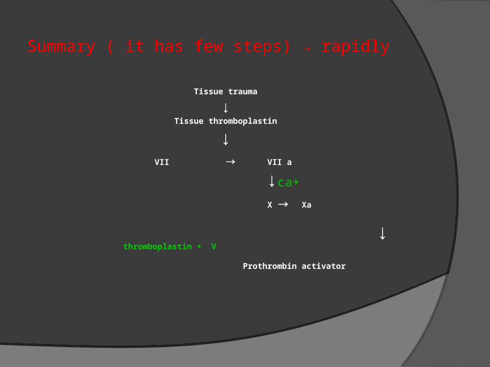
Summary ( it has few steps) → rapidly
Tissue trauma
↓Tissue thromboplastin
↓VII → VII a
↓ca+
X → Xa
↓ thromboplastin + V
Prothrombin activator
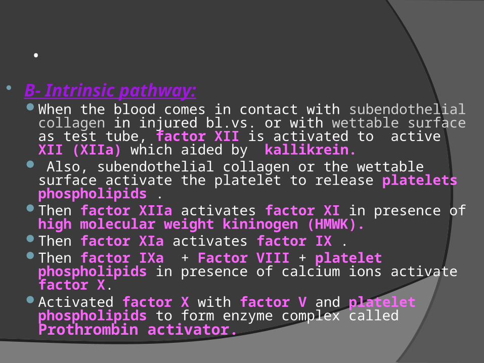
. B- Intrinsic pathway:
When the blood comes in contact with subendothelial collagen in injured bl.vs. or with wettable surface as test tube, factor XII is activated to active XII (XIIa) which aided by kallikrein.
Also, subendothelial collagen or the wettable surface activate the platelet to release platelets phospholipids .
Then factor XIIa activates factor XI in presence of high molecular weight kininogen (HMWK).
Then factor XIa activates factor IX .Then factor IXa + Factor VIII + platelet phospholipids in
presence of calcium ions activate factor X.Activated factor X with factor V and platelet
phospholipids to form enzyme complex called Prothrombin activator.
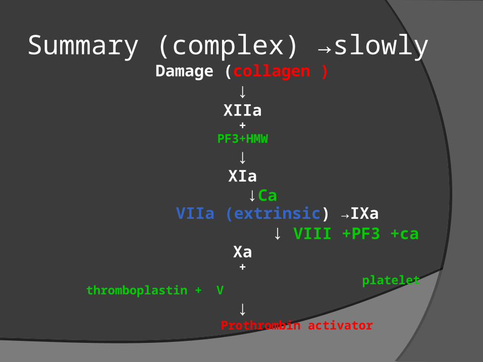
Summary (complex) →slowlyDamage (collagen )
↓XIIa
+PF3+HMW
↓XIa
↓Ca VIIa (extrinsic) →IXa
↓ VIII +PF3 +caXa+
platelet thromboplastin + V
↓ Prothrombin activator
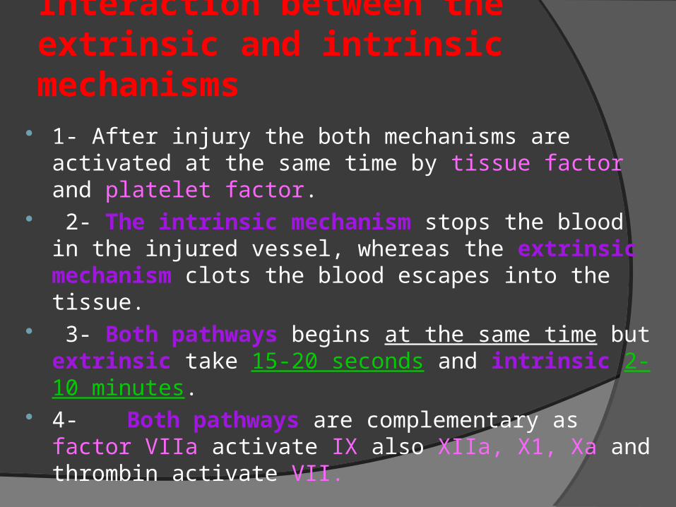
Interaction between the extrinsic and intrinsic mechanisms
1- After injury the both mechanisms are activated at the same time by tissue factor and platelet factor.
2- The intrinsic mechanism stops the blood in the injured vessel, whereas the extrinsic mechanism clots the blood escapes into the tissue.
3- Both pathways begins at the same time but extrinsic take 15-20 seconds and intrinsic 2-10 minutes.
4- Both pathways are complementary as factor VIIa activate IX also XIIa, X1, Xa and thrombin activate VII.
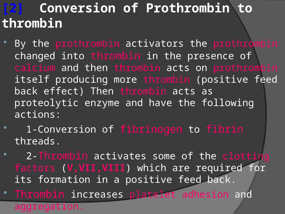
[2] Conversion of Prothrombin to thrombin By the prothrombin activators the prothrombin
changed into thrombin in the presence of calcium and then thrombin acts on prothrombin itself producing more thrombin (positive feed back effect) Then thrombin acts as proteolytic enzyme and have the following actions:
1-Conversion of fibrinogen to fibrin threads. 2-Thrombin activates some of the clotting factors
(V,VII,VIII) which are required for its formation in a positive feed back.
Thrombin increases platelet adhesion and aggregation.
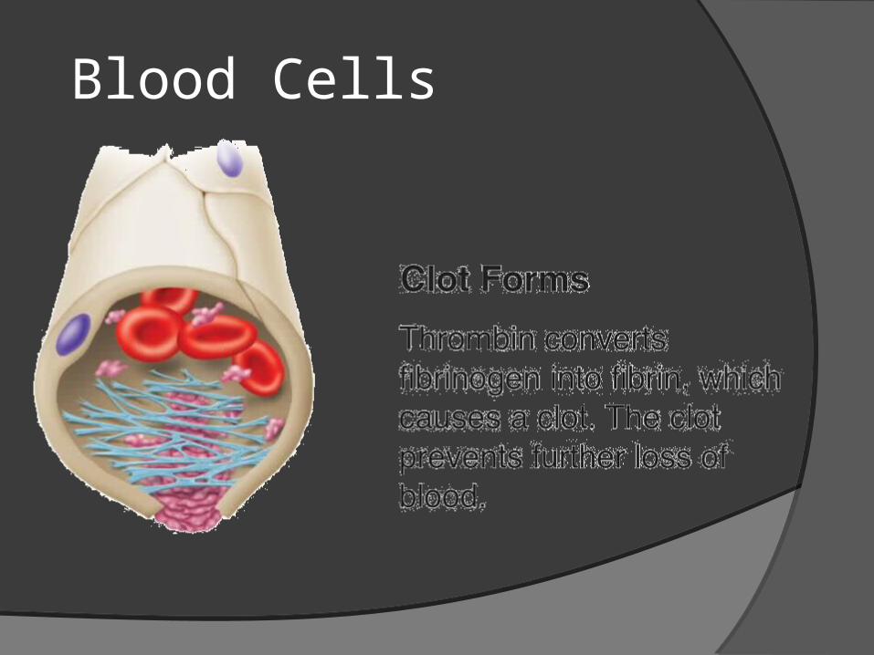
[3] Conversion of fibrinogen to fibrin (formation of fibrin clot)
Thrombin splits small negatively charged peptide fragments from the fibrinogen molecules (removing the repulsive forces from the molecules) and allowing the remainder to polymerize to form fibrin polymers or threads.
These threads adhere to the injured blood vessel wall forming loose net like meshwork that traps the blood cells forming red soft blood clot.
Stabilization of blood clot by activated factor XIII which activated by thrombin.
Factor XIIIa strengthens the fibrin clot by forming strong bonds between fibrin polymers.
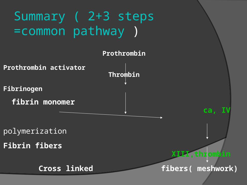
Summary ( 2+3 steps =common pathway )
Prothrombin
Prothrombin activatorThrombin
Fibrinogen fibrin monomer ca, IV
polymerization Fibrin fibers
XIII,thrombin Cross linked
fibers( meshwork)
Clot retractionClot retraction: the fibrin clot contains large
number of platelets which release factor XIII and attached to the fibrin threads ,also their contractile protein (actin, myosin and thrombosthenin) contract leading to the fibrin meshwork shrinks and becomes stronger and clear yellowish serum is squeezed from the clot.
This step is stimulated by thrombin and calcium ions released from the platelets.
This step makes the clot more dense and strong and also pulls the edges of the wound together facilitating the repair of the injury.
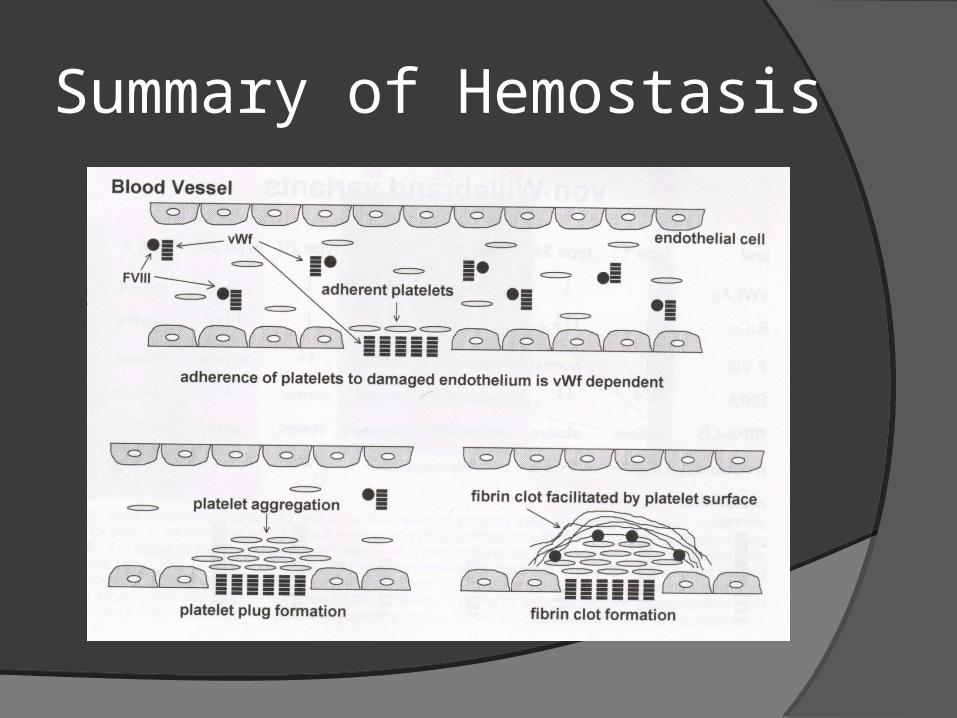
Summary of Hemostasis