Hemostasis & Coagulation Ahmad Sh. Silmi Unit 1 : Primary Hemostasis.
71
Hemostasis & Coagulation Ahmad Sh. Silmi Unit 1 : Primary Hemostasis
-
date post
20-Dec-2015 -
Category
Documents
-
view
274 -
download
6
Transcript of Hemostasis & Coagulation Ahmad Sh. Silmi Unit 1 : Primary Hemostasis.
Hemostasis & Coagulation
Ahmad Sh. Silmi
Unit 1 : Primary Hemostasis
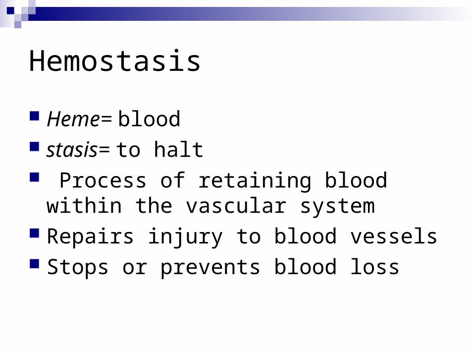
Hemostasis
Heme= blood stasis= to halt Process of retaining blood within the
vascular system Repairs injury to blood vessels Stops or prevents blood loss
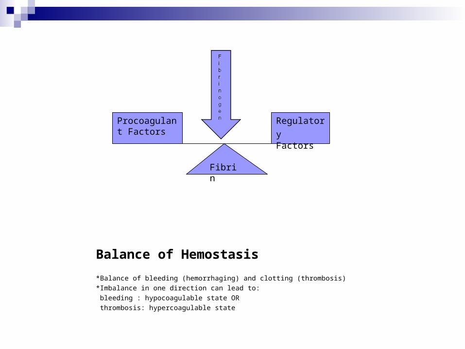
Balance of Hemostasis
*Balance of bleeding (hemorrhaging) and clotting (thrombosis)
*Imbalance in one direction can lead to:
bleeding : hypocoagulable state OR
thrombosis: hypercoagulable state
Procoagulant Factors
Regulatory Factors
Fibrin
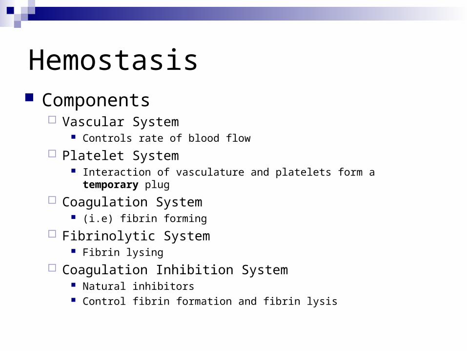
Hemostasis Components
Vascular System Controls rate of blood flow
Platelet System Interaction of vasculature and platelets form a temporary plug
Coagulation System (i.e) fibrin forming
Fibrinolytic System Fibrin lysing
Coagulation Inhibition System Natural inhibitors Control fibrin formation and fibrin lysis
Failure or deficiencies in any of these five systems can leads to varying degrees of uncontrolled hemorrhaging or clotting
Hemostasis
The hemostatic components remain inert in the presence of intact vascular tissue or endothelium
Following injury, each component must function optimally.
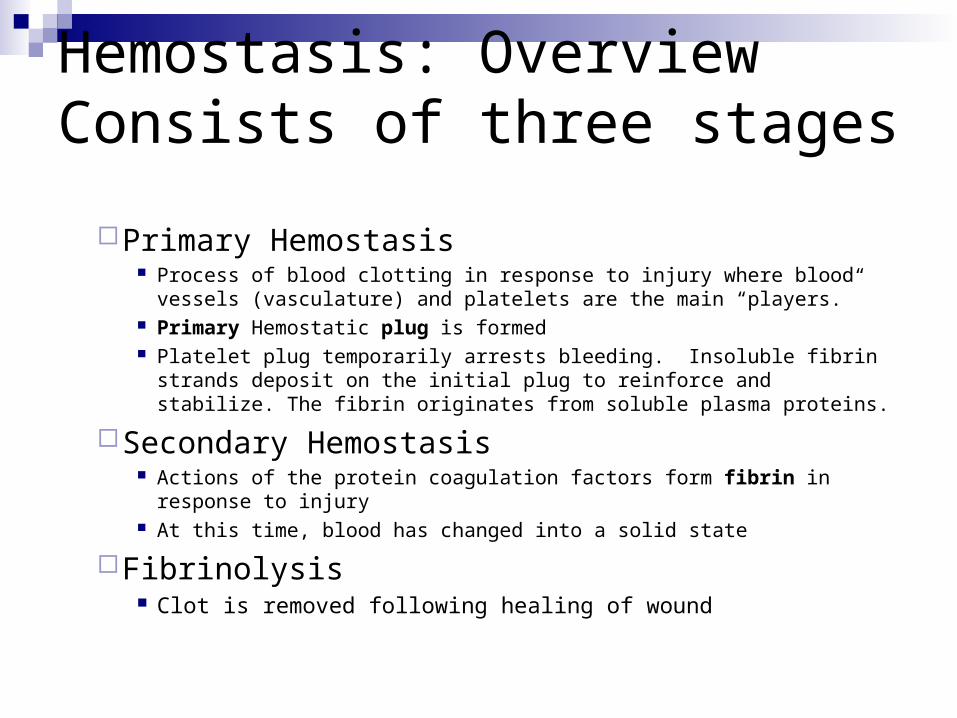
Hemostasis: OverviewConsists of three stages
Primary Hemostasis Process of blood clotting in response to injury where blood vessels
(vasculature) and platelets are the main “players.” Primary Hemostatic plug is formed Platelet plug temporarily arrests bleeding. Insoluble fibrin strands
deposit on the initial plug to reinforce and stabilize. The fibrin originates from soluble plasma proteins.
Secondary Hemostasis Actions of the protein coagulation factors form fibrin in response
to injury At this time, blood has changed into a solid state
Fibrinolysis Clot is removed following healing of wound
http://health.howstuffworks.com/adam-200077.htm
Vascular System
Blood Vessels Arteries
Carry blood from the heart to capillaries Thickest walls of the vasculature
Veins Return blood from capillaries to the heart Thinnest walls of vasculature
Capillaries No vessel wall Do not contribute to hemostasis
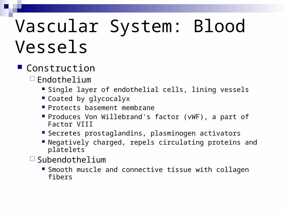
Vascular System: Blood Vessels Construction
Endothelium Single layer of endothelial cells, lining vessels Coated by glycocalyx Protects basement membrane Produces Von Willebrand's factor (vWF), a part of Factor
VIII Secretes prostaglandins, plasminogen activators Negatively charged, repels circulating proteins and
platelets Subendothelium
Smooth muscle and connective tissue with collagen fibers
Vascular System: Blood Vessels
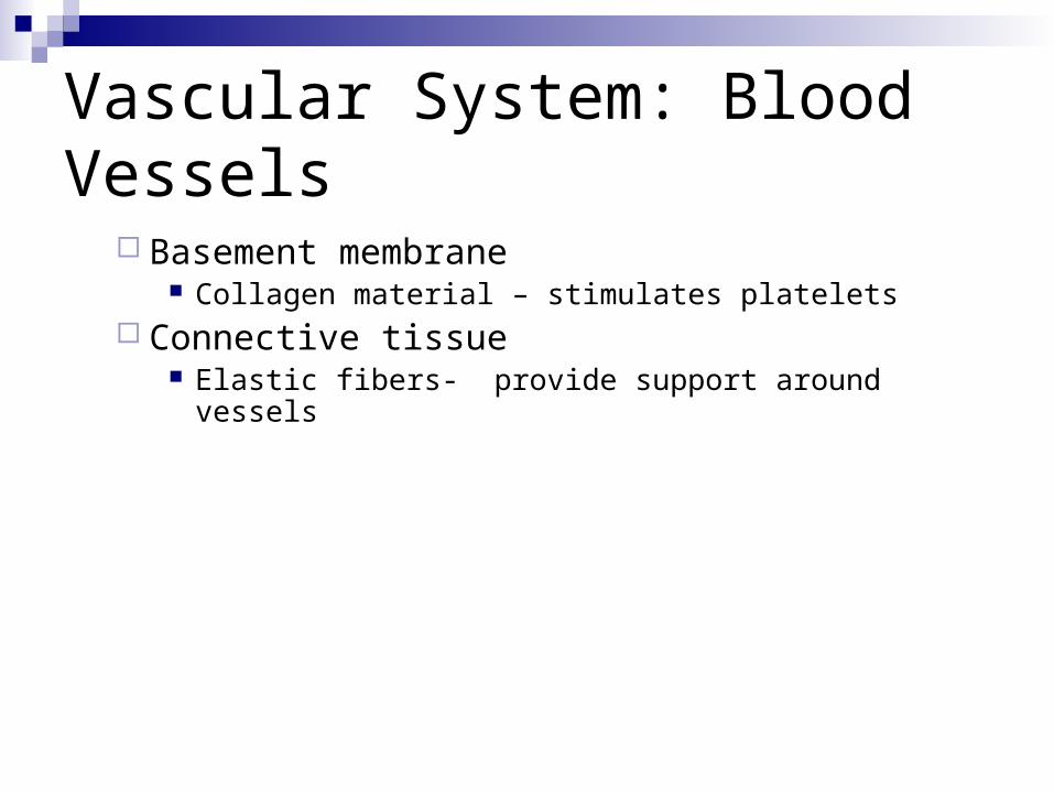
Basement membrane Collagen material – stimulates platelets
Connective tissue Elastic fibers- provide support around vessels

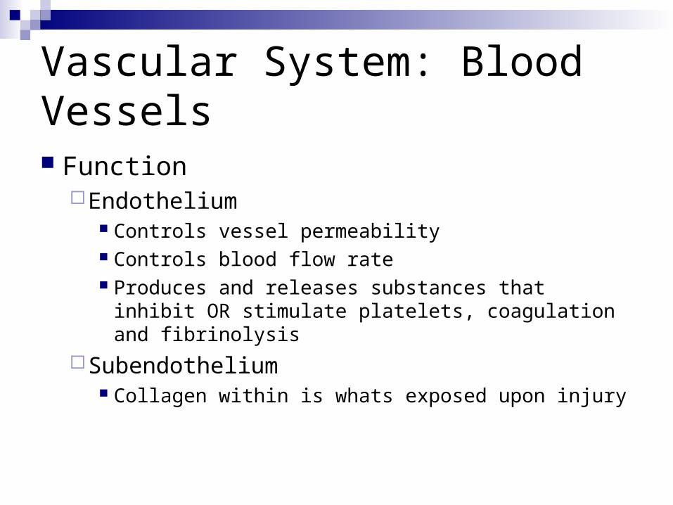
Vascular System: Blood Vessels Function
Endothelium Controls vessel permeability Controls blood flow rate Produces and releases substances that inhibit OR
stimulate platelets, coagulation and fibrinolysis
Subendothelium Collagen within is whats exposed upon injury
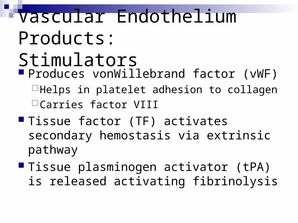
Vascular Endothelium Products:Stimulators Produces vonWillebrand factor (vWF)
Helps in platelet adhesion to collagen Carries factor VIII
Tissue factor (TF) activates secondary hemostasis via extrinsic pathway
Tissue plasminogen activator (tPA) is released activating fibrinolysis
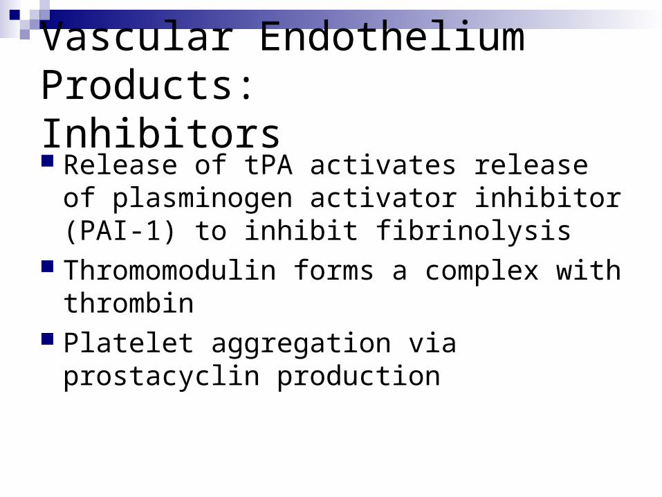
Vascular Endothelium Products:Inhibitors Release of tPA activates release of
plasminogen activator inhibitor (PAI-1) to inhibit fibrinolysis
Thromomodulin forms a complex with thrombin
Platelet aggregation via prostacyclin production
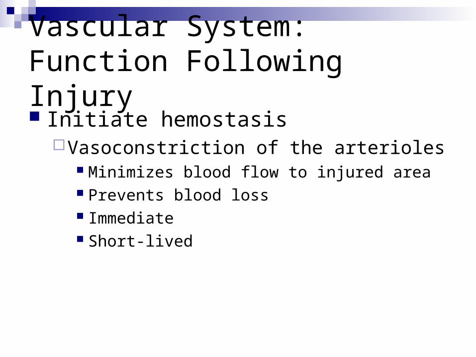
Vascular System: Function Following Injury Initiate hemostasis
Vasoconstriction of the arterioles Minimizes blood flow to injured area Prevents blood loss Immediate Short-lived
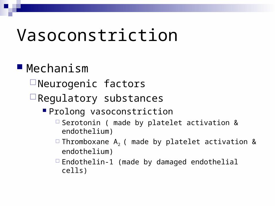
Vasoconstriction
MechanismNeurogenic factorsRegulatory substances
Prolong vasoconstriction Serotonin ( made by platelet activation &
endothelium) Thromboxane A2 ( made by platelet activation &
endothelium) Endothelin-1 (made by damaged endothelial cells)
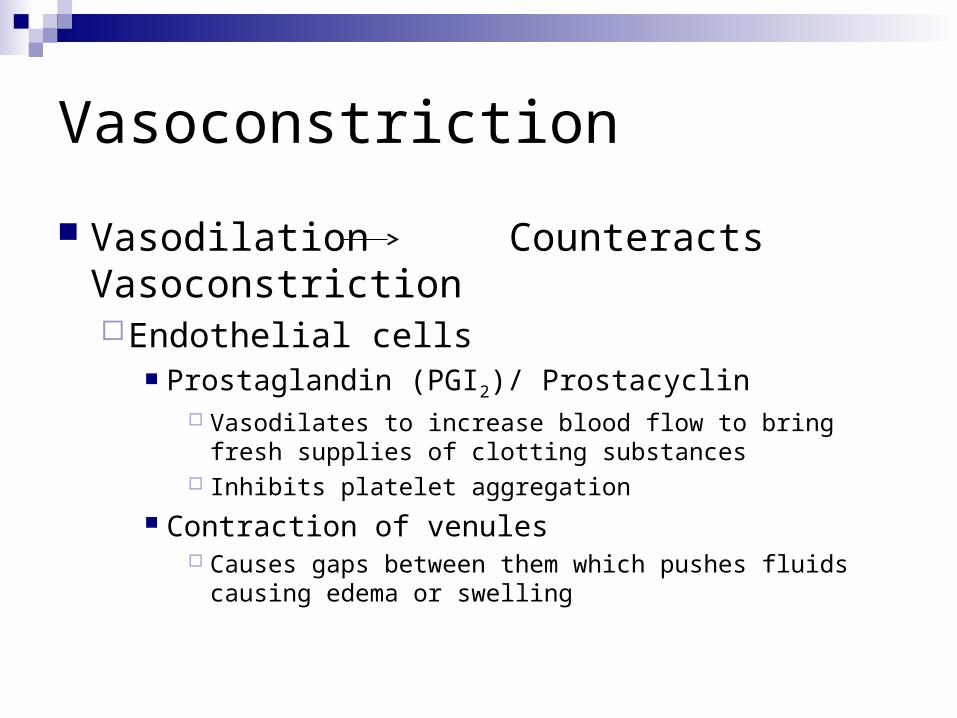
Vasoconstriction
Vasodilation Counteracts VasoconstrictionEndothelial cells
Prostaglandin (PGI2)/ Prostacyclin Vasodilates to increase blood flow to bring fresh
supplies of clotting substances Inhibits platelet aggregation
Contraction of venules Causes gaps between them which pushes fluids
causing edema or swelling
Thought question…
Think about the last time you cut your finger with a piece of paper. Did your finger bleed immediately?
If not, what might have prevented the bleeding?
Answer..
No, the finger probably did not bleed immediately, due to vasoconstriction of the blood vessels
Discussion
What actions of the endothelial cells prevent clotting from occurring within the blood vessels?
Answers..
Since the endothelial lining has a negative charge, it normally repels coagulation proteins and platelets in the circulation. It synthesizes products that help to inhibit fibrin formation.
All About Platelets…
Second major component of the hemostatic system
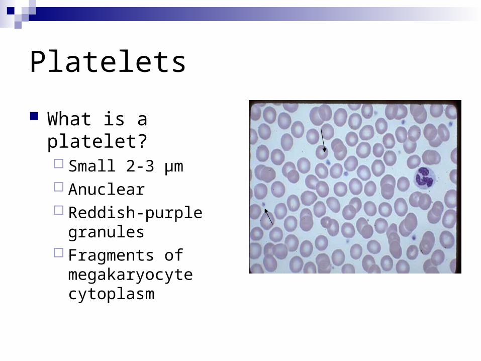
Platelets
What is a platelet? Small 2-3 µm Anuclear Reddish-purple
granules Fragments of
megakaryocyte cytoplasm
Platelets
Life span9-10 days
Normal Range150-440 x 109 /L
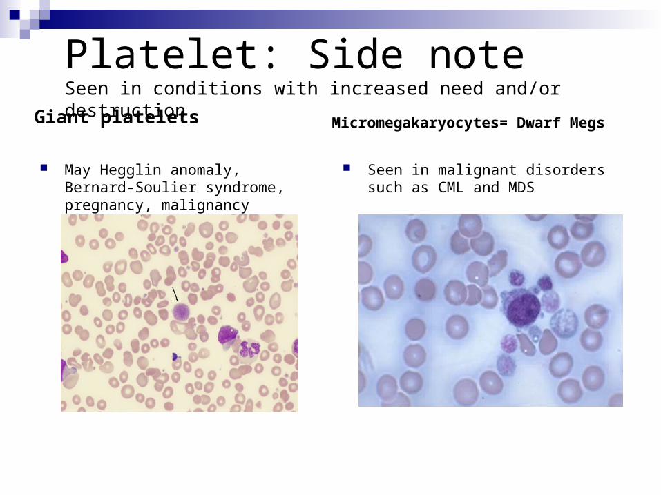
Platelet: Side noteSeen in conditions with increased need and/or destruction
Giant platelets
May Hegglin anomaly, Bernard-Soulier syndrome, pregnancy, malignancy
Micromegakaryocytes= Dwarf Megs
Seen in malignant disorders such as CML and MDS
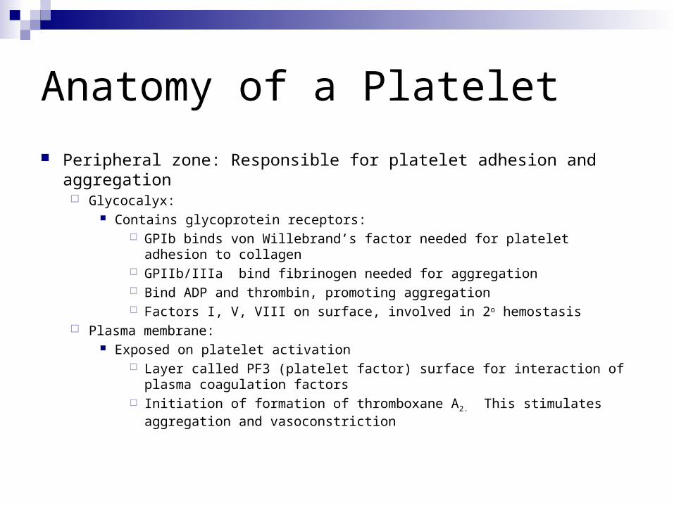
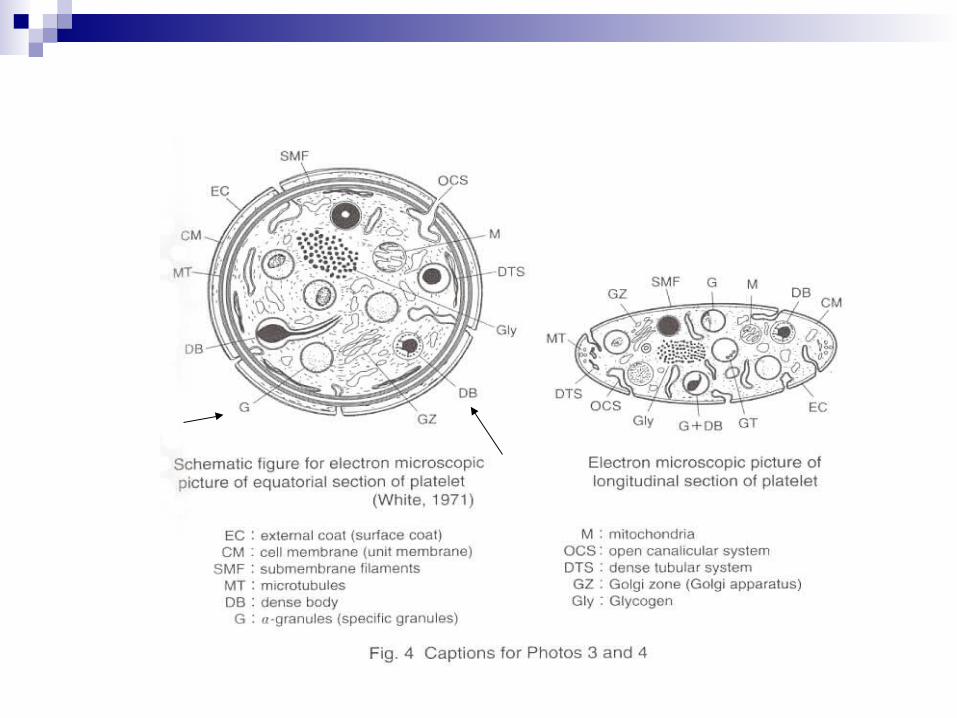
Anatomy of a Platelet
Peripheral zone: Responsible for platelet adhesion and aggregation Glycocalyx:
Contains glycoprotein receptors: GPIb binds von Willebrand’s factor needed for platelet adhesion to
collagen GPIIb/IIIa bind fibrinogen needed for aggregation Bind ADP and thrombin, promoting aggregation Factors I, V, VIII on surface, involved in 2o hemostasis
Plasma membrane: Exposed on platelet activation
Layer called PF3 (platelet factor) surface for interaction of plasma coagulation factors
Initiation of formation of thromboxane A2. This stimulates aggregation and vasoconstriction
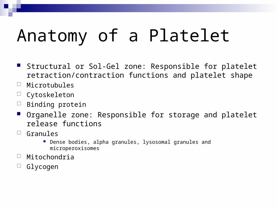
Anatomy of a Platelet
Structural or Sol-Gel zone: Responsible for platelet retraction/contraction functions and platelet shape
Microtubules Cytoskeleton Binding protein Organelle zone: Responsible for storage and platelet release
functions Granules
Dense bodies, alpha granules, lysosomal granules and microperoxisomes
Mitochondria Glycogen
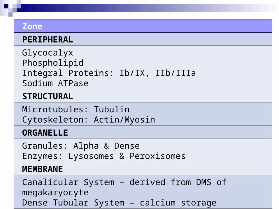
Zone
PERIPHERAL
GlycocalyxPhospholipidIntegral Proteins: Ib/IX, IIb/IIIaSodium ATPase
STRUCTURAL
Microtubules: TubulinCytoskeleton: Actin/Myosin
ORGANELLE
Granules: Alpha & DenseEnzymes: Lysosomes & Peroxisomes
MEMBRANE
Canalicular System – derived from DMS of megakaryocyteDense Tubular System – calcium storage
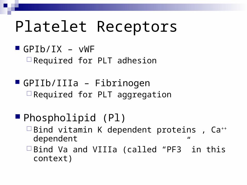
Platelet Receptors GPIb/IX – vWF
Required for PLT adhesion
GPIIb/IIIa – Fibrinogen Required for PLT aggregation
Phospholipid (Pl) Bind vitamin K dependent proteins , Ca++
dependent Bind Va and VIIIa (called “PF3” in this context)
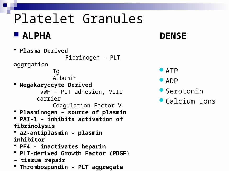
Platelet Granules ALPHA Plasma Derived Fibrinogen – PLT aggrgation
Ig Albumin
Megakaryocyte Derived vWF – PLT adhesion, VIII carrier
Coagulation Factor V Plasminogen – source of plasmin PAI-1 – inhibits activation of fibrinolysis a2-antiplasmin – plasmin inhibitor PF4 – inactivates heparin PLT-derived Growth Factor (PDGF) – tissue repair Thrombospondin – PLT aggregate stabilizer Fibronectin – PLT binding
DENSE
ATPADPSerotoninCalcium Ions
Diagrammatic Representation of the Platelet
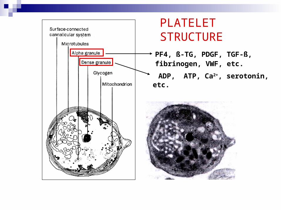
PF4, ß-TG, PDGF, TGF-ß,fibrinogen, VWF, etc.
ADP, ATP, Ca2+, serotonin, etc.
PLATELET STRUCTURE
Production of Platelets
Made in Bone marrow Need dictates the amount of platelets produced. Stimulus for production is the platelet mass in
circulating blood ~ 80 % and megakaryocyte mass in bone marrow
Originate from CFU-GEMM to form CFU-Meg Cytokines and growth factors such as IL-3 and
GM-CSF influences progenitor stages
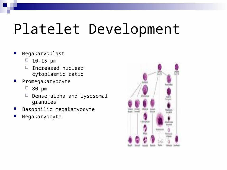
Platelet Development
Megakaryoblast 10-15 µm Increased nuclear:
cytoplasmic ratio Promegakaryocyte
80 µm Dense alpha and lysosomal
granules Basophilic megakaryocyte Megakaryocyte
Production of Platelets
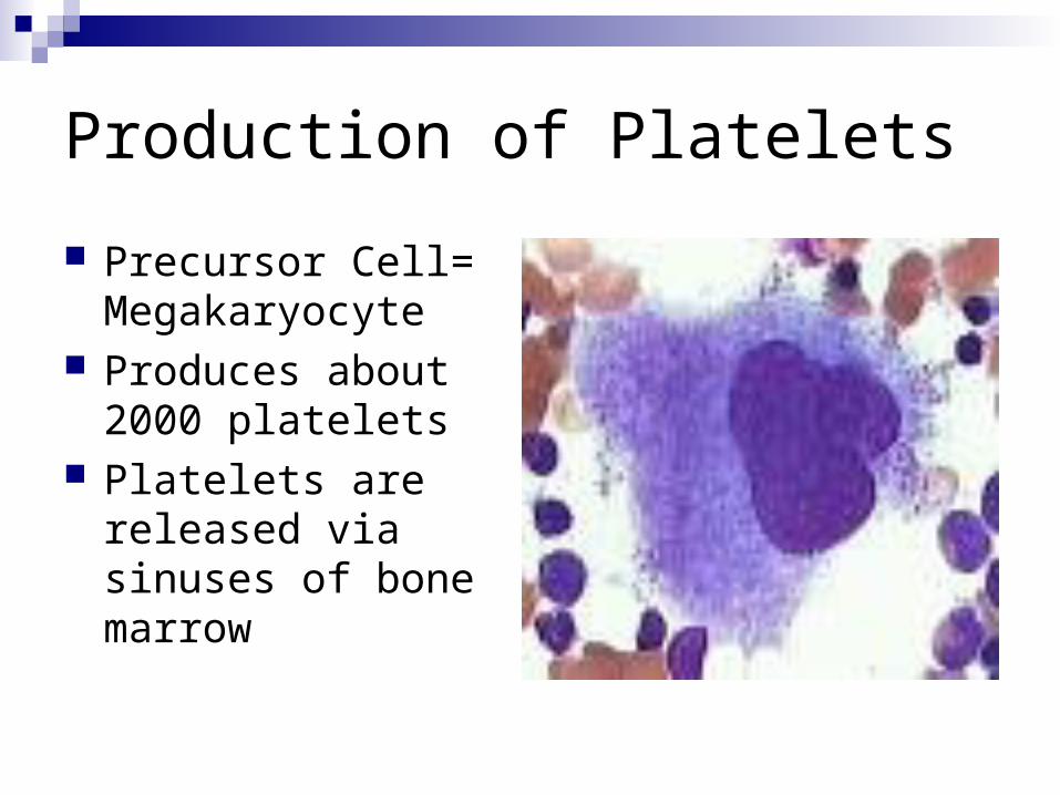
Precursor Cell= Megakaryocyte
Produces about 2000 platelets
Platelets are released via sinuses of bone marrow
Production of Platelets
Thrombopoietin (TPO)Regulates platelet development Influences all stages of megakaryocyte
productionProduced in the liver, kidney and spleen
Production of Platelets
How does TPO work?Maintains a constant number of
platelets in peripheral blood by binding Mp1 (platelet receptor). Bound TPO can not stimulate proliferation of bone marrow progenitor cells
The higher the platelet count, the more TPO is bound and stimulation of bone marrow is decreased.
Thought question…
If a patient had a low platelet count what will happen?
Answer…
TPO increases the number of megakaryocytes in the bone marrow, increases size and DNA count of megakaryocytes and increases maturation rate
Function of Platelets
Surveillance of blood vessel continuity Checks endothelial lining for gaps and breaks Fill-in small gaps caused by separation of
endothelial cells Formation of primary hemostatic plug Surface for coagulation factors to make
secondary hemostatic plug Aid in healing injured tissue
Formation of Primary Hemostatic Plug Once the platelets “normal” environment is
changed, they become activated or adhesive
Three stages of plug formation
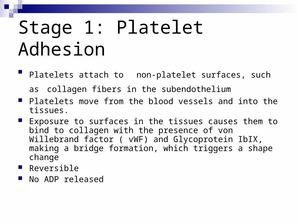
Stage 1: Platelet Adhesion
Platelets attach to non-platelet surfaces, such as collagen fibers in the subendothelium
Platelets move from the blood vessels and into the tissues. Exposure to surfaces in the tissues causes them to bind to
collagen with the presence of von Willebrand factor ( vWF) and Glycoprotein IbIX, making a bridge formation, which triggers a shape change
Reversible No ADP released
Stage 1: Platelet Activation
Platelets undergo a shape change from disc to spiny sphere with projections
Activation required for 1O hemostatic plug formation
Activation continues until Ca ++ threshold met Outcome
Activation of GPIIb/IIIa receptors for fibrinogen Secretion of granules within platelets into
tissues
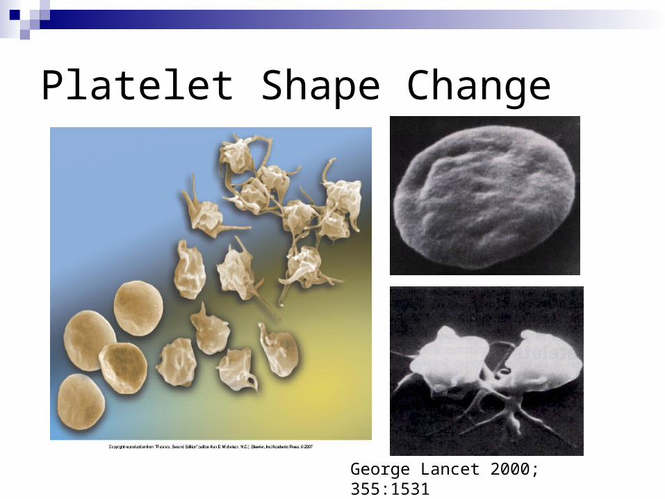
Platelet Shape Change
George Lancet 2000; 355:1531
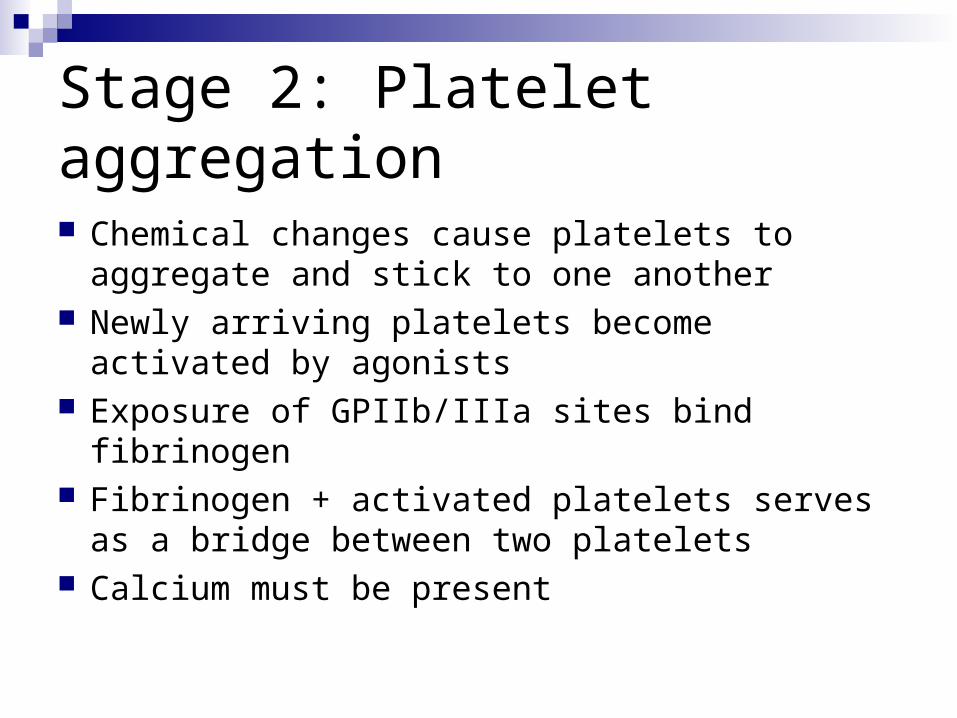
Stage 2: Platelet aggregation Chemical changes cause platelets to
aggregate and stick to one another Newly arriving platelets become activated
by agonists Exposure of GPIIb/IIIa sites bind fibrinogen Fibrinogen + activated platelets serves as
a bridge between two platelets Calcium must be present
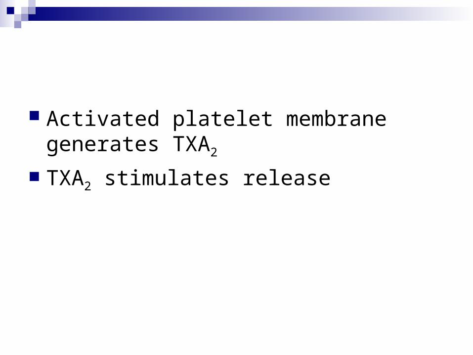
Activated platelet membrane generates TXA2
TXA2 stimulates release
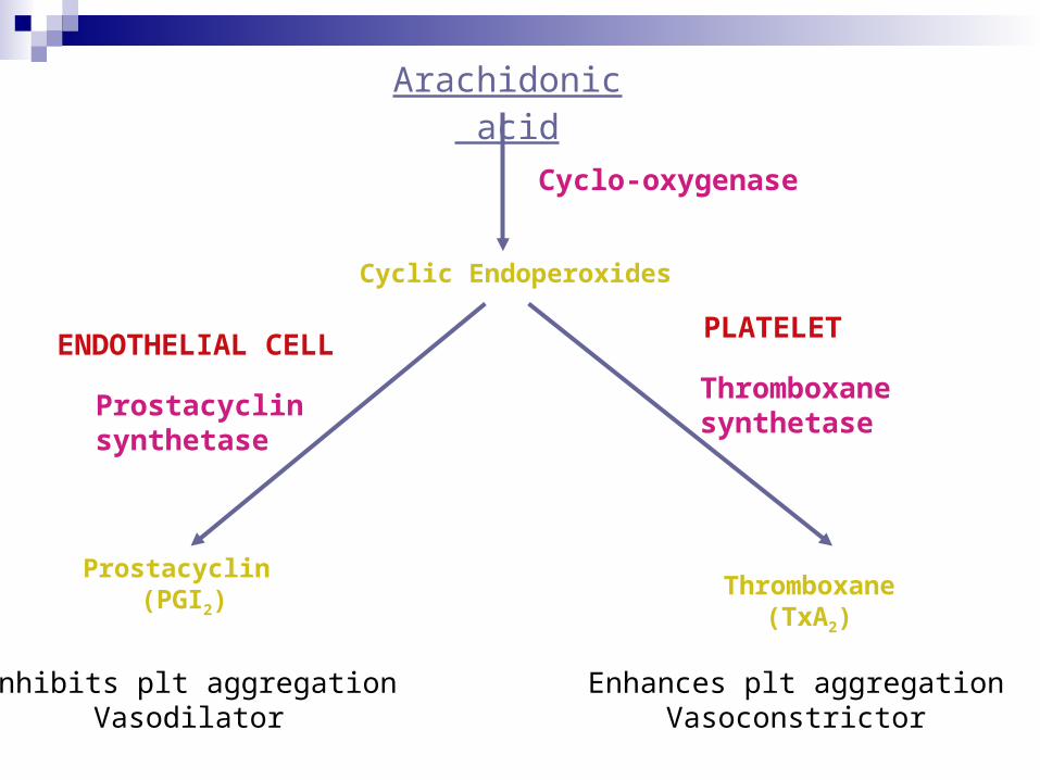
Arachidonic acid
ENDOTHELIAL CELLPLATELET
Thromboxane synthetase
Prostacyclinsynthetase
Cyclo-oxygenase
Prostacyclin (PGI2)
Thromboxane(TxA2)
Inhibits plt aggregationVasodilator
Enhances plt aggregationVasoconstrictor
Cyclic Endoperoxides
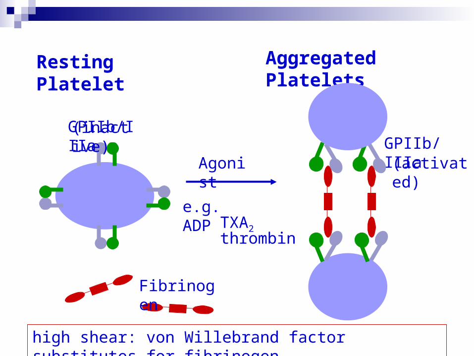
GPIIb/IIIa(inactive)
Agonist
e.g. ADPTXA2thrombin
Fibrinogen
Aggregated PlateletsResting Platelet
GPIIb/IIIa(activated)
high shear: von Willebrand factor substitutes for fibrinogen
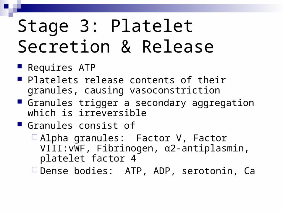
Stage 3: Platelet Secretion & Release Requires ATP Platelets release contents of their granules,
causing vasoconstriction Granules trigger a secondary aggregation which
is irreversible Granules consist of
Alpha granules: Factor V, Factor VIII:vWF, Fibrinogen, α2-antiplasmin, platelet factor 4
Dense bodies: ATP, ADP, serotonin, Ca
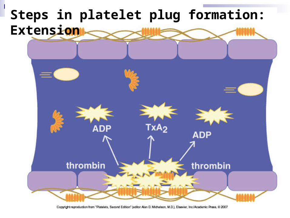
Steps in platelet plug formation: Extension
Granules con’t
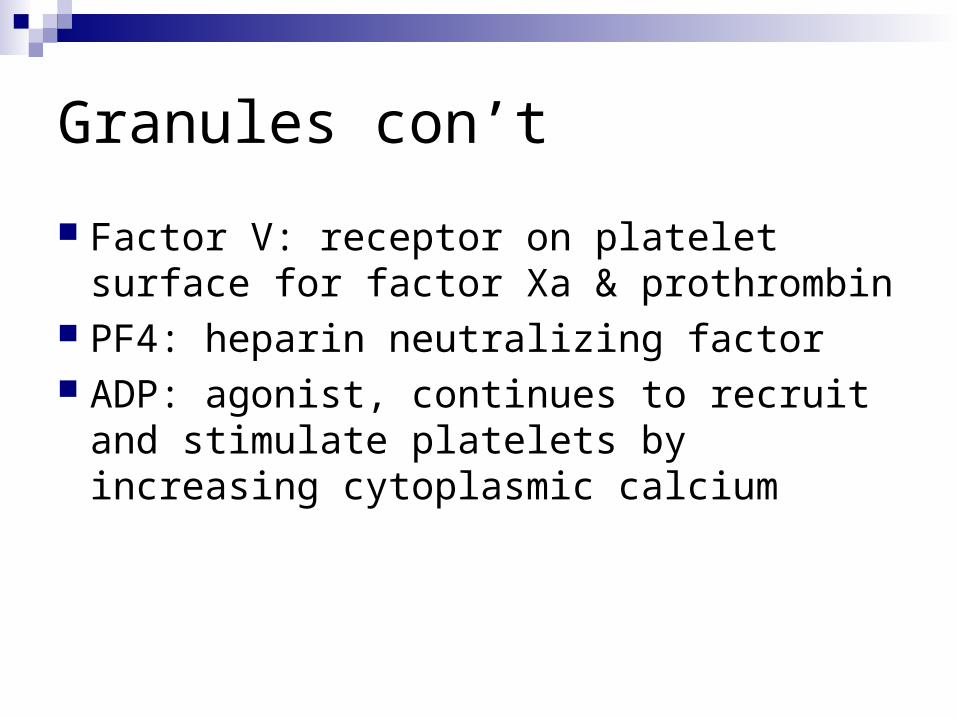
Factor V: receptor on platelet surface for factor Xa & prothrombin
PF4: heparin neutralizing factor ADP: agonist, continues to recruit and
stimulate platelets by increasing cytoplasmic calcium
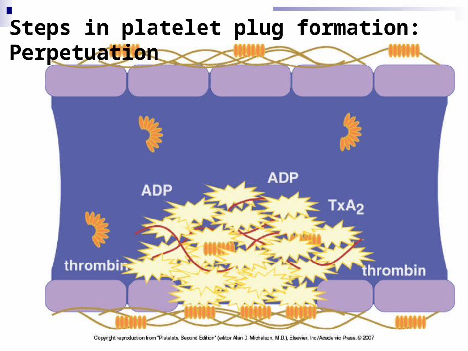
Steps in platelet plug formation: Perpetuation
Lets have a close view about the mechanism
Adhesion
GpIIb/IIIa
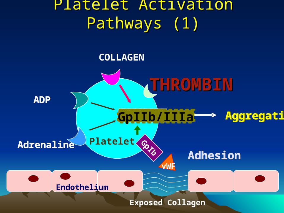
Platelet Activation Pathways (1)Platelet Activation Pathways (1)
GpIIb/IIIaGpIIb/IIIa Aggregation
ADP
Adrenaline Platelet GpIb
Exposed Collagen
Endothelium
vWF
COLLAGEN
GpIIb/IIIaGpIIb/IIIa AggregationGpIIb/IIIaGpIIb/IIIa Aggregation
AdhesionAdhesion
ADP
Adrenaline
THROMBINTHROMBIN
Platelet
Coagulation factors…
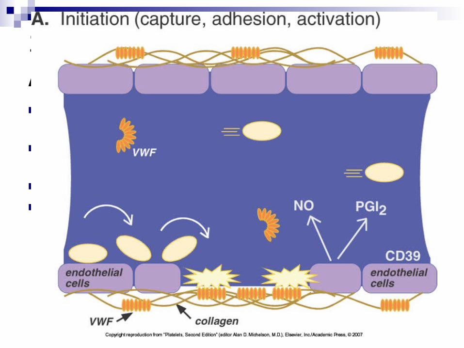
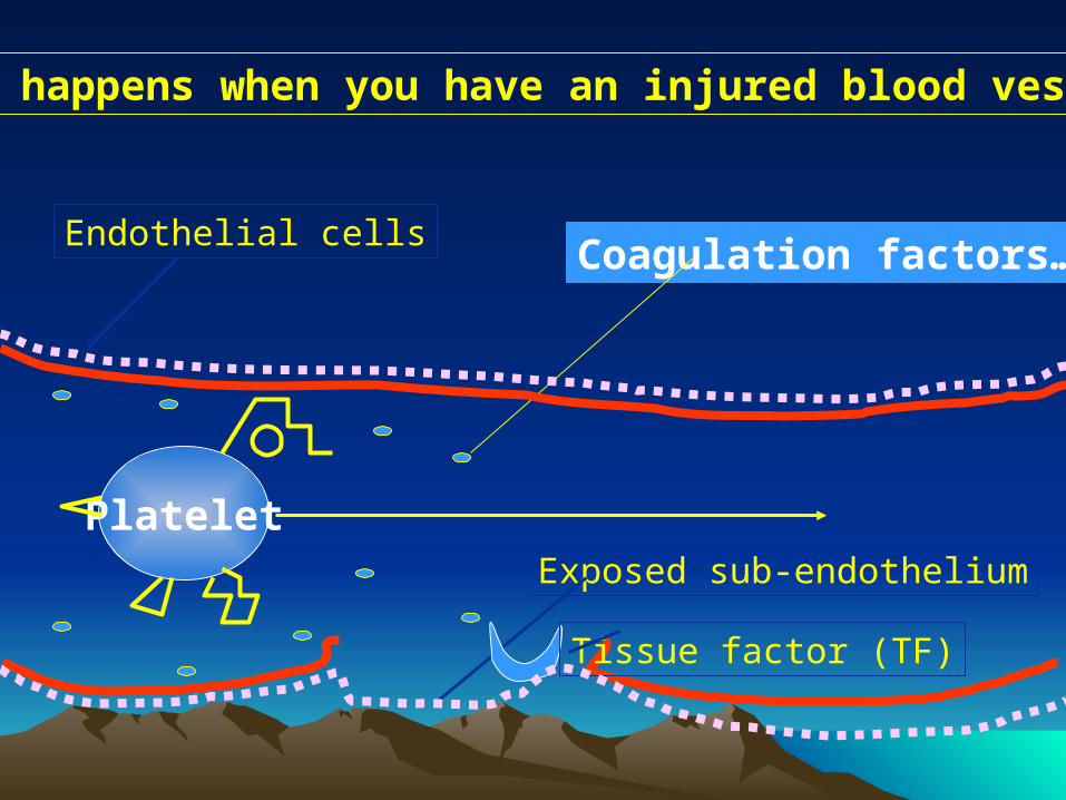
What happens when you have an injured blood vessel?
Exposed sub-endothelium
Endothelial cells
Tissue factor (TF)
Platelet
vWF
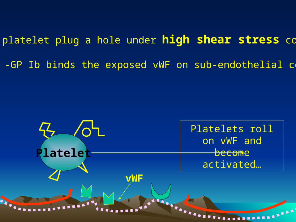
How does a platelet plug a hole under high shear stress conditions?
Platelets roll on vWF and become activated…
-GP Ib binds the exposed vWF on sub-endothelial cells
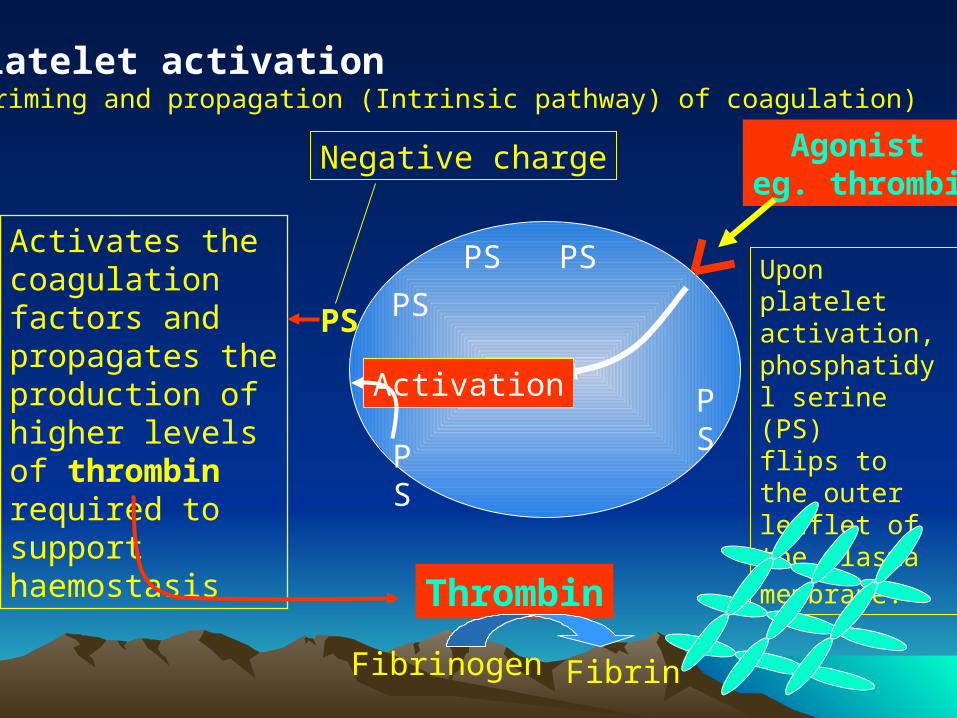
Platelet activation(Priming and propagation (Intrinsic pathway) of coagulation)
Upon platelet activation, phosphatidyl serine (PS)flips to the outer leaflet of the plasma
membrane.
Agonist eg. thrombin
PS
PS
PSPS
Activation
PS
PS
Negative charge
Activates the coagulation factors and propagates the production of higher levels of thrombin required to support haemostasis
Thrombin
Fibrinogen Fibrin
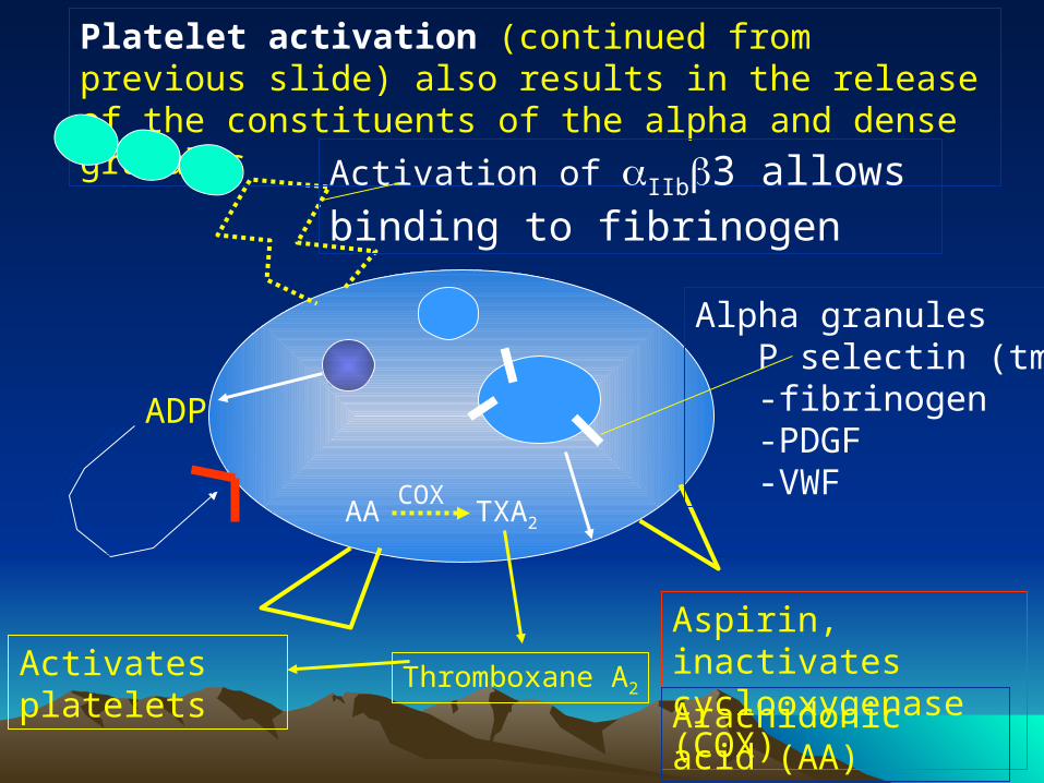
Platelet activation (continued from previous slide) also results in the release of the constituents of the alpha and dense granules
Alpha granules P selectin (tm) -fibrinogen -PDGF -VWF
Activation of IIb3 allows binding to fibrinogen
ADP
Thromboxane A2
Aspirin, inactivates cyclooxygenase (COX)
AA
Activates platelets
TXA2
COX
Arachidonic acid (AA)
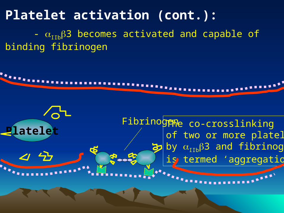
Platelet activation (cont.):
- IIb3 becomes activated and capable of binding fibrinogen
PlateletFibrinogen The co-crosslinking
of two or more plateletsby IIb3 and fibrinogenis termed ‘aggregation’
Side note
Heparin is used on patients who clot excessively. Endothelial cells make heparin-like molecules and expose them on their surface. PF4 binds these substances. Heparin can complex with bound PF4 and heparin will be neutralized.
Final Stage : Stabilization of Clot AKA: primary hemostatic plug
formation Thrombus formation Platelets release Factor V Expose factor III, accelerating
coagulation cascade Promote activation of clotting factors
Platelet System: Additional Functions Provides the reaction surface for some coagulation
system reactions, as well as platelet factor 3 (PF3) which is platelet phospholipid
Supports and maintains endothelial lining Defective hemostasis can occur due to
decreased number of platelets (quantitative) abnormally functioning platelets (qualitative)
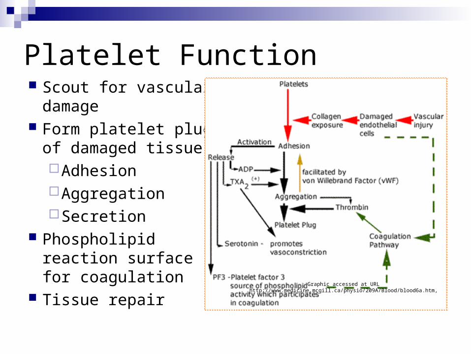
Platelet Function Scout for vascular
damage Form platelet plug of
damaged tissueAdhesionAggregationSecretion
Phospholipid reaction surface for coagulation
Tissue repair Graphic accessed at URL http://www.medicine.mcgill.ca/physio/209A/Blood/blood6a.htm,
Agonists“a term used to describe substances that
can attach to a platelet membrane receptor and activate platelets causing them to aggregate .”
McKenzie, SB. Clinical Hematology, Glossary, p. 919
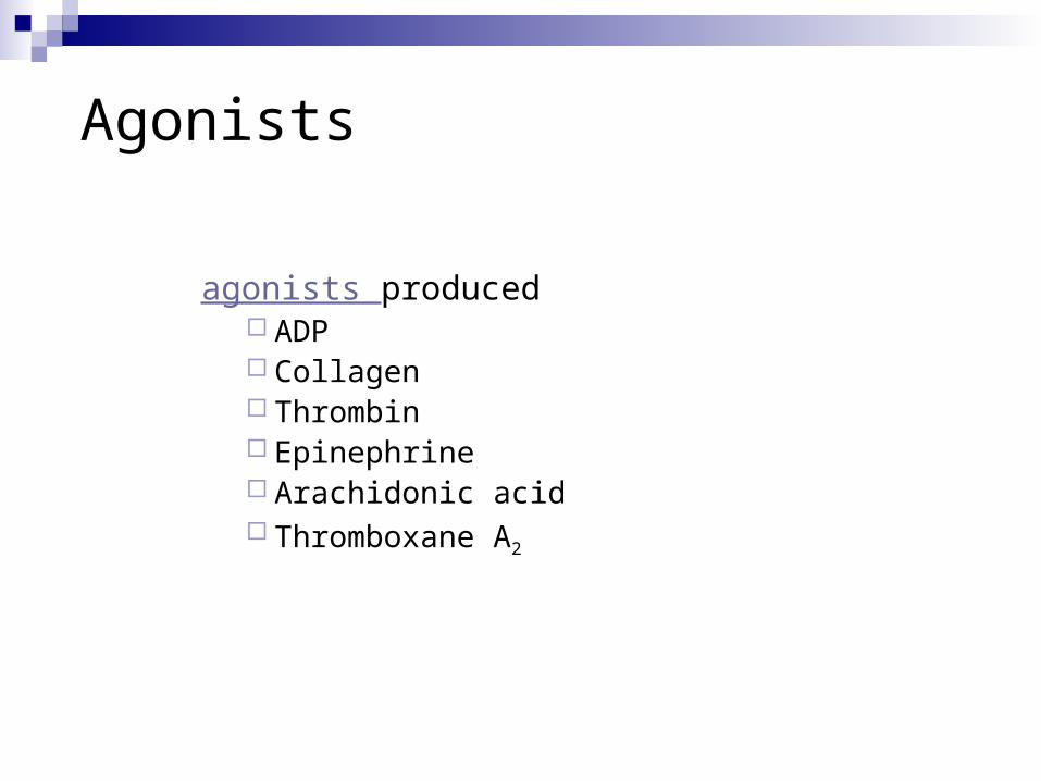
agonists produced ADP Collagen Thrombin Epinephrine Arachidonic acid Thromboxane A2
Agonists
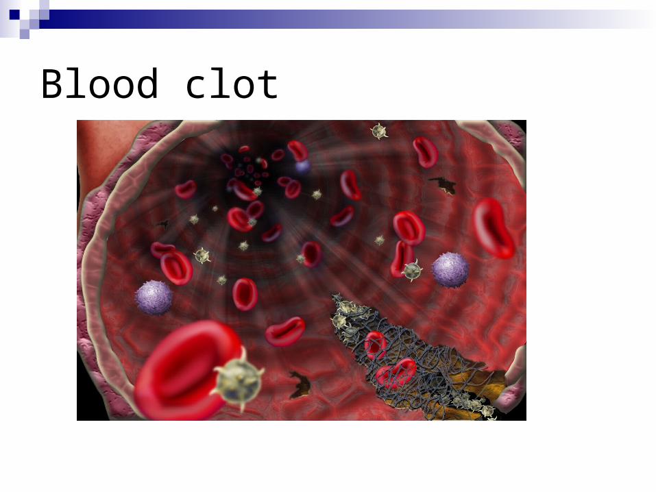
Blood clot
Coagulation System
Composed of 14 coagulation factors (serine proteases) which are interdependent (Factors I through XIII – there is no Factor VI – and PK and HMWK) Inactive form of each is an enzyme precursor which is usually designated by a Roman
numeral but also given a name – Ex. Factor I fibrinogen. Numbers correspond to order of discovery NOT order in cascade.
Active forms are usually designated by the letter “a” after the Roman numeral and may also have a different name – Ex. Ia Fibrin
Cofactors are needed for many reactions in the cascade – Ex. Calcium, platelet factor 3 (PF3)
Each molecule must be present in sufficient quantity as well as functioning normally
Final product is fibrin mesh or clot which completely stops bleeding Secondary hemostasis
Slow contraction and lysis of the clot occurs
Fibrinolytic System
Plasminogen is converted to plasmin Plasmin enzymatically attacks the fibrin molecule
producing fibrin degradation products (FDPs, sometimes called FSPs) that are cleared from the circulation by macrophages
Fibrin is a product formed during hemostasis, tissue repair or inflammation
Fibrin plays a temporary role Once injury heals, the fibrin clot is lysed
Coagulation Inhibition System
Provides balance and control of clotting mechanisms Natural inhibitors and anticoagulants circulate in the
plasma to: Prevent clotting when it’s not needed Limit or localize the clotting that is needed
Examples: Protein C and S, antithrombin III


![Hemostasis & Coagulation Disorders(Ringkas II) - Dicky [Compatibility Mode]](https://static.fdocuments.us/doc/165x107/577cc4cd1a28aba7119a7e52/hemostasis-coagulation-disordersringkas-ii-dicky-compatibility-mode.jpg)