PULMONARYCOLLAPSE IN PERTUSSIS - adc.bmj.comadc.bmj.com/content/archdischild/24/117/29.full.pdf ·...
12
PULMONARY COLLAPSE IN PERTUSSIS BY DAVID P. NICHOLSON, M.D., M.R.C.P. Late Registrar to the Paediatric Unit, the County Hospital, Farnborough, Kent; Clinical Assistant to Out-patients, Brompton Hospital, London It is recognized that whooping-cough, or as it is better termed pertussis, is a frequent cause of chronic lung disease, though the actual nature of the morbid process is not always clearly understood. Articles on bronchiectasis frequently refer to pertussis as an antecedent illness, and this is also true of many articles on catarrhal lung, chronic bronchitis, and recurrent pneumonia, occurring in children. Bronchiectasis is ascribed to pertussis in a variable percentage of cases but there is less information available as to how many cases of pertussis develop bronchiectasis. This article deals with the case histories and radiographs of forty-four children who developed pertussis and who were followed in out-patients until free from symptoms and signs or until they were lost sight of. In this way an attempt has been made to follow the respiratory complications of pertussis to resolution, or until such changes as had occurred might be considered permanent. As it is not everywhere accepted that collapse of a lobe is a common complication of pertussis, and because there is as yet no general agreement that lobar collapse is the main cause of bronchiectasis, the theoretical considerations are first reviewed. Pertussis Pertussis was first described by Guillaume de Baillou in 1578. He named the disease Quinta or Quintana to indicate the tendency for the bouts of coughing to recur at five-hourly intervals. Cruick- shank (1942) has reviewed the epidemiology of the disease and has given the overall mortality as 1*26 per cent. with a maximum mortality in the age group 0-2 years of 3 * 7 per cent. In the five-year period 1943-7 notifications for England and Wales averaged 87,652 a year, and deaths 894, or a mortality of a little over 1 per cent. In hospitals the mortality is higher; the L.C.C. hospital service recorded a mortality of 8 47 per cent. for the years 1929-32, whilst the combined Glasgow hospitals recorded a mortality of 27 per cent. for the years 1931-5. In this small series the mortality was 9* 1 per cent. In the years 1929-32 pertussis was the fourth major cause of death in London in the age group 0-5 years, and Dingman (1946) in the U.S.A. gives pertussis as the third major cause of death in the age group 0-2 years. Mortality apart, the morbidity of the disease is high though there are few figures available to demonstrate this. Oswald (1947) states that serious pulmonary complications are to be expected in not less than 10 per cent. of cases. The pulmonary collapse so often associated with pertussis has been variously ascribed to the enlarge- ment of the mediastinal glands or to the plugging of the distal small bronchi and bronchioli with viscid sputum. Engel (1947) favours enlarged glands, but Kohn et al. (1944) could find enlarged glands in Qnly five of 154 cases, and Oswald (1947) considers that enlarged glands are rare and play little part in the production of collapse associated with pertussis. Kohn et al. (1944), Feyrter (1927), Erb (1933), and Erwin (1939) have noted blocking of the terminal bronchioli by plugs of viscid sputum at autopsy, and Pospischill (1921) recorded the presence of bronchiectasis in practically all fatal cases of pertussis. Vining (1943) considers that pertussis is liable to leave behind a prolonged cough; of one hundred children with a chronic cough, ninety-two started their symptoms before the age of 5, and in thirty-five the cough dated from an attack of pertussis. Vining (1943) and Oswald (1947) think that the main cause of such symptoms is collapse caused by blocking of the terminal bronchioli by plugs of viscid sputum. The latter author found that collapse was more common in the left than the right lower lobe, that re-expansion was unlikely after six months, and that of forty cases where the lobe failed to re-expand bronchiectasis developed in thirty-eight. Ogilvie (1941) has observed that the collapse associated with pertussis may clear after the expectoration of a quantity of sputum. Lapin (1943) considers that bronchiectasis is a common complication, and that the obstruction of the bronchial tree is maximal at the bases, giving rise to cord-like and triangular shadows on the radiograph. Harries and Mitman (1947) agree, and stress the importance of the prolonged inspiration following the bout of coughing in displacing the sputum further into the lung. Hinshaw and Schmidt (1944) think that persistence of radio- graphic shadows indicates incipient bronchiectasis and that they should be watched until they dis- appear. Kohn et al. (1944) thought that the pulmonary complications of pertussis were due to consolidated ' atelectatic ' lung, and described the radiographic 29 A3 on 26 July 2018 by guest. Protected by copyright. http://adc.bmj.com/ Arch Dis Child: first published as 10.1136/adc.24.117.29 on 1 March 1949. Downloaded from
Transcript of PULMONARYCOLLAPSE IN PERTUSSIS - adc.bmj.comadc.bmj.com/content/archdischild/24/117/29.full.pdf ·...
PULMONARY COLLAPSE IN PERTUSSISBY
DAVID P. NICHOLSON, M.D., M.R.C.P.Late Registrar to the Paediatric Unit, the County Hospital, Farnborough, Kent; Clinical Assistant
to Out-patients, Brompton Hospital, London
It is recognized that whooping-cough, or as it isbetter termed pertussis, is a frequent cause ofchronic lung disease, though the actual nature ofthe morbid process is not always clearly understood.Articles on bronchiectasis frequently refer topertussis as an antecedent illness, and this is alsotrue of many articles on catarrhal lung, chronicbronchitis, and recurrent pneumonia, occurring inchildren. Bronchiectasis is ascribed to pertussis ina variable percentage of cases but there is lessinformation available as to how many cases ofpertussis develop bronchiectasis. This article dealswith the case histories and radiographs of forty-fourchildren who developed pertussis and who werefollowed in out-patients until free from symptomsand signs or until they were lost sight of. In thisway an attempt has been made to follow therespiratory complications of pertussis to resolution,or until such changes as had occurred might beconsidered permanent. As it is not everywhereaccepted that collapse of a lobe is a commoncomplication of pertussis, and because there is asyet no general agreement that lobar collapse is themain cause of bronchiectasis, the theoreticalconsiderations are first reviewed.
PertussisPertussis was first described by Guillaume de
Baillou in 1578. He named the disease Quinta orQuintana to indicate the tendency for the bouts ofcoughing to recur at five-hourly intervals. Cruick-shank (1942) has reviewed the epidemiology of thedisease and has given the overall mortality as1*26 per cent. with a maximum mortality in theage group 0-2 years of 3 * 7 per cent. In the five-yearperiod 1943-7 notifications for England and Walesaveraged 87,652 a year, and deaths 894, or a mortalityof a little over 1 per cent. In hospitals the mortalityis higher; the L.C.C. hospital service recorded amortality of 8 47 per cent. for the years 1929-32,whilst the combined Glasgow hospitals recorded amortality of 27 per cent. for the years 1931-5. Inthis small series the mortality was 9* 1 per cent.In the years 1929-32 pertussis was the fourth majorcause of death in London in the age group 0-5 years,and Dingman (1946) in the U.S.A. gives pertussisas the third major cause of death in the age group
0-2 years. Mortality apart, the morbidity of thedisease is high though there are few figures availableto demonstrate this. Oswald (1947) states thatserious pulmonary complications are to be expectedin not less than 10 per cent. of cases.The pulmonary collapse so often associated with
pertussis has been variously ascribed to the enlarge-ment of the mediastinal glands or to the pluggingof the distal small bronchi and bronchioli withviscid sputum. Engel (1947) favours enlargedglands, but Kohn et al. (1944) could find enlargedglands in Qnly five of 154 cases, and Oswald (1947)considers that enlarged glands are rare and playlittle part in the production of collapse associatedwith pertussis. Kohn et al. (1944), Feyrter (1927),Erb (1933), and Erwin (1939) have noted blockingof the terminal bronchioli by plugs of viscid sputumat autopsy, and Pospischill (1921) recorded thepresence of bronchiectasis in practically all fatalcases of pertussis. Vining (1943) considers thatpertussis is liable to leave behind a prolonged cough;of one hundred children with a chronic cough,ninety-two started their symptoms before the ageof 5, and in thirty-five the cough dated from anattack of pertussis. Vining (1943) and Oswald(1947) think that the main cause of such symptomsis collapse caused by blocking of the terminalbronchioli by plugs of viscid sputum. The latterauthor found that collapse was more common inthe left than the right lower lobe, that re-expansionwas unlikely after six months, and that of forty caseswhere the lobe failed to re-expand bronchiectasisdeveloped in thirty-eight. Ogilvie (1941) hasobserved that the collapse associated with pertussismay clear after the expectoration of a quantity ofsputum. Lapin (1943) considers that bronchiectasisis a common complication, and that the obstructionof the bronchial tree is maximal at the bases, givingrise to cord-like and triangular shadows on theradiograph. Harries and Mitman (1947) agree, andstress the importance of the prolonged inspirationfollowing the bout of coughing in displacing thesputum further into the lung. Hinshaw andSchmidt (1944) think that persistence of radio-graphic shadows indicates incipient bronchiectasisand that they should be watched until they dis-appear.Kohn et al. (1944) thought that the pulmonary
complications of pertussis were due to consolidated' atelectatic ' lung, and described the radiographic
29 A3
on 26 July 2018 by guest. Protected by copyright.
http://adc.bmj.com
/A
rch Dis C
hild: first published as 10.1136/adc.24.117.29 on 1 March 1949. D
ownloaded from

ARCHIVES OF DISEASE IN CHILDHOODappearances in 222 children. In group one, con-sisting of 154 children who had little or no fever,coarse linear basal striations were seen in forty-eight,or 31 6 per cent., and consolidated lung in thirty-eight, or 24 per cent. In this group fifty-eightchildren were under the age of two; thirty-one hadnormal and twenty-seven abnormal radiographs.Ninety-six children were over the age of two, andthe radiograph was normal in thirty-seven andabnormal in fifty-nine. The highest incidence ofx-ray abnormalities was in the age group 3-5 years.In group two, children who had a fever of up to1010 F., thirteen out of fifteen, or 86-6 per cent.,had abnormal radiographs. Group three, theremainder, consisted of children who had a feverof over 1010 F., and forty-three of fifty-three, or85 per cent., had abnormal radiographs. Re-expan-sion of collapsed areas usually occurred withinfourteen days, and the majority of the collapsedareas were thought to be middle lobes.
BronchiectasisCollapse is probably a common complication of
pertussis, and there is evidence that collapse maygiyVA to bronchiectasis, a condition that may bedefined as any pathological dilatation of the bronchiand bronchioles beyond their normal size (Warner,1935). Bronchiectasis was described in 1823 byLaennec, and in 1836 by Andral; both these authorsconmnented upon its association with whooping-cough. Since then many theories have beenpostulated to explain the development of thedisease, but until 1920 there was sufficient confusionbetween tuberculosis, fibroid lung, chronic pneu-monia, and bronchiectasis, to render such specula-tions unprofitable. It is only in the last decades thatthe frequency of collapse and bronchiectasis hasbeen recognized.
Louis in 1826- differentiated collapse fromconsolidation, and Alderson (1820), Bailey andLegendre (1846), and Gairdner (1850) commentedupon the frequency of collapse in respiratorydiseases. Heller in 1885 was perhaps the first todescribe congenital atelectatic bronchiectasis and toprovide a clear link between these two conditions.That collapse could be produced experimentally byblocking the bronchi was demonstrated by Traubein 1846, Mendelssohn in 1845, Lictheim in 1878.Tlhis has been confirmed more recently by Tannen-berg and Pinner (1942), Lee-Lander and Davidson(1938), Adams and Escudero (1938), and Weinberg(1937).The argument as to the ultimate cause of
bronchiectasis still continues. Laennec (1823)thought that the dilation was due to the pressure ofthe retained secretions, and Andral (1836) incrimin-ated the chronic cough, which in turn producednutritional changes in the bronchial wall. Neitherof these two theories bears close scrutiny today,though Adams and Escudero (1938), while discussingthe importance of bronchial obstruction, concludethat this merely serves to dam up secretions, which
produce the dilatation. They found experimentalythat incomplete and not complete bronchial obstruc-tion was associated with dilatation, a findingsupported by the work of Tannenberg and Pinner(1942), and that the increased negative intra-pleuralpressure associated with the collapse only affectedthe actual degree of dilatation and was not thebasic cause. This view is difficult to uphold, andin any case complete bronchial obstruction wouldproduce a better dam. It is unlikely that the weightof the secretions is sufficient to give rise to dilatation,and Wall and Hoyle (1932) have described cases ofdry or uninfected bronphiectasis. Raymond in. 1835suggested that the strain of the constant coughwould be sufficient of itself, and this view has beenextended by Lemon (1926) to include the strainimposed by coughing upon a wall weakened byinfection, a view supported by Thoxpe (1929) andMarcy (1937). It is now agreed that the strain ofinspiration and coughing is insufficient to causedilatation unless there is a rise in intrapleuralpressure from coincident lobar collapse. Thebronchi are subject to atmospheric pressure on-bothsides of their lumen and the only variable factor isthe intrapleural pressvre.
Stokes in 1882 considered that the dilatation wasa result of the neurom7uscular paralysis of thebronchial wall secondary to infection. Corriganin 1838 introduced the conception of traction uponthe bronchial wall by fibrous tissue, a view supportedby Findlay and Graham (1927). Warner (1935)disposes of fibrosis as a potential cause becausedilatation is often seen without fibrosis, and becausefibrosis might serve equally to compress as to dilatethe bronchi. In 1843 Rilliet and Barthez hadrecorded a case of bronchitis that developedbronchiectasis within twenty-one days, and Perryand King (1940) consider that a collapsed lobe willalways show bronchial dilatation long beforefibrosis could have arisen.The idea that hereditary influences may play a
part in the causation of bronchiectasis was intro-duced by Hoffman, and recently Engel (1947) hasgone to some lengths to produce evidence in favourof such a view. He considers- that certain bronchiare constitutionally weak and are more likely todilate. There is some evidence that vitamindeficiencies in pregnancy may lead to a maldevelop-ment of the finer bronchioles, and it would bepremature to discard the influence of pre-natalfactors altogether.Leys (1927) writing on 'Chronic Pulmonary
Catarrh,' reviews some of the literature and addsthat the prevailing views at the time were either thoseof Laennec or of Corrigan. Leys (1927) describeschronic cough occurring after pertussis, and statesthat the disease lacks a name since it lacks a knownetiology. In such cases he describes physical signsthat might well be interpreted as those of collapse,yet he disputes the claim of Davidson that thecondition is one of collapse associated with sinusinfection, and he prefers to incriminate the
30
on 26 July 2018 by guest. Protected by copyright.
http://adc.bmj.com
/A
rch Dis C
hild: first published as 10.1136/adc.24.117.29 on 1 March 1949. D
ownloaded from

PULMONARY COLLAPSE IN PERTUSSISH. influenzae of Pfeiffer as the causal agent, a viewwhich he has almost certainly abandoned since.At the present time opinions are divided as to the
relative importance of collapse and infection incausing bronchiectasis. McNeil et al. (1929) havedescribed a process of acute interstitial pneumoniaof the bronchi that leads to necrosis and excavationfollowed by relining with epithelium and dilatation.Hedblom (1931) thinks that infection weakens thewall and that then mechanical factors dilate it.The infection precedes the collapse, which is themost important mechanical factor. Moll (1932)reviews the whole subject and decides in,favour ofinfection being the causal agent. Infection destroysthe wall which is then permanently dilated, thoughhe does suggest that the first stage is one of loss oftone that may be temporary. Bronchial obstructionfavours the infection. He describes fifty-fiveautopsies and thirty-seven patients, and as he oftennoted more extensive disease at autopsy than wasevident during life he concludes that the diseaseis capable of spread. The infective theory is upheldby Erb (1933), Boyd (1931), and Robinson (1933).This last author examined sixteen lobectomyspecimens and found that collapse played no partin the production of -the dilatation, but that itfavoured infection. Other writers, whilst agreeingthat infection is the basic cause, consider that thecollapse and the increase in negative intra-pleuralpressure actually cause the dilatation (Vining,, 1943;Hinshaw and Schmidt, 1944; Warner, 1935 and1934; Wearing, 1948). In their comprehensivemonograph on bronchiectasis Lisa and Rosenblatt(1943) find that the one factor common to all thediseases that cause bronchiectasis is infection, andthat bronchial obstruction whether partial orcomplete is not the basic cause.
Gairdner in 1850 had suggested that obstructionof the bronchi by sputum might produce collapse.That this is possible has been confirmed in recentyears by Kohn et al. (1944), Feyrter (1927), Erwin(1939), Ogilvie (1941), Jackson (1925, 1927, 1930),Archibald and Brown (1928), Lloyd (1931), Walland Hoyle (1932), Anspach (1943), Warner (1934),and Lee-Lander and Davidson (1938). Oswald(1947) stresses the importance of this type ofcollapse and also the possibility of the sputum beingsucked further in during inspiration. That this isa frequent cause of collapse is now realized, andconcurrently the effects of bronchial obstructionhave been more accurately defined. Completeobstruction of a main bronchus leads to distalcollapse, since the gas tension in venous blood is54 mm. Hg. less than the alveolar gas tension, andthe air distal to the block is slowly absorbed. Inthis type of obstruction there is collapse but nodilatation. Incomplete obstruction of a bronchuscauses no collapse, but if the obstruction is ballvalve then, depending on whether it is inlet or outletin type, so emphysema or collapse will develop.Again there is usually no dilatation. If the obstruc-tion is distal and many small bronchi and bronchioli
are blocked, then the total anatomical effect is thesame as in main bronchial obstruction, and the lobecollapses partly or totally. As a result of thecollapse the intrapleural negative pressure increasesand the bronchi proximal to the block are subjectedto this increased force which, as the bronchi arenow open to the atmosphere, dilates them. Theweaker the walls from coincident infection, the morelikely are they to dilate. In massive collapse theintrapleural negative pressure may reach-423 cm.ofwater (Jennings, 1937). It is as well to rememberthat the bronchi are incapable of dilating unlessthey are open to the atmosphere, and this accountsfor the experimental findings previously noted thatincomplete and not complete main bronchialobstruction is associated with dilatation. Theoretic-ally incomplete main bronchial obstruction is notassociated with dilatation, but there is always thepossibility that particles ofthe obstructing agent maysplit off, be inspired deeper into the lung, and causemultiple distal bronchial obstructions. It is interest-ing to observe that in lipiodol bronchograms ofdilated areas alveolar filling is not seen, presumablybecause the terminal bronchi are always obstructed.This leads us to a consideration of the remaining
theory of origin of bronchiectasis. Collapse iscommon and it is often caused by plugging of thesmall bronchi with sputum. Lobar collapse resultsin an increase of the negative intrapleural pressurethat is capable of causing-bronchial dilatation. Infact a collapsed lobe probably always containsbronchial dilatations. If the collapsed area shouldre-expand before irreparable damage has been doneto the bronchial wall by infection, then the dilatationmay disappear; such a sequence of events wouldaccount for the cases of reversible or ' pseudo- 'bronchiectasis. If the collapse is maintained, so isthe dilatation; and with chronic infection super-imposed the dilatation becomes permanent to suchan extent that it does not disappear if the collapsedarea should now re-expand. Oswald (1947) statesthat re-expansion of a collapsed lobe is unlikelyafter six months. Anspach (1933) observed fortychildren with bronchiectasis all of whom hadtriangular basal shadows on the radiographs, and hethought that the collapse was the main cause of thedilatation. Wall and Hoyle (1932) described casesof dry bronchiectasis which they thought were dueto the dilating force of inspiration when the smallbronchi were blocked. Anspach (1934) states thatthe longer the duration of the collapse the morelikely is bronchiectasis to develop. Ellis (1933)describes the sequence as collapse then dilatation,and Miller (1934) quotes Schneider (1927) as sayingthat bronchiectasis is preceded by a purely dilatedstate in which there is a simple atrophy of the wall.King (1937) observed dilatation in a lobe five daysafter operation -and discards collapse as a causesince the dilatation occurred too quickly, anoccurrence that is not impossible if collapse is thecause of the dilatation. Lee-Lander and Davidson(1938), by blocking the distal bronchi, were able to
31
on 26 July 2018 by guest. Protected by copyright.
http://adc.bmj.com
/A
rch Dis C
hild: first published as 10.1136/adc.24.117.29 on 1 March 1949. D
ownloaded from

ARCHIVES OF DISEASE IN CHILDHOODproduce bronchiectasis in a cat. The degree ofdilatation was less if an artificial pneumothoraxwas subsequently induced. In twenty patients withbronchiectasis they noted that the calibre of thebronchi varied more on inspiration and expirationthan did that of a normal bronchus. Greenfield(1940) did not agree with this last finding, whichLee-Lander and Davidson had given as a point infavour of the bronchial wall being intact but dilated.Erwin (1939) supports the collapse theory andagrees that in the early stages the condition may4bereversible. Andrus (1940) regards any chronic basalpneumonia as suspicious of bronchiectasis, as areincreased basal striations if vascular congestion beexcluded. Displacement of organs toward thediseased area occurred in 90 per cent. of cases andconfirmed the association with collapse; majoratelectasis, if chronic, is virtually diagnostic ofbronchiectasis. He continues by saying thatpneumonia regularly undergoes resolution so longas there is no collapse; if there is collapse thendilatation results from the enhanced pull of the lung.Oswald (1947) and Wearing (1948) agree, and Korn-blum (1944), whilst adhering to the infective theoryof origin, states that should it be demonstrated thatthe disease is reversible then our whole concept ofthe disease must be altered, presumably in favourof collapse as the main etiological agent. Wearing(1948) reports such an instance of reversiblebronchiectasis but complains that the literaturecontains few references.
Reversible bronchiectasis. If bronchiectasis isreversible up to a certain stage in the disease process,.then it is reasonable to assume that the bronchial
wall has not been permanently damaged by infectionand that the muscle and elastic layers are inta'ct.Lee-Lander and Davidson (1938) have given reasonsfor believing that in fact the wall is intact. In someinstances a collapsed area is seen to re-expand andstill leave dilatation: this would be possible if,subsequent to the dilatation, infection had ensuedand irreparably damaged the wall. Dilatation maybe observed in a collapsed lobe or segment at anearly date, before it would' be possible for infectionto have destroyed the wall. If infection followedby destruction is the cause of the dilatation then thecondition must surely be irreversible. There areseveral references in the literature to reversible or' pseudo ' bronchiectasis (Erwin, 1939; Ogilvie,1941; Moll, 1932; Robinson, 1933; Greenfield,1940; Andrus, 1940; Kornblum, 1944). Lisa andRosenblatt (1943) and Hinshaw and Schmidt (1944)regard such reversible dilatation as an illusioncreated by the temporary crowding and fore-shortening of the bronchi in a collapsed lobe. Sucha criticism eradicates bronchiectasis as a diseaseentity and can hardly be taken seriously. Oschner(1929) mentions four cases of reversible bronchi-ectasis but gives no details. Wearing (1948)describes one case and asks, ' How long does it takereversible bronchiectasis to become irreversible ?'Oswald (1947) suggests that the time is six months:Several examples of reversible bronchiectasis havebeen gleaned from the literature, and a summary ofthese appears in table 1. Blades and Dugan'(1944)discuss four other cases but give no details exceptthat the bronchi returned to normal in three months,and they suggest that this may still happen after
TABLE 1
SURVEY OF CASES OF REVERSIBLE BRONCHIECTASIS OCCURRING IN THE LITERATURE
Year Author Lobe involved Duration Cause
1927 Findlay and Graham 1. Rt. L.L. Less than 5 yrs. Pertussis2. Rt. L.L. Less than 3 yrs. Pertussis3. Lt. L.L. Less than 3 yrs. Bronchopneumonia
1933 Warner and Graham 1. Rt. L.L. Persistent, but variedwith the degree ofcollapse
1935 Fletcher and Dimson 1. Lt. L.L. Less than 5 months Measles and pneumonia1937 Jennings 1. Lt. L.L. 1 month1935 Findlay 1. Rt. M. and L.L. 7-15 months Measles1938 Raia 1. Rt. L.L. 4 months
2. Rt. L.L. 6 months3. Rt. M. and Lt. L.L. Less than 3 yrs.
1939 Wilson 1. Rt. L.L. Less than 7 months1941 Fleischner 1. Lt. M. and L.L. Indefinite1942 Diamond and VanLoon 1. Rt. L.L. 2 months
2. Lt. M. and L.L. andRt. L.L. 2 months
1943 Campbell et al. 1. Rt. L.L. 3 months Atypical pneumonia1944 Blades and Dugan 1. Lt. L.L. 9-18 weeks1946 Lee-Lander 1. Rt. L.L. 6-7 months
2. Lt. L.L. 6-7 weeks3. Rt. M. and Lt. L.L. 2-15 months
1948 Wearing 1. Lt. L.L. 4-12 months Pneumonia
32
on 26 July 2018 by guest. Protected by copyright.
http://adc.bmj.com
/A
rch Dis C
hild: first published as 10.1136/adc.24.117.29 on 1 March 1949. D
ownloaded from

PULMONARY COLLAPSE IN PERTUSSIS
FIG. 1. FIG. 2.
FIG. 1.-Case 6: radiograph taken on Feb. 5, 1948,of a boy aged five years. The illness was inthe acute stage and there was bilateral lowerand right middle lobe collapse.
FIG. 2.-The same case on March 19, 1948, showingcollapse of the right middle lobe.
FIG. 3.-The same case: a bronchogram taken onApril 1, 1948, showing collapse and dilatationof the right middle lobe.
FIG. 3.
33
on 26 July 2018 by guest. Protected by copyright.
http://adc.bmj.com
/A
rch Dis C
hild: first published as 10.1136/adc.24.117.29 on 1 March 1949. D
ownloaded from
ARCHIVES OF DISEASE IN CHILDHOOD
FIG. 4
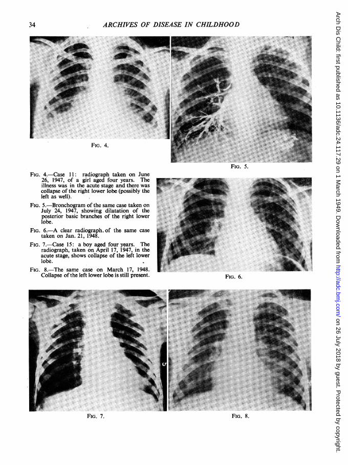
FIG. 5.FIG. 4.-Case 11: radiograph taken on June
26, 1947, of a girl aged four years. Theillness was in the acute stage and there wascollapse of the right lower lobe (possibly theleft as well).
FIG. 5.-Bronchogram of the same case taken onJuly 24, 1947, showing dilatation of theposterior basic branches of the right lowerlobe.
FIG. 6.-A clear radiograph. of the same casetaken on Jan. 21, 1948.
FIG. 7.-Case 15: a boy aged four years. Theradiograph, taken on April 17, 1947, in theacute stage, shows collapse of the left lowerlobe.
FIG. 8.-The same case on March 17, 1948.Collapse of the left lower lobe is still present.
FIG. 8.
34
FIG. 6.
FIG. 7.
on 26 July 2018 by guest. Protected by copyright.
http://adc.bmj.com
/A
rch Dis C
hild: first published as 10.1136/adc.24.117.29 on 1 March 1949. D
ownloaded from
PULMONARY COLLAPSE IN PERTUSSIS
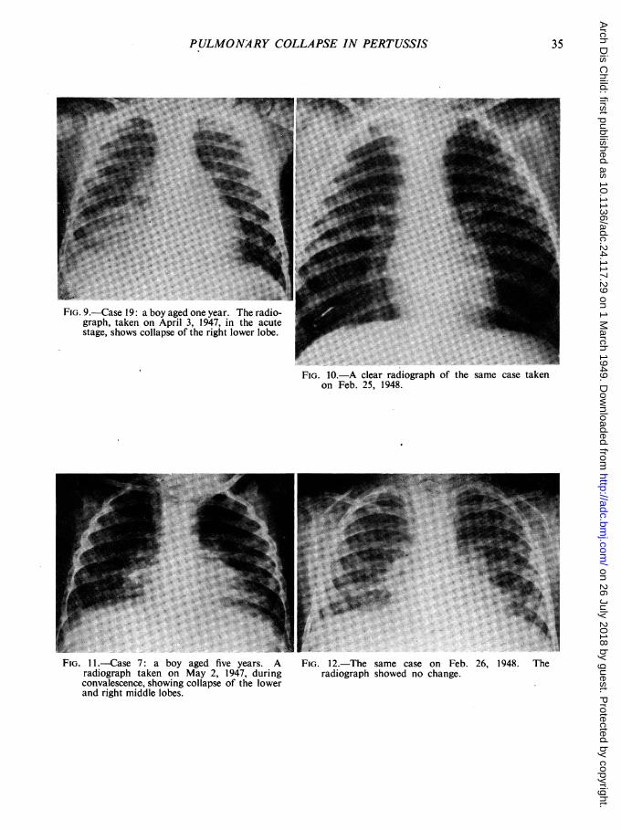
FIG. 9.-Case 19: a boy aged one year. The radio-graph, taken on April 3, 1947, in the acutestage, shows collapse of the right lower lobe.
FIG. 10.-A clear radiograph of the same case takenon Feb. 25, 1948.
FIG. 11.-Case 7: a boy aged five years. A FIG. 12.-The same case on Feb. 26, 1948. Theradiograph taken on May 2, 1947, during radiograph showed no change.convalescence, showing collapse of the lowerand right middle lobes.
35
on 26 July 2018 by guest. Protected by copyright.
http://adc.bmj.com
/A
rch Dis C
hild: first published as 10.1136/adc.24.117.29 on 1 March 1949. D
ownloaded from

ARCHIVES OF DISEASE IN CHILDHOODthree months. In these examples, where a secondbronchogram was done reasonably soon after thefirst, the dilatation had lasted roughly six monthsor less. This suggests that if the collapse is morepermanent the dilatation becomes irreversible.
If collapse is the cause of bronchiectasis thenspread of the process should not arise unless anintercurrent disease occurs that is capable ofinitiating further collapse. Churchill and Belsey(1939) and Perry and King (1940) consider thatspread is unusual unless this should occur.
Clinical observation of cases of postoperativemassive collapse lends further support to the viewthat viscid sputum may lead to blocking of thebronchi and collapse (Pasteur, 1908, 1914; Brad-ford, 1918; Briscoe, 1914, 1919; Sante, 1927;Findlay, 1932; Scott, 1925; Elliot and Dingley,1914; Corrylos and Birnbaum, 1928). XAnatomy. A detailed knowledge of the anatomy
of the bronchi is important if the full effects ofbronchial obstruction are to be understood. Theanatomy of the bronchi has been described byEwart (1889), Kramer and Glass (1932), Neil et al.(1937a and bI), Nelson (1934), and more recently byBrock (1942-3-4) and Foster-Carter (1942). Thesize of the bronchi has been estimated by Engel(1947) and Miller (1947). The diameter of thebronchi at birth is given as 5 - 7 mm. in the coronal,and 6 0 mm. in the sagittal plane. By adult lifethese measurements are 16-5 mm., and 14 4 mm.respectively. The terminal bronchioles measure0 12 mm. at three months and 0 20 mm. in diameterby adult life. Kohn and others (1944) found thatin pertussis respiratory complications were mostfrequent in the age group 3-5 years. If size alonewere the only consideration then it would be expectedthat the bronchi of infants would be blocked morefrequently. This does not appear to be so, and itmay be that the bronchial glands, which areimmature at birth, are incapable of secretingsufficient mucus until early childhood. In childhoodthe goblet cells are well developed but they are neverpresent in bronchi with a diameter of less than044mm.
Radiography. Ewart in 1889 introduced the ideaof constant broncho-pulmonary segments, areascomposed of a framework of small bronchi whosetrunk is an offshoot of a lobar bronchus. Thisconception has been amplified by Kramer and Glass(1932) and Foster-Carter and Hoyle (1945). Collapseof a broncho-pulmonary segment throws a shadowon the radiograph that is relativelyconstant, and withthe aid of lateral views the area may be defined.Interpretation of radiographs plays an importantpart in the control and assessment of cases ofpertussis. It has been assumed that the triangularbasal shadows so frequently seen on radiographyrepresent collapsed lower lobes. In the past theseshadows have been variously interpreted as indicat-ing pneumonia, mediastinal pleurisy, or fibrosis.In 1926 Singer and Graham noted that these
shadows were often associated with bronchiectasis,and at operation they found that the lower lobeswere collapsed. This has been confirmed by Warner(1935), Warner and Graham (1933), Ogilvie (1941),Riggins (1941), Findlay (1935), Anspach (1934),and Andrus (1940), and it has been observed thatin these cases the distal small bronchi are oftenplugged with sputum (Anspach, 1939; Warner andGraham, 1933). Triangular basal shadows werenoted in 6 per cent. of cases of bronchiectasis byWarner and Graham (1933), in twenty-seven ofsixty-eight cases by Ogilvie (1941), and in 17 percent. of cases by Joress and Robins (1944). If thecollapse is lobar then compensatory emphysema ofthe adjacent healthy lung, and mediastinal shift,occur.
Relation of Pertussis to BronchiectasisArticles on bronchiectasis often refer to pertussis
as an antecedent and causal illness. Table 2 givesa list of such references with the percentage incidencegiven, pertussis being an antecedent illness in anaverage of 14 per cent. of cases.
TABLE 2-ARTICLES ON BRONCHIECTASIS REFERRINGTO PERT'USSIS AS AN ANTECEDENT ILLNESS
Authors
Warner (1935)Diamond andvan Loon (1942)Thorpe (1929)Raia (1938)Perry and King (1940)Oswald (1947)Ogilvie (1941)Lemon (1926)Moll (1932)Joress and Robins (1944)Hedblom (1931)Graham et al. (1935)Findlay and Graham (1927)Boyd (1931)
Average
Percent. ofcasesaffected
48
26219148
46989
11
99
14I~~~~~~~~
In this discussion it has become evident thatcollapse is a common complication of respiratorydiseases, and that it is almost certainly the forerunnerif not the actual cause of bronchial dilatation.
In uncomplicated pertussis with few or no signs ofsecondary respiratory infection the radiograph isoften normal and enlargement of the mediastinalglands is not seen. If such glandular enlargementis seen it is highly suspicious of a coincident primarytuberculous infection. If there are signs andsymptoms of secondary infection of the respiratorytract, then abnormal x-ray shadows becomeincreasingly frequent. The abnormal shadow isoften typical of lobar or segmental collapse.Clinically the physical signs usually support such a
36
on 26 July 2018 by guest. Protected by copyright.
http://adc.bmj.com
/A
rch Dis C
hild: first published as 10.1136/adc.24.117.29 on 1 March 1949. D
ownloaded from

PULMONARY COLLAPSE IN PERTUSSISdiagnosis, and as a rule neither the signs nor theradiographic appearances respond to chemotherapyin the manner of a classical- lobar or broncho-pneumonia, a fact borne out by the investigation ofKohn, Rubin, and Hobart (1940). Pyrexia andcough tend to continue until the radiograph is clear,and it is probable that the majority of respiratorycomplications in pertussis are due to lobar orsegmental collapse. Collapse will be more likelyif there is secondary infection and excess secretionof mucus, particles of which tend to be inspireddeeper into the lung and to block the small bronchiand bronchioli. The collapse is due to the blockingof the terminal bronchi, and this was evident inthree of the autopsies in this series. The alveolibecome airless, the lobe diminishes in size, thenegative intrapleural pressure increases, and thebronchi proximal to the block undergo compensatorydilatation; this is reversible in the first instance butbecomes permanent if the area fails to re-expand orif infection supervenes. If the collapsed area shouldre-expand then the dilatation may disappear. Aftersix months or so this becomes increasingly unlikely,and such a possibility probably depends upon thedegree of infection present.
Present InvestigationThe present series consists of forty-four children
suffering from pertussis who were in most instancessufficiently ill to warrant admission to hospital.There were twenty-three girls and twenty-one boys.The duration of symptoms before admission wasless than one week in thirteen, or 29 5 per cent.,and more than one week in thirty-one, or 70 5 percent. The age incidence is shown jn table 3. This
TABLE 3AGE INCIDENCE IN FORTY-FOUR CHILDRENWITH PERTUSSIS ADMITTED TO HOSPITAL
0-6 6-12 1-2 2-3 3-4 4-5 5-8mths. mths. yrs. -yrs. yrs. yrs. yrs.
No. 8 4 5 9 5 7 6deaths I 0 0 1 2 0 0
gives an overall mortality of 9-1 per cent. During-their stay in hospital, and subsequently as out-patients these children were examined frequentlyboth clinically and by radiograph. Five children,or 11 * 4 per cent. (three boys and two girls) revealedno x-ray abnormality at any time. The rest hadat some time abnormal signs both physically andradiographically. It is, ofcourse, possible that sometransient radiographic changes occurred unnoticed.The shadows seen on the radiographs were usually
typical of lobar or segmental collapse, mainly of themiddle and lower lobes. Evidence in favour of theshadows representing collapsed lobe was as follows:(1) physical signs of collapse and lack of responseto chemotherapy; (2) association of mediastinal
shift and compensatory emphysema; (3) thepresence in some radiographs of visibly dilatedbronchi in the shadow; (4) the similarity of theshadows with the triangular basal shadows pre-viously discussed.As far as possible the children were divided into
three groups. Group one was composed of mildcases with few or no signs of respiratory infectionand little or no pyrexia. Group two, cases ofmoderate severity with signs of secondary infectionand pyrexia of up to 1010 F. Group three, severewith marked signs of secondary infection andpyrexia of over 1010 F., and a prolonged course torecovery. Four children died, and all were classifiedas severe cases. Case 13, a girl aged two, showedcollapse of both lower and the right middle lobeson radiography, and this was confirmed at autopsywhen the distal small bronchi were seen to be blockedby plugs of sputum. A similar state of affairs wasseen in a girl aged three, Case 23, who in life showedbilateral lower-lobe collapse, which was confirmedat autopsy, and in Case 26, a boy aged seven weeks,who had shown bilateral lower-lobe collapse as well.Case 31, a girl aged three, died soon after admissionand autopsy revealed an extensive bilateral broncho-pneumonia. Four cases were mild, sixteen, or36 * 4 per cent. were classified as moderate, andtwenty-four, or 54 5 per cent., as severe (table 4).
TABLE 4DEGREE OF SEVERITY IN THE PRESENT SERIES
Age No. Mild Moderate Severe Deaths
0-6 mths. 8 1 4 3 16-12 ,, 4 0 3 1 01-2 yrs. 5 0 1 4 02-3 ,, 9 2 2 5 13-4 ,, 5 0 2 3 24-5 ,, 7 1 3 3 05-8 ,, 6 0 1 5 0
Total 44 4 1 16 24 4
There was no evidence of collapse in one out of fourmild cases, three out of sixteen moderate cases, andone out of twenty-four severe cases; on the otherhand collapse of over two months' duration wasseen in one out of four mild cases, nine out ofsixteen moderate cases, and thirteen out ofseventeensevere cases, that were followed for a sufficientlength of time. The collapse was bilateral ineighteen: the right upper lobe was involved once,the right middle lobe ten times, the right lower lobethirty-five times, the left lingula five times, and theleft lower lobe nineteen times. This preponderanceof right- over left-sided collapse-is unusual, thoughit will be appreciated that left lower-lobe collapseis more difficult to diagnose. Oswald (1947) notedleft lower-lobe collapse twenty-eight times toeighteen times on the right, Diamond and Van Loon(1942)an incidence of fifteen to twelve, andCampbell
37
on 26 July 2018 by guest. Protected by copyright.
http://adc.bmj.com
/A
rch Dis C
hild: first published as 10.1136/adc.24.117.29 on 1 March 1949. D
ownloaded from
ARCHIVES OF DISEASE IN CHILDHOODet al. (1943) an equal incidence in seventy-eightcases. The reasons offered for the greater frequencyof left-sided collapse are that the left main bronchusarises at a more acute angle and that it is in intimaterelationship with the arch of the aorta and thepulmonary artery. From the foregoing theoreticalconsiderations it would be expected that collapsewould have an equal incidence on both sides, andit is difficult to explain this apparent anomaly.
TABLE 5INCIDENCE OF COLLAPSE IN PRESENT SERIES
Collapse of overAge two months No collapse
0- 6 mths. 2 out of 7 2 out of 86-12 ,, 2 out of 4 2 out of 41-2 yrs. 2outof3 Ooutof52-3 ,, 4 out of 8 1 out of 93-4 ,, 2 out of 3 Oout of 54-5 ,, 7outof7 Ooutof75-8 ,, 4 out of 6 out of 6
Collapse of a serious nature seems to be morefrequent in childhood than in infancy, a factcorroborated by Kohn et al. (1944). In allcollapse occurred in thirty-nine out of forty-fourcases, a high incidence (table 5).
Lee-Lander (1936) says that in the collapsefollowing the infectious fevers no prognosis can begiven since our knowledge of the condition 'isinsufficient. The duration of the collapse varies;the more severe examples tend to last longer. Ofthe cases that were followed until clear or for longerthan a year, thirty-five of forty-eight collapsed lobeshad re-expanded within nine months; thereafter nofurther re-expansion was seen during the time ofobservation. It is probable that no furtherre-expansion will occur after nine months; thecollapse is now permanent and dilatation estab-lished. Thirty-three children were followed formore than one year, and seven still had a collapsedlobe. If bronchiectasis is a possible end result in21 per cent. of cases of severe pertussis then treat-ment must be started early. Only three out offourteen cases admitted in the first -week of thedisease developed serious pulmonary complications,whereas nineteen out of twenty-seven cases admittedafter the first week developed such complications.
Treatment. The early diagnosis of pertussis isdifficult, and in any case there is no specific treat-ment. Aerosporin, a new drug, is obtained fromthe B. Aerosporus, and it will inhibit H. Pertussisin a minimal dilution of 0 * 04 ,ug per ml. (Ainsworthand others, 1947; Brownlee and Bushby, 1948).In some cases in this series Swift (1948) gave0 8 mg. per kg. four-hourly, and from his serieshe concluded that a response was usual withintwenty-four hours, but that the ultimate benefitseemed to depend on the duration of symptomsbefore the start of treatment rather than upon the
severity of the disease itself. If aerosporin is givenbefore secondary infection has caused collapse thenthe pulmonary complications might be averted,though in this series the small number of cases whohad aerosporin differed in no way from the others.Excluding specific treatment, sulphonamides and/orpenicillin should be given to avert or reducesecondary infection, and such measures may preventfurther collapse. In a few cases inhalations ofpenicillin were tried, using either a ' Deedon ' or'Aerosol ' inhaler and a mixture of carbon dioxideand oxygen. Results were unsatisfactory as childrendo not tolerate a mask or tent well, but further trialswould be justified.
If collapse is still present after the acute stage isover further treatment is imperative, and should bedirected at the collapsed lobe. Postural drainage inthe appropriate position should be given (Nelson,1934), and this should be combined with percussionover the affected lobe in an attempt to induce thechild to cough up the particles of sputum. Treat-ment in this manner can be given several times a dayand must be given until all hope of re-expansion isabandoned, after six or nine months. It may evenbe continued at home under the guidance of theparents.
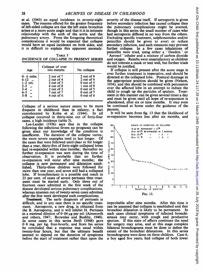
It will be seen from fig. 13 that the likelihood ofre-expansion becomes less after six months, and
45.
40-
035.
300
250
, 200
15-
z 10O
5.
GRAPH OF DURATION OF COLLAPSE13 of 48 PERMANENT = 27%27or 563% RE-EXPANDED IN 3 MONTHS
x29or 58 3%% 6 **
" 35or 73 % ,, 9
XX~~~~~~~
x_Kx--x--x- -x
2 3 4 5 6 7 8 9 10 it 12'DURATION IN MONTHS
FIG. 13.
improbable after nine months. After this time itcan be assumed that collapse is established and thatbronchial dilatation is likely to be permanent. Insuch cases clinical symptoms of infected bronchi-ectasis may occur, with cough and productivesputum. If this state of affairs continues the needfor surgery may arise, and at this stage completebilateral bronchograms must be done to define theextent of the bronchial dilatations. In this seriesbronchograms were done on three children. Case 6,a boy aged five years, had collapse of both lower
0 s , . . . . I
38
c.o_
on 26 July 2018 by guest. Protected by copyright.
http://adc.bmj.com
/A
rch Dis C
hild: first published as 10.1136/adc.24.117.29 on 1 March 1949. D
ownloaded from

PULMONARY -COLLAPSE IN PERTUSSIS 39
lobes and the right middle lobe. After ten weeksthe middle lobe was still collapsed and a broncho-gram showed dilatation and crowding of the middlelobe branches (fig. 3). - Case 11, a girl aged fouryears, had a collapsed right lower lobe; after threeweeks this had not completely re-expanded and abronchogram revealed dilatation of the posteriorbasic branches (fig. 5). Subsequently the lobere-expanded completely. Case 36, a girl aged fouryears, had bilateral basal collapse that cleared ineight weeks; a subsequent bronchogram wasnormal.
Following an attack of pertussis a child maycontinue to suffer from cough, which may beparoxysmal, and attacks ofbronchitis. The examina-tion and radiograph of these children usually revealsa collapsed lobe. If treatment is inadequate andfails to produce re-expansion, the cough continuesand eventually may merge with the symptoms ofbronchiectasis. Many of these cases still gounrecognized, and all too often the treatmentconsists of an ineffective cough mixture.
-SummaryThe literature relating to pertussis and bronchi-
ectasis has been reviewed. It has been concludedthat pulmonary collapse is a common feature ofpertussis, and that if re-expansion does not occurwithin nine months it is unlikely to do so, andbronchiectasis may supervene. Treatment of thesecases must be prolonged and efficient. Post-pertussis pulmonary collapse is often still unrecog-nized.My thanks are due to Dr. F. H. Hackwood,
Surgeon Superintendent, for permission to publishthis article, and to Dr. D. Leys and Dr. P. N. Swiftfor their advice and help.
REFERENCES
Adams, W. E., and Escudero, L. (1938). Tuberck, 19,351.
Ainsworth, G. C., Brown, A. M., and Brownlee, G.(1947). Nature, 160, 263.
Alderson, J. (1820). Medico-Chirurgical Trans., 16, 78.Andral (1836). Clinique Medicale. Trans. D. Spillane,
London.Andrus, P. M. (1940). Amer. Rev. Tuberc., 41, 87, 99,
104.Anspach, W. E. (1934). Amer. J. Dis. Child., 47, 1011.
(1939). Amer. J. Roentgen., 41, 173.(1943). Dis. Chest, 9, 24.
Archibald, E., and Brown, A. L. (1928). Arch. Surg.,16, 322.
Bailey, P, and Legendre, F. L. (1846). Obstet. Soc.,Paris.
Blades, B., and Dugan, D. J. (1944). J. Thorac. Surg.,13, 40.
Boyd, G. L. (1931). Canad. med. Ass. J., 25, 174.Bradford, J. R. (1918). Quart. J. Med., 12, 127.
(1914). Lancet, 1, 1502.Briscoe, J. C. (1919). Quart. J. Med., 13, 293.
(1914). Lancet, 1, 1502.
Brock, R. C.-(1942). Guy's Hosp. Rep., 91, 111.(1943). Ibid., 92, 26; 92, 123.(1944). Ibid., 93, 90.
Brown, A. L. (1931). Arch. Surg., 22, 976.Brownlee, G., and Bushby, S. R. M. (1948). Lancet,
1, 127.Campbell, T. A., Strong, P. S., Grier, G. S., and Lutz,
R. J. (1943). J. Amer. med. Ass., 122, 723.Churchill, E. D., and Belsey, R. (1939). Ann. Surg.,
109, 481.Corrigan, D. J. (1838). Dublin. J. med. Sci., 13, 266.Corrylos, P. N., and Bimbaum, G. L. (1928). Arch.
Surg., 16, 501.Cruickshank, R. (1942). Control of Common Fevers.
Lancet. London.Dingman, H. (1946). Risk Appraisal. National Under-
writer Corporation. Cincinnati.Diamond, S., and van Loon, E. L. (1942). J. Amer. med.
Ass., 118, 771.Elliot, T. R., and Dingley, L. A. (1914). Lancet, 1,
1305.Ellis, R. W. B. (1933). Arch. Dis. Childh., 8, 35.Engel, S. (1947). The Child's Lungs. 'First Edit.
London.'Erb, I. H. (1933). Arch. Path., 15, 357.Erwin, G. S. (1939). Brompton Hos. Rep., 8, 43.Ewart, W. (1889). The Bronchi and Pulmonary Blood
Vessels. London.Findlay, L. (1935). Arch. Dis. Childh., 10, 61.
(1932). Proc. R. Soc. Med., 25, 407.and Graham, S. (1927). Arch. Dis. Childh., 2, 71.
- , - (1931). Ibid., 6, 1.Feyrter, F. (1927). Frankfurt. Z. Path., 35, 213.Fleischner, F. G.. (1941). Amer. J. Roentgen., 46, 166.Fletcher, E., and Dimson, S. B. (1935). Lancet, 1, 987.Foster-Carter, A. F. (1942). Brit. J. Tuberc., 36, 19.- , and Hoyle, C. (1945). Dis. Chest., 11, 511.Gairdner, W. T. (1850). Monthly J. med. Sci., 11, 135.Graham, E. A., Singer, J. J., and Ballon, H. C. (1935).
Surgical Diseases of the Chest. London.Greenfield, J. (1940). J. clin. Invest., 19, 723.Harries, E. H. R., and Mitman, M. (1947). Clinical
Practice in Infectious Diseases. Third Edit.Livingstone. Edinburgh.
Hedblom, C. A. (1931). Surg. Gynec. Obstet., 52, 406.Heller, A. (1885). Dtch. Arch. Klin. Med., 36- 189.Hinshaw, H. C., and Schmidt, H. W. (1944). Dis.
,Chest, 10, 115.Holinger, P., and Andrews, A. H. (1941). Amer. J.
Surg., 54, 193.Jackson, C. (1925). Ann. Surg., 72, 364.- (1927). Bronchoscopy and Oestphagoscopy.
Second Edit., p. 301. Philadelphia.(1930). J. Amer. med. Ass., 95, 639.
Jennings, G. H. (1937). Brit. med. J., 2, 963.Joress, M. H., and Robins, S. A. (1944). Dis. Chest,
10, 489.King, D. S. (1937). Internat. Clin., 1, 130.Kohn, J. L., Schwartz, I., Greenbaum, J., and Daly,
M. M. I. (1944). Amer. J. Dis. Child., 67, 463.Rubin, H. T., and Hobart, H. M. (1940). Arch.Paediat., 57, 410.
Kornblum, K. (1944). Amer; J. Roentgen., 51, 292.Kramer, R., and Glass, A. (1932). Ann. Otol. Laryng.
etc. St. Louis, 41, 1210.Laennec, R. T. H. Treatise on Diseases of the Chest
and on Mediate Auscultation. 1823. London.Lapin, J. H. (1943). Whooping Cough. C. C. Thomas.
Springfield, Illinois.
on 26 July 2018 by guest. Protected by copyright.
http://adc.bmj.com
/A
rch Dis C
hild: first published as 10.1136/adc.24.117.29 on 1 March 1949. D
ownloaded from

40 ARCHIVES OF DISEASE IN CHILDHOODLee Lander, F. P., and Davidson, M. (1938). Brit. J.
Radiol., 11, 65.(1946). Thorax, 1, 198.(1936). Proc. R. Soc. Med., 29, 1383.
Lemon, W. S. (1926). Med. Clin. N. Amer., 10, 531.Leys, D. G. (1927). Chronic Pulmonary Catarrh.
H. K. Lewis. London.Lictheim, L. (1878). Arch. exp. Path. Pharmokol.,
10, 54.Lisa, J. R., and Rosenblatt, M. B. (1943). Bronchiec-
tasis. Oxford University Press. London.Lloyd, M. S. (1931). Amer. Rev. Tuberc., 23, 476.Louis, P. C. A. (1826). Memoires, ou Recherches
Anatomico-pathologique, etc. Paris.Marcy, C. H. (1937). Internat. Clinic., 1, 144.McNeil, C., Macgregor, A. R., and Alexander, W. A.
(1929). Arch. Dis. Childh., 4, 170.Mendelssohn, A. (1845). Der Mechanismus der Respira-
tion. Berlin.Miller, J. A. (1934). J. thorac. Surg., 3, 246.Miller, W. S. (1947). The Lung. C. C. Thomas.
Springfield.Moll, H. H. (1932). Quart. J. Med. n.s., 1, 457.Neil, J. H., Gilmour, W., and Gwynne, F. J. (1937a).
Ann. Otol. etc. St. Louis, 46, 338.(1937b). Med. J. Aust., 2, 165.
Nelson, H. P. (1934). Brit. med. J., 2, 251.Ogilvie, A. G. (1941). Arch. int. Med., 68, 395.Oswald, N. C. (1947). Proc. R. Soc. Med., 40, 736.Ochsner, A. (1929). J. Amer. med. Ass., 93, 188.Pasteur, W. (1908). Lancet, 2, 1351.
(1914). Ibid., 1, 1428.(1914). Brit. J. Surg., 1, 587.
Perry, K. M. A., and King, D. S. (1940). Amer. Rev.Tuberc., 41, 531.
Pospischill, D. (1921). Uber Klinik und Epidemiologieder Pertussis. Berlin.
Punch, A. L. (1940). Lancet, 1, 5.Raia, A. (1938). Amer. Jour. Dis. Cbildh., 56, 852.Raymond, A. (1835). Mem. Acad. Roy. Med. Paris,
14, 117.Riggins, H. M. (1941). Amer. J. Surg., 54, 50.Rilliet, F., and Barthez, E. (1843). Traitd Cliniques et
Pratiques des Maladies des Enfants. Paris.Vol. 1.
Robinson, W. L. (1933). Brit. J. Surg., 21, 302.Rubin, E. H. (1947). Diseases of the Chest. W. B.
Saunders. Philadelphia.Sante, L. R. (1927). Radiology, 8, 1.Scott, W. J. M. (1925). Arch. Surg., 10, 73.Schneider, H. (1927). Erwokene. Bronchiectasie. Ziegler.
Beitr. path. anat., 79,466.Singer, J. J., and Graham, E. A. (1926). Amer. J.
Roentgen., 15, 54.Stokes, W. (1882). A Treatise on Diagnosis and Treat-
ment of Diseases of the Chest, p. 129.Swift, P. N. (1948). Lancet, 1, 133.Tannenberg, J. R., and Pinner, M. (1942). J. thorac.
- Surg., 11, 571;Thorpe, E. S. (1929). Amer. J. med. Sci., 177, 759.Traube, L. (1846). Beitr. z. exper. Path. Physiol., 1, 65.Vining, C. W. (1943). Practitioner, 4, 205.Wall, C. and Hoyle, J. C. (1932). Brit. med. J., 1,
597.Warner, W. P. (1935). Amer. med. Ass., 105, 1666.- (1934). Quart. J. med. n.s., 3, 401.
Warner, W. P., and Graham, D. (1933). Arch. int. Med.,52, 888.
Wearing, J. D. H. (1948). Lancet, 1, 822.Weinberg, J. (1937). J. thorac. Surg., 6, 402.Wilson, R. W. (1939). J. Paediat., 14, 368.
on 26 July 2018 by guest. Protected by copyright.
http://adc.bmj.com
/A
rch Dis C
hild: first published as 10.1136/adc.24.117.29 on 1 March 1949. D
ownloaded from