Principles of Neural Science - weizmann.ac.il
19
Back 42 The Cerebellum Claude Ghez W. Thomas Thach THE CEREBELLUM (Latin, little brain) constitutes only 10% of the total volume of the brain but contains more than half of all its neurons. These neurons are arranged in a highly regular manner as repeating units, each of which is a basic circuit module. Despite its structural regularity the cerebellum is divided into several distinct regions, each of which receives projections from different portions of the brain and spinal cord and projects to different motor systems. These features suggest that regions of the cerebellum perform similar computational operations but on different inputs. The cerebellum influences the motor systems by evaluating disparities between intention and action and by adjusting the operation of motor centers in the cortex and brain stem while a movement is in progress as well as during repetitions of the same movement. Three aspects of the cerebellum's organization underlie this function. First, the cerebellum is provided with extensive information about the goals, commands, and feedback signals associated with the programming and execution of movement. The importance of this input is evident in the fact that 40 times more axons project into the cerebellum than exit from it. Second, the output projections of the cerebellum are focused mainly on the premotor and motor systems of the cerebral cortex and brain stem, systems that control spinal interneurons and motor neurons directly. Third, synaptic transmission in P.833 the circuit modules can be modified, a feature that is crucial for motor adaptation and learning. Figure 42-1 Gross features of the cerebellum, including the nuclei, cerebellar peduncles, lobes, folia, and fissures. (Adapted from Nieuwenhuys et al. 1988.) A. Dorsal view. Part of the right hemisphere has been cut out to show the underlying cerebellar peduncles. B. Ventral view of the cerebellum detached from the brain stem. C. Midsagittal section through the brain stem and cerebellum, showing the branching structures of the cerebellum. Removal of the cerebellum does not alter sensory thresholds or the strength of muscle contraction. Thus the cerebellum is not necessary to basic elements of perception or movement. Rather, damage to the cerebellum disrupts the spatial accuracy and temporal coordination of movement. It impairs balance and reduces muscle tone. It also markedly impairs motor learning and certain cognitive functions. In this chapter we first consider briefly the functional organization of the cerebellum into regions with different inputs and outputs. We then examine how these regions are connected to see how information is processed within the cerebellum. Finally, we describe in detail the contributions of each region to sensorimotor processing and the disorders that result from damage to each region. The Cerebellum Has Three Functionally Distinct Regions The cerebellum occupies most of the posterior cranial fossa. It is composed of an outer mantle of gray matter (the cerebellar cortex), internal white matter, and three
Transcript of Principles of Neural Science - weizmann.ac.il

Back
42
The Cerebellum
Claude Ghez
W. Thomas Thach
THE CEREBELLUM (Latin, little brain) constitutes only 10% of the total volume of the brain but contains more than half of all its neurons. These neurons are arranged in a highly regular manner as repeating units, each of which is a basic circuit module. Despite its structural regularity the cerebellum is divided into several distinct regions, each of which receives projections from different portions of the brain and spinal cord and projects to different motor systems. These features suggest that regions of the cerebellum perform similar computational operations but on different inputs.
The cerebellum influences the motor systems by evaluating disparities between intention and action and by adjusting the operation of motor centers in the cortex and brain stem while a movement is in progress as well as during repetitions of the same movement. Three aspects of the cerebellum's organization underlie this function. First, the cerebellum is provided with extensive information about the goals, commands, and feedback signals associated with the programming and execution of movement. The importance of this input is evident in the fact that 40 times more axons project into the cerebellum than exit from it. Second, the output projections of the cerebellum are focused mainly on the premotor and motor systems of the cerebral cortex and brain stem, systems that control spinal interneurons and motor neurons directly. Third, synaptic transmission in
P.833
the circuit modules can be modified, a feature that is crucial for motor adaptation and learning.
Figure 42-1 Gross features of the cerebellum, including the nuclei, cerebellar peduncles, lobes, folia, and fissures. (Adapted from Nieuwenhuys et al.
1988.)
A. Dorsal view. Part of the right hemisphere has been cut out to show the underlying cerebellar peduncles.
B. Ventral view of the cerebellum detached from the brain stem.
C. Midsagittal section through the brain stem and cerebellum, showing the branching structures of the cerebellum.
Removal of the cerebellum does not alter sensory thresholds or the strength of muscle contraction. Thus the cerebellum is not necessary to basic elements of perception or movement. Rather, damage to the cerebellum disrupts the spatial accuracy and temporal coordination of movement. It impairs balance and reduces muscle tone. It also markedly impairs motor learning and certain cognitive functions.
In this chapter we first consider briefly the functional organization of the cerebellum into regions with different inputs and outputs. We then examine how these regions are connected to see how information is processed within the cerebellum. Finally, we describe in detail the contributions of each region to sensorimotor processing and the disorders that result from damage to each region.
The Cerebellum Has Three Functionally Distinct RegionsThe cerebellum occupies most of the posterior cranial fossa. It is composed of an outer mantle of gray matter (the cerebellar cortex), internal white matter, and three
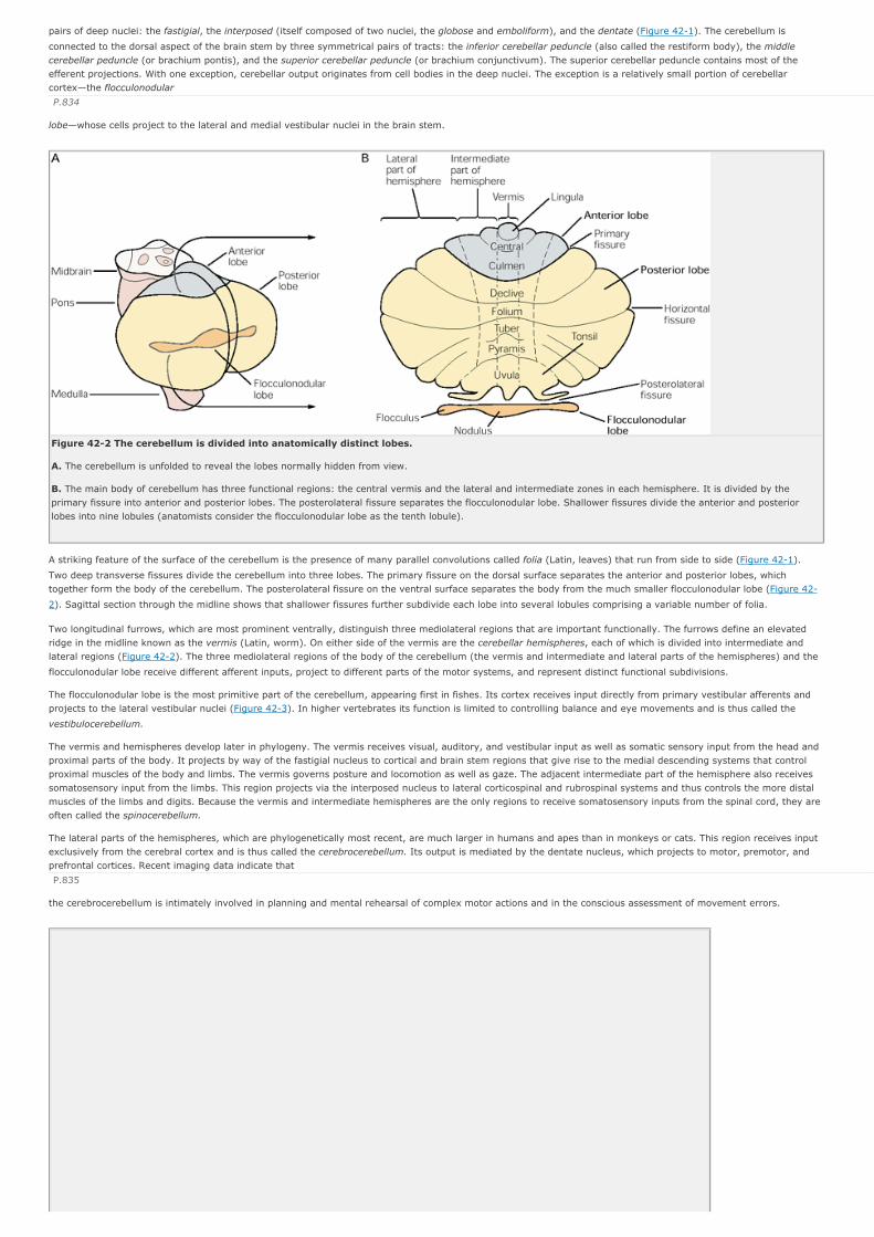
pairs of deep nuclei: the fastigial, the interposed (itself composed of two nuclei, the globose and emboliform), and the dentate (Figure 42-1). The cerebellum is
connected to the dorsal aspect of the brain stem by three symmetrical pairs of tracts: the inferior cerebellar peduncle (also called the restiform body), the middle cerebellar peduncle (or brachium pontis), and the superior cerebellar peduncle (or brachium conjunctivum). The superior cerebellar peduncle contains most of the efferent projections. With one exception, cerebellar output originates from cell bodies in the deep nuclei. The exception is a relatively small portion of cerebellar cortex—the flocculonodular
P.834
lobe—whose cells project to the lateral and medial vestibular nuclei in the brain stem.
Figure 42-2 The cerebellum is divided into anatomically distinct lobes.
A. The cerebellum is unfolded to reveal the lobes normally hidden from view.
B. The main body of cerebellum has three functional regions: the central vermis and the lateral and intermediate zones in each hemisphere. It is divided by the primary fissure into anterior and posterior lobes. The posterolateral fissure separates the flocculonodular lobe. Shallower fissures divide the anterior and posterior lobes into nine lobules (anatomists consider the flocculonodular lobe as the tenth lobule).
A striking feature of the surface of the cerebellum is the presence of many parallel convolutions called folia (Latin, leaves) that run from side to side (Figure 42-1).
Two deep transverse fissures divide the cerebellum into three lobes. The primary fissure on the dorsal surface separates the anterior and posterior lobes, which together form the body of the cerebellum. The posterolateral fissure on the ventral surface separates the body from the much smaller flocculonodular lobe (Figure 42-
2). Sagittal section through the midline shows that shallower fissures further subdivide each lobe into several lobules comprising a variable number of folia.
Two longitudinal furrows, which are most prominent ventrally, distinguish three mediolateral regions that are important functionally. The furrows define an elevated ridge in the midline known as the vermis (Latin, worm). On either side of the vermis are the cerebellar hemispheres, each of which is divided into intermediate and lateral regions (Figure 42-2). The three mediolateral regions of the body of the cerebellum (the vermis and intermediate and lateral parts of the hemispheres) and the
flocculonodular lobe receive different afferent inputs, project to different parts of the motor systems, and represent distinct functional subdivisions.
The flocculonodular lobe is the most primitive part of the cerebellum, appearing first in fishes. Its cortex receives input directly from primary vestibular afferents and projects to the lateral vestibular nuclei (Figure 42-3). In higher vertebrates its function is limited to controlling balance and eye movements and is thus called the
vestibulocerebellum.
The vermis and hemispheres develop later in phylogeny. The vermis receives visual, auditory, and vestibular input as well as somatic sensory input from the head and proximal parts of the body. It projects by way of the fastigial nucleus to cortical and brain stem regions that give rise to the medial descending systems that control proximal muscles of the body and limbs. The vermis governs posture and locomotion as well as gaze. The adjacent intermediate part of the hemisphere also receives somatosensory input from the limbs. This region projects via the interposed nucleus to lateral corticospinal and rubrospinal systems and thus controls the more distal muscles of the limbs and digits. Because the vermis and intermediate hemispheres are the only regions to receive somatosensory inputs from the spinal cord, they are often called the spinocerebellum.
The lateral parts of the hemispheres, which are phylogenetically most recent, are much larger in humans and apes than in monkeys or cats. This region receives input exclusively from the cerebral cortex and is thus called the cerebrocerebellum. Its output is mediated by the dentate nucleus, which projects to motor, premotor, and prefrontal cortices. Recent imaging data indicate that
P.835
the cerebrocerebellum is intimately involved in planning and mental rehearsal of complex motor actions and in the conscious assessment of movement errors.

Figure 42-3 The three functional regions of the cerebellum have different inputs and outputs.
Cerebellar Circuits Consist of a Main Excitatory Loop and an Inhibitory Side-LoopThe cerebellar cortex is a simple three-layered structure consisting of only five types of neurons: the inhibitory stellate, basket, Purkinje, and Golgi neurons; and the excitatory granule cells.
Neurons in the Cerebellar Cortex Are Organized into Three LayersThe outermost or molecular layer of the cerebellar cortex contains the cell bodies of two types of inhibitory inter-neurons, the stellate and basket cells, dispersed among the excitatory axons of granule cells and the dendrites of inhibitory Purkinje cells, whose cell bodies lie in deeper layers (Figure 42-4). The axons of the granule
cells in this layer run parallel to the long axis of the folia and therefore are called parallel fibers. The dendrites of Purkinje neurons are oriented perpendicular to these axons.
Beneath the molecular layer is the Purkinje cell layer, consisting of a single layer of Purkinje cell bodies. Purkinje neurons have large cell bodies (50-80 µm) and fan-like dendritic arborizations that extend upward into the molecular layer. Their axons project into the underlying white matter to the deep cerebellar or vestibular nuclei
and provide the output of the cerebellar cortex. This output is entirely inhibitory and mediated by the neurotransmitter γ-aminobutyric acid (GABA).
The innermost or granular layer contains a vast number (estimated at 1011) of granule cells (so called because they appear as small and densely packed darkly stained nuclei in histological sections) and a few larger Golgi interneurons. The mossy fibers, the major source of afferent input to the cerebellum (see below), terminate in this layer. The bulbous terminals of the mossy fibers contact granule cells and Golgi neurons in synaptic complexes called cerebellar glomeruli (Figure 42-
4).
The Purkinje Cells Receive Excitatory Input From Two Afferent Fiber Systems and Are Inhibited by Three Local InterneuronsThe cerebellum receives two main types of afferent inputs, mossy fibers and climbing fibers. Both groups of
P.836
P.837
fibers form excitatory synapses with cerebellar neurons, but the two groups terminate differently in the cerebellar cortex and produce different patterns of firing in the Purkinje neurons.

Figure 42-4 The cerebellar cortex is organized into three layers and contains five types of neurons. A vertical section of a single cerebellar folium, in both longitudinal and transverse planes, illustrates the general organization of the cerebellar cortex. The detail of a cerebellar glomerulus in the granular layer is also shown. A glomerulus is a clear space where the bulbous terminal of a mossy fiber makes synaptic contact with Golgi and granule cells.
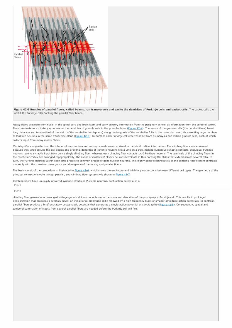
Figure 42-5 Bundles of parallel fibers, called beams, run transversely and excite the dendrites of Purkinje cells and basket cells. The basket cells then inhibit the Purkinje cells flanking the parallel fiber beam.
Mossy fibers originate from nuclei in the spinal cord and brain stem and carry sensory information from the periphery as well as information from the cerebral cortex. They terminate as excitatory synapses on the dendrites of granule cells in the granular layer (Figure 42-4). The axons of the granule cells (the parallel fibers) travel
long distances (up to one-third of the width of the cerebellar hemisphere) along the long axis of the cerebellar folia in the molecular layer, thus exciting large numbers of Purkinje neurons in the same transverse plane (Figure 42-5). In humans each Purkinje cell receives input from as many as one million granule cells, each of which
collects input from many mossy fibers.
Climbing fibers originate from the inferior olivary nucleus and convey somatosensory, visual, or cerebral cortical information. The climbing fibers are so named because they wrap around the cell bodies and proximal dendrites of Purkinje neurons like a vine on a tree, making numerous synaptic contacts. Individual Purkinje neurons receive synaptic input from only a single climbing fiber, whereas each climbing fiber contacts 1-10 Purkinje neurons. The terminals of the climbing fibers in the cerebellar cortex are arranged topographically; the axons of clusters of olivary neurons terminate in thin parasagittal strips that extend across several folia. In turn, the Purkinje neurons within each strip project to common groups of deep nuclear neurons. This highly specific connectivity of the climbing fiber system contrasts markedly with the massive convergence and divergence of the mossy and parallel fibers.
The basic circuit of the cerebellum is illustrated in Figure 42-6, which shows the excitatory and inhibitory connections between different cell types. The geometry of the
principal connections—the mossy, parallel, and climbing fiber systems—is shown in Figure 42-7.
Climbing fibers have unusually powerful synaptic effects on Purkinje neurons. Each action potential in a
P.838
P.839
climbing fiber generates a prolonged voltage-gated calcium conductance in the soma and dendrites of the postsynaptic Purkinje cell. This results in prolonged depolarization that produces a complex spike: an initial large-amplitude spike followed by a high-frequency burst of smaller-amplitude action potentials. In contrast, parallel fibers produce a brief excitatory postsynaptic potential that generates a single action potential or simple spike (Figure 42-8). Consequently, spatial and
temporal summation of inputs from several parallel fibers are needed before the Purkinje cell will fire.

Figure 42-6 Synaptic organization of the basic cerebellar circuit module. Mossy and climbing fibers convey output from the cerebellum via a main excitatory loop through the deep nuclei. This loop is modulated by an inhibitory side-loop passing through the cerebellar cortex. This figure shows the excitatory (+) and inhibitory (-) connections among the cell types. Figures 42-4, 42-5, and 42-7 show the geometry of the divergence and convergence of these basic connections.

Figure 42-7 The geometry of the mossy and parallel fiber system contrasts with that of the climbing fiber system. Mossy fibers excite granule cells whose parallel fibers branch transversely to excite hundreds of Purkinje cells several millimeters from the branch point, both medially and laterally. By contrast, climbing fibers branch in the sagittal dimension to excite 10 or so Purkinje cells anterior and posterior to the branch point. The transverse connections of the parallel fibers and the sagittal connections of the climbing fibers thus form an orthogonal matrix.
Figure 42-8 Simple and complex spikes recorded intracellularly from cerebellar Purkinje cells. Complex spikes (right bracket) are evoked by climbing fiber synapses, while simple spikes (left bracket) are produced by mossy fiber input. (From Martinez et al. 1971.)
The activity of the Purkinje neurons is inhibited by the stellate, basket, and Golgi interneurons. The short axons of stellate cells contact the nearby dendrites of Purkinje cells, and the long axons of basket cells run perpendicular to the parallel fibers and form synapses with Purkinje neurons anterior and posterior to the parallel fiber beam (Figure 42-4). Stellate and basket cells are facilitated by parallel fibers. This arrangement—facilitation of a central array of neurons and inhibition of
surrounding cells by local input—resembles the center-surround antagonism in visual and somatosensory pathways.
The Golgi cell has an elaborate dendritic tree in the overlying molecular layer. The GABA-ergic terminals of Golgi cells form axodendritic synapses with the granule cells in the glomeruli (Figure 42-4). Golgi cell firing, initiated by firing in the parallel fibers, suppresses mossy fiber excitation of the granule cells and thus tends to
shorten the duration of bursts in the parallel fibers.
Mossy and Climbing Fibers Encode Peripheral and Descending Information DifferentlyMossy and climbing fibers respond very differently to sensory stimulation and during motor activity. Spontaneous activity in mossy fibers produces a steady stream of simple spikes in Purkinje cells. Somatosensory, vestibular, or other sensory stimuli change the frequency of the simple spikes, which may reach several
P.840
hundred spikes per second. Voluntary eye or limb movements are also associated with a marked change in frequency. Thus the frequency of simple spikes can readily encode the magnitude and duration of peripheral stimuli or centrally generated behaviors.
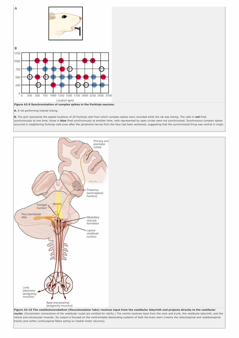
Figure 42-9 Synchronization of complex spikes in the Purkinje neurons.
A. A rat performing trained licking.
B. The grid represents the spatial locations of 29 Purkinje cells from which complex spikes were recorded while the rat was licking. The cells in red fired synchronously at one time; those in blue fired synchronously at another time; cells represented by open circles were not synchronized. Synchronous complex spikes occurred in neighboring Purkinje cells even after the peripheral nerves from the face had been sectioned, suggesting that the synchronized firing was central in origin.
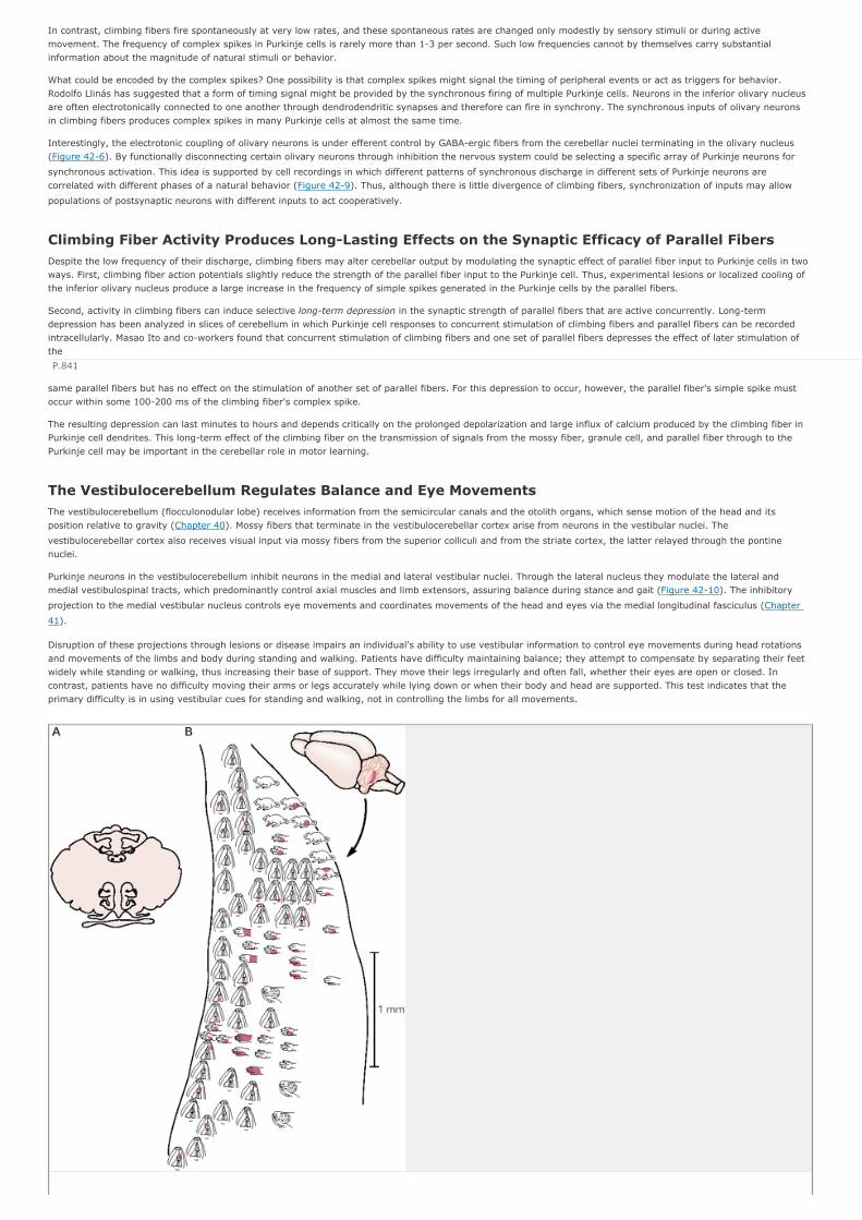
Figure 42-10 The vestibulocerebellum (flocculonodular lobe) receives input from the vestibular labyrinth and projects directly to the vestibular nuclei. (Oculomotor connections of the vestibular nuclei are omitted for clarity.) The vermis receives input from the neck and trunk, the vestibular labyrinth, and the retinal and extraocular muscles. Its output is focused on the ventromedial descending systems of both the brain stem (mainly the reticulospinal and vestibulospinal tracts) and cortex (corticospinal fibers acting on medial motor neurons).
In contrast, climbing fibers fire spontaneously at very low rates, and these spontaneous rates are changed only modestly by sensory stimuli or during active movement. The frequency of complex spikes in Purkinje cells is rarely more than 1-3 per second. Such low frequencies cannot by themselves carry substantial information about the magnitude of natural stimuli or behavior.
What could be encoded by the complex spikes? One possibility is that complex spikes might signal the timing of peripheral events or act as triggers for behavior. Rodolfo Llinás has suggested that a form of timing signal might be provided by the synchronous firing of multiple Purkinje cells. Neurons in the inferior olivary nucleus are often electrotonically connected to one another through dendrodendritic synapses and therefore can fire in synchrony. The synchronous inputs of olivary neurons in climbing fibers produces complex spikes in many Purkinje cells at almost the same time.
Interestingly, the electrotonic coupling of olivary neurons is under efferent control by GABA-ergic fibers from the cerebellar nuclei terminating in the olivary nucleus (Figure 42-6). By functionally disconnecting certain olivary neurons through inhibition the nervous system could be selecting a specific array of Purkinje neurons for
synchronous activation. This idea is supported by cell recordings in which different patterns of synchronous discharge in different sets of Purkinje neurons are correlated with different phases of a natural behavior (Figure 42-9). Thus, although there is little divergence of climbing fibers, synchronization of inputs may allow
populations of postsynaptic neurons with different inputs to act cooperatively.
Climbing Fiber Activity Produces Long-Lasting Effects on the Synaptic Efficacy of Parallel FibersDespite the low frequency of their discharge, climbing fibers may alter cerebellar output by modulating the synaptic effect of parallel fiber input to Purkinje cells in two ways. First, climbing fiber action potentials slightly reduce the strength of the parallel fiber input to the Purkinje cell. Thus, experimental lesions or localized cooling of the inferior olivary nucleus produce a large increase in the frequency of simple spikes generated in the Purkinje cells by the parallel fibers.
Second, activity in climbing fibers can induce selective long-term depression in the synaptic strength of parallel fibers that are active concurrently. Long-term depression has been analyzed in slices of cerebellum in which Purkinje cell responses to concurrent stimulation of climbing fibers and parallel fibers can be recorded intracellularly. Masao Ito and co-workers found that concurrent stimulation of climbing fibers and one set of parallel fibers depresses the effect of later stimulation of the
P.841
same parallel fibers but has no effect on the stimulation of another set of parallel fibers. For this depression to occur, however, the parallel fiber's simple spike must occur within some 100-200 ms of the climbing fiber's complex spike.
The resulting depression can last minutes to hours and depends critically on the prolonged depolarization and large influx of calcium produced by the climbing fiber in Purkinje cell dendrites. This long-term effect of the climbing fiber on the transmission of signals from the mossy fiber, granule cell, and parallel fiber through to the Purkinje cell may be important in the cerebellar role in motor learning.
The Vestibulocerebellum Regulates Balance and Eye MovementsThe vestibulocerebellum (flocculonodular lobe) receives information from the semicircular canals and the otolith organs, which sense motion of the head and its position relative to gravity (Chapter 40). Mossy fibers that terminate in the vestibulocerebellar cortex arise from neurons in the vestibular nuclei. The
vestibulocerebellar cortex also receives visual input via mossy fibers from the superior colliculi and from the striate cortex, the latter relayed through the pontine nuclei.
Purkinje neurons in the vestibulocerebellum inhibit neurons in the medial and lateral vestibular nuclei. Through the lateral nucleus they modulate the lateral and medial vestibulospinal tracts, which predominantly control axial muscles and limb extensors, assuring balance during stance and gait (Figure 42-10). The inhibitory
projection to the medial vestibular nucleus controls eye movements and coordinates movements of the head and eyes via the medial longitudinal fasciculus (Chapter
41).
Disruption of these projections through lesions or disease impairs an individual's ability to use vestibular information to control eye movements during head rotations and movements of the limbs and body during standing and walking. Patients have difficulty maintaining balance; they attempt to compensate by separating their feet widely while standing or walking, thus increasing their base of support. They move their legs irregularly and often fall, whether their eyes are open or closed. In contrast, patients have no difficulty moving their arms or legs accurately while lying down or when their body and head are supported. This test indicates that the primary difficulty is in using vestibular cues for standing and walking, not in controlling the limbs for all movements.

Figure 42-11 The spinocerebellum contains two somatotopic neural maps of the body.
A. Two regions of the cerebellar surface contain somatotopic maps of the entire body. In both maps the head and trunk are located in the vermis, which also receives input from vestibular, visual, and auditory receptors. The limb representations are located on either side of the midline, in the intermediate part of the cerebellar hemispheres.
B. Recordings of the receptive fields of granule cells in the rat cerebellar cortex reveal multiple representations of the same body parts in different locations, an arrangement referred to as fractured somatotopy. The receptive fields of individual granule cells are indicated by the red areas on body parts. (Adapted from Shambes et al. 1978.)
The Spinocerebellum Regulates Body and Limb Movements
Somatosensory Information Reaches the Spinocerebellum Through Direct and Indirect Mossy Fiber PathwaysCerebellar afferents from the spinal cord—mainly from somatosensory receptors—are distributed exclusively to the spinocerebellum (see Figure 42-3). Somatosensory
information is conveyed to the spinocerebellum through several direct and indirect pathways.
Figure 42-12 Neurons in the intermediate and lateral parts of the cerebellar hemisphere project to the contralateral red nucleus and motor cortex. The intermediate zone (spinocerebellum) receives sensory information from the limbs and controls the dorsolateral descending systems (rubrospinal and corticospinal tracts) acting on the ipsilateral limbs. There is a modest projection from the fastigial nucleus to the ventrolateral nucleus in the thalamus and the primary motor cortex. The lateral zone (cerebrocerebellum) receives cortical input via the pontine nuclei and influences the motor and premotor cortices via the ventrolateral nucleus of the thalamus.
P.842
Direct pathways originate from interneurons in the spinal gray matter and terminate as mossy fibers in the vermis or intermediate cortex. Two important pathways are the ventral and dorsal spinocerebellar tracts. These pathways from spinal interneurons provide the cerebellum with somatic sensory information from the legs— notably from muscle and joint proprioceptors—and with information about descending commands reaching the interneurons.
Recordings from neurons in the dorsal and ventral spinocerebellar tracts of decerebrate cats walking on a treadmill show that both systems are modulated rhythmically and in phase with the step cycle. However, when the dorsal roots are cut, preventing spinal neurons from receiving phase-dependent peripheral excitation, dorsal spinocerebellar neurons fall silent while ventral spinocerebellar neurons continue to be modulated. This finding demonstrates that the ventral tract carries internally generated information about the central locomotor rhythm as well as the rhythmic discharge of somatic sensory receptors, while the dorsal tract provides the cerebellum with sensory feedback only during evolving movements. Other direct pathways provide comparable information from the upper extremities.
Direct pathways from the spinal cord to the cerebellum synapse first with neurons in one of several so-called precerebellar nuclei in the brain stem reticular formation (the lateral reticular nucleus, reticularis tegmenti pontis, and paramedian reticular nucleus). These inputs provide the cerebellum with different versions of the changing state of the organism and its environment and permit comparisons between such signals. Similar monitoring of outgoing commands is as crucial for perception as for movement, since the internal sensory signals resulting from movement must be distinguished from the external sensory signals in the environment.

The Spinocerebellum Contains Sensory MapsThe initial mapping studies of the spinocerebellum by Edgar Adrian and Ray Snider in the 1940s revealed two inverted somatotopic maps. In both maps the head is represented in the posterior vermis, and the repre-sentations of the neck and trunk extend on either side along the dorsal and ventral portions of the vermis. Arms and legs are represented adjacent to the vermis over the intermediate cortex of the hemispheres (Figure 42-11A). Visual input from the superior colliculi and visual
cortex is distributed to both vermal and paravermal portions of the posterior lobe (Figure 42-3).
This early mapping was based on recordings of surface potentials, which reflect the predominant input and provide only a coarse representation of somatotopic connections. More refined mapping studies of the cerebellar cortex based on single-cell recordings reveal that input from a given peripheral site, such as a local area of skin, diverges to multiple discrete patches of granule cells, an arrangement called a fractured somatotopy (Figure 42-11B).
P.843
Recent anatomical studies of primates show that the deep cerebellar nuclei are also organized somatotopically. They are arranged to receive projections from the two maps on the dorsal and ventral surfaces of the inter-mediate and lateral zones of the cerebellar cortex and project to the magnocellular red nucleus and primary motor cortex via the thalamus (Figure 42-12).
The Spinocerebellum Modulates the Descending Motor Systems in the Brain Stem and Cerebral CortexPurkinje neurons in the spinocerebellum project somatotopically to different deep nuclei that control various components of the descending motor pathways. Neurons in the vermis in both the anterior and posterior lobes send projections to the fastigial nucleus, which in turn projects bilaterally to the brain stem reticular formation and lateral vestibular nuclei. The latter areas project directly to the spinal cord (Figure 42-10). Axons of the fastigial nucleus also cross to the contralateral side and
project to the area's primary motor cortex controlling proximal muscles via a synapse in the ventrolateral nucleus of the thalamus (Figure 42-12). Thus the medial
region of the cerebellum controls mainly the cortical and brain stem components of the medial descending systems. This control affects primarily the head and neck and proximal parts of the limb, rather than the wrist and digits. It is therefore important for movements of the face, mouth, and neck and for balance and postural control during voluntary motor tasks.
Purkinje neurons in the intermediate part of the cerebellar hemisphere project to the interposed nucleus (Figure 42-12). Some axons of this nucleus exit through the
superior cerebellar peduncle and cross to the contralateral side to terminate in the magnocellular portion of the red nucleus, whose axons cross back and descend to the spinal cord. Other axons from the interposed nucleus continue rostrally and terminate in the ventrolateral nucleus of the thalamus. This cerebellar receiving area (in ventral lateral thalamus) is located posterior to the area that receives input from the basal ganglia (the ventral anterior nuclei) and anterior to the area recieving from the lemniscal sensory system (ventral posterior lateral nucleus) (see Figure 18-5).
These thalamic neurons project to the limb control areas of the primary motor cortex. By acting on the neurons that give rise to the rubrospinal and corticospinal systems, the intermediate cerebellum focuses its action on limb muscles and axial musculature. Because the axons of the interposed nucleus cross to the contralateral side and the rubrospinal and corticospinal tracts cross back (Figure 42-12), cerebellar lesions can disrupt movements of ipsilateral limbs.
The Spinocerebellum Uses Feed-Forward Mechanisms to Regulate MovementsBecause deep nuclear neurons are tonically active and produce powerful excitatory postsynaptic potentials in their target neurons, damage to the interposed nucleus reduces the activity of rubrospinal and corticospinal neurons through disfacilitation. This in turn reduces the excitability of motor neurons themselves and results in a reduction in muscle tone (cerebellar hypotonia). Experimental lesions of the interposed nucleus also disrupt the accuracy of reaching movements because of increased errors in timing the components of movements and systematic errors in direction and extent, a clinical sign called dysmetria (Greek, abnormal measure). Joint motions are poorly coordinated or ataxic (Greek, loss of order) so that the path of the hand in reaching is curved rather than straight. Attempts by patients to correct such movements are associated with new errors, and the hand oscillates irregularly around the target, with a characteristic terminal tremor. Another deficit is seen in stretch reflexes: Although tendon reflexes may be strong, the limb tends to oscillate as it returns to its initial position (pendular reflexes).
Are these abnormalities the result of an impairment in correcting movement errors or in programming the movement itself? This question was addressed by Tutis Vilis and Jonathan Hore, who trained monkeys to hold a motor-driven handle in a fixed position and to resist perturbing forces applied unpredictably to the handle. Once an animal was trained, Vilis and Hore compared its performance in circumstances in which the dentate and interposed nuclei were reversibly inactivated with a cooling probe implanted in the nuclei.
When a normal animal is attempting to keep its arm in a fixed position, the application of a transient force to extend the elbow evokes a short-latency stretch reflex in the biceps; the arm then returns rapidly and precisely to its initial position. The precision of the return movement depends on the contraction of the extensor triceps muscle, which prevents the elbow from overshooting. Activation of the triceps occurs shortly after that of the biceps (Figure 42-13A). At this point the perturbation
still extends the elbow and shortens the triceps. This extensor contraction is therefore an anticipatory or feed-forward response rather than a stretch reflex.
When the dentate and interposed nuclei are inactivated by cooling, the elbow shows a pronounced oscillation after the perturbation instead of returning
P.844
precisely to its initial position. The triceps is no longer activated during the initial shortening phase, but only after it has been stretched, when the flexion produced by the biceps contraction overshoots its mark (Figure 42-13A). This delayed contraction of the triceps represents a feedback correction to excessive flexion rather than an
anticipatory response. Moreover, active triceps force is now superimposed with the elastic recoil of the limb and extends the limb excessively, evoking a new flexor response in the biceps and triggering another cycle of flexion-extension. The same mechanism accounts for the oscillations in the pendular knee jerks observed in humans who have cerebellar diseases.

Figure 42-13 Inactivation of the interposed and dentate nuclei disrupts the precisely timed sequence of agonist and antagonist activation that follows external perturbations during normal rapid movements. (Courtesy of Vilis and Hore 1977.)
A. The records show position, velocity, and electromyographic responses in biceps and triceps of a trained monkey after the forearm was suddenly displaced from a held stationary position. Prior to inactivation of the deep nuclei, through local cooling, the limb returns to its original position after the external torque is stopped; only minimal overshooting is evident on the position trace. While the nuclei are cooled the limb returns with marked overshoot and sequential corrections produce oscillations. (From Vilis and Hore 1977.)
B. With inactivation (cooling) of the nuclei, agonist (biceps) activation becomes slower and more prolonged; activation of the antagonist (triceps), which is needed to stop the movement at the correct location, is delayed and prolonged so that the initial movement overshoots its appropriate extent. Similar delays in successive phases of the movement produce oscillations similar to the terminal tremor seen in patients with cerebellar damage.
Vilis and Hore also examined whether the same mechanism might account for the terminal tremor after movement. Rapid single-joint movements are initially accelerated by the contraction of an agonist muscle and decelerated by an appropriately timed contraction of the antagonist (Chapter 33). When the dentate and
interposed nuclei are inactivated by cooling, contraction of the antagonist muscle is delayed until the limb has overshot the target (Figure 42-13B). The anticipatory
contraction has been replaced by a feedback correction. This correction is itself dysmetric and results in another error, necessitating a new adjustment. Thus both the oscillatory response to an external perturbation and the terminal tremor at the end of a voluntary reaching movement result from defective anticipatory control of limb motion.
The failure to decelerate the limb at the correct time reflects defective adaptation of motor commands to the aim of the movement. Specifically, the sequence of muscle commands is not matched correctly to the inertial and viscoelastic properties of the limb. As we saw in Chapter 33, multi-joint movements of a limb are more
complicated than single-joint movements because motions at several joints of the limb produce interaction torques that vary with time at each joint. We normally learn to anticipate these forces and continuously recalibrate the internal representation of our limbs. This ability, however, depends on cerebellar processing of proprioceptive information from the limb. The inherent difficulty in controlling the inertial interactions among the multiple segments of a limb accounts for the greater inaccuracy of multi-joint movements in cerebellar ataxia.

Figure 42-14 Activity in the dentate nucleus is significantly greater when the subject is mentally active during movement. A functional magnetic resonance image (color) overlaid on an anatomical image (gray) shows activation of the dentate nucleus during two pairs of tests. In one pair, subjects first passively experienced sandpaper rubbed across the fingers (A) and then were asked to discriminate the degree of roughness of the sandpaper (B). In the other pair, subjects were asked to lift and drop a series of objects (C) and then had to identify the felt object from a similar group near the other hand (D).
P.845
The Cerebrocerebellum Is Involved in Planning Movement and Evaluating Sensory Information for ActionClinical observations of neurologists and neurosurgeons initially suggested that, like the rest of the cerebellum, the lateral hemispheres were primarily concerned with motor function. However, recent studies of patients with lesions of the lateral hemisphere and experiments using functional brain imaging indicate that the lateral hemispheres, or cerebrocerebellum, also have a variety of perceptual and cognitive functions. In addition, the lateral hemispheres are much larger in humans than in monkeys, consistent with a role in higher cognitive functions.
The Cerebrocerebellum Is Part of a High-Level Internal Feedback Circuit That Regulates Cortical Motor ProgramsIn contrast to other regions of the cerebellum, which receive sensory information more directly, the lateral hemispheres receive input exclusively from the cerebral cortex. This cortical input originates mainly in the pontine nuclei and projects through the middle cere- bellar peduncle to the contralateral dentate nucleus and terminate as mossy fibers in the lateral cerebellar cortex.
Purkinje neurons in the lateral cerebellar cortex project to the dentate nucleus. Most dentate axons exit the cerebellum via the superior cerebellar peduncle and have two main terminations. One termination is in the
P.846
contralateral ventrolateral thalamus, in the same area receiving input from the interposed nucleus. These thalamic cells project to premotor and primary motor areas of the cerebral cortex (Figure 42-12). The second main termination of dentate neurons is in the contralateral parvocellular red nucleus, a portion of the red nucleus
that is distinct from the part receiving input from the interposed nucleus. These neurons project to the inferior olivary nucleus, which in turn projects back to the contralateral cerebellum in the climbing fibers, thus forming a feedback loop. In addition to receiving input from the dentate nucleus, parvocellular neurons also receive input from the lateral premotor areas. The intriguing suggestion has been made, based on brain imaging, that this premotor-cerebello-rubrocerebellar loop is involved in the mental rehearsal of movements and perhaps with motor learning.
Lesions of the Cerebrocerebellum Disrupt Motor Planning and Prolong Reaction TimeIn the first half of the twentieth century Gordon Holmes and Jean Babinski identified two characteristic motor disturbances in patients with localized damage in the cerebrocerebellum: variable delays in initiating movements and irregularities in the timing of movement components. The same defect is seen in primates with lesions of the dentate nucleus.
Many motor acts are made up of multiple components, each of which is initiated before the preceding one is completed. An example is prehension, in which the shaping of the hand to the object to be grasped begins during the transport phase. During each component of movement the motions at each joint are coordinated precisely one with another (see Chapter 33). Lateral cerebellar lesions disrupt the timing of the various components, which appear to take place sequentially rather
than being coordinated smoothly, a defect known as decomposition of movement. One of Holmes's patients, who had a lesion of his right cerebellar hemisphere, reported that “movements of my left arm are done subconsciously, but I have to think out each movement of the right arm. I come to a dead stop in turning and have to think before I start again.” In humans and primates lesions of the dentate nucleus in particular impair the coordination of distal and proximal components of prehensile movements and the independent use of the digits in manipulatory tasks.
These increases in reaction time and abnormalities in hand paths suggest that the cerebrocerebellum has a role in the planning and programming of hand movements. The activity patterns of single dentate neurons in primates support this idea. Recordings from primates show that dentate nucleus neurons fire some 100 ms before a movement begins. This firing occurs before the discharge of neurons in either the primary motor cortex or interposed nuclei, which are more directly concerned with the execution of movement itself. Hore and his colleagues inactivated the dentate nucleus by localized cooling. Inactivation of the early output from the dentate nucleus delayed the onset of firing in the primary motor cortex, which delayed the onset of movement. Because movement was eventually initiated, the dentate nucleus is not absolutely necessary for initiation.
The Cerebrocerebellum Also Has Purely Cognitive FunctionsWhen patients with cerebellar lesions attempt to make regular tapping movements with their hands or fingers, the rhythm is irregular and the motions are variable in duration and force. Richard Ivry and Steven Keele first sought to determine whether this defect results from a motor deficit or from a more fundamental defect in the timing of serial events. Based on a theoretical model of how such tapping movements are generated, Ivry and Keele found that medial cerebellar lesions interfered only with accurate execution of the response, whereas lateral cerebellar lesions interfered with the timing of serial events. This timing defect was not limited to motor events. It also affected the patient's ability to judge elapsed time in purely mental or cognitive tasks, as in the ability to distinguish whether one tone was longer or shorter than another or whether the speed of one moving object was greater or less than another.
This demonstration that the cerebellum is responsible for a cognitive computation independent of motor execution prompted other researchers to investigate purely cognitive functions of the cerebellum. Steve Petersen, Julie Fiez, and Marcus Raichle used positron emission tomography to image the brain activity of people during silent reading, reading aloud, and speech. As expected, cerebral cortical areas known to be involved in the control of mouth movements were more active when subjects read words aloud than when they read silently. Brain activity during the generation of language was assessed using a verb association task in which subjects had to identify the actions corresponding to certain nouns (eg, a subject might respond with “bark” if he saw the word “dog”). Compared with the brain activity associated with reading aloud, verb generation produced an expected increase in activity in the left frontal lobe, corresponding to Broca's area (see Chapter 59), as
well as a pronounced increase within the right lateral cerebellum. Further support of the conclusion that the cerebellum has cognitive functions independent
P.847
of motor functions comes from the observation that a patient damaged in the right cerebellum (blocked posterior inferior cerebellar artery) could not learn a word association task.
Functional magnetic resonance imaging data have provided evidence for the role of the lateral cerebellum in other cognitive activities. For example, Peter Strick and co-workers showed that solving a pegboard puzzle involves greater activity in the dentate nucleus and lateral cerebellum than does the simple motor task of moving the pegs on the board. Interestingly, the area activated in the dentate nucleus corresponds to the area receiving input from the part of cortex (area 46) involved in working memory (Chapter 62). Another example in which cognitive activity could be isolated from sensory stimulation or movement is shown in Figure 42-14. Activity
in the dentate nucleus increased dramatically when subjects were required to evaluate sensory information consciously.
Thus the dentate nucleus appears to be particularly important in acquiring and processing sensory information for tasks requiring complex spatial and temporal judgments, which are essential for programming complex motor actions and sequences of movements. However, the specific contribution of the cerebrocerebellum to sensory discrimination remains to be determined.

The Cerebellum Participates in Motor LearningOn the basis of mathematical modeling of cerebellar function, David Marr and James Albus independently suggested in the early 1970s that cerebellar cortical circuits might be used in learning motor skills. Specifically, they proposed that the climbing fiber input to Purkinje neurons modifies the response of the neurons to mossy fiber inputs and does so for a prolonged period of time. Experimental evidence discussed earlier in the chapter supports this idea: The climbing fiber weakens the parallel fiber-Purkinje cell synapse in a process called long-term depression.
According to the theories of Marr and Albus, altering the strength of certain parallel fiber-Purkinje cell synapses would select specific Purkinje cells to program or correct eye or limb movements. During a movement the climbing fibers would provide an error signal that would depress parallel fibers that are active concurrently and allow “correct” movements (with no error) to emerge. With successive movements the effects of parallel fiber inputs associated with a flawed central command would increasingly be suppressed and a more appropriate pattern of activity would emerge over time. In accordance with this idea, climbing fibers detect differences between expected and actual sensory inputs rather than simply monitoring afferent information. Also, as is reviewed below, motor learning is often impaired following cerebellar damage. It is not yet clear, however, whether climbing fibers can, under natural conditions, also produce the appropriate and specific long-term changes in parallel fiber activation required by the theory.
Initial studies by Masao Ito and his colleagues focused on the vestibulo-ocular reflex, a coordinated response that maintains the eyes on a fixed target when the head is rotated (Chapter 39). In this short-latency reflex motion of the head in one direction is sensed by the vestibular labyrinth, which initiates eye movements in the
opposite direction in order to maintain the image in the same position on the retina. When humans and experimental animals wear prism glasses that reverse the left and right visual fields, the vestibulo-ocular reflex is initially maladaptive because the reflex accentuates the motion of the visual field on the retina rather than stabilizing it. After the glasses have been worn continuously for several days the direction of the reflex becomes progressively reduced and eventually reverses direction. This adaptation can be blocked by lesions of the vestibulocerebellum in experimental animals, indicating that the cerebellum has an important role in mediating this form of learning.
Control of limb movements also adapts when subjects wear prisms for an extended period. A striking example is the adjustment of eye-hand coordination in throwing darts. When people throw darts they normally fixate the target visually, and the direction of the throw matches the direction of gaze. When people wear prisms, which bend the light path sideways, the initial throw in the direction of gaze misses the target to the side by an amount proportional to the diopter of the prism (how much the light is bent by the prism). The prisms thus require subjects to shift their gaze to the opposite side, along the bent light path, if they are to fixate the target. With repeated throws aimed at the perceived target, subjects gradually increase the angle between the direction of gaze and the direction of throw, so that the darts land on target within 10-30 throws (Figure 42-15).
At that point the subjects have learned to aim their throw in a direction different from the direction of gaze. When the prisms are removed, gaze is now on target but the widened angle between the direction of gaze and the direction of throw persists: hits miss the target to the side opposite by roughly the same distance as the initial prisminduced error (this is called an after-effect).
Patients with a damaged cerebellar cortex or inferior olive (the source of climbing fibers to the cerebellar cortex) are severely impaired or unable to adapt at
P.848
P.849
all. Experiments have shown that the kind of motor adaptation and learning with which the cerebellum is concerned requires trial-and-error practice. Once the behavior becomes adapted as learned, it is performed automatically.

Figure 42-15 Readjustment of eye-hand coordination during adaptation to prism glasses. (Adapted from Martin et al. 1996a and 1996b.)
A. Laterally displacing prisms bend the optic path to the subject's right. The subject looks to the left along the bent light path to see the target directly in front of her. Her hand is held in position ready to throw a dart at the target in front of her.
B. While wearing the prisms (gaze shifted to the left), the first hit is displaced left of center: The hand throws to where the eyes are looking toward the target. Thereafter, hits trend rightward away from where the eyes are looking. After removing the prisms the gaze is centered on the target, and the first throw hits right of center, away from where the eyes are looking. Thereafter hits trend toward the target where the eyes are looking. Data during and after prism use have been fit with exponential curves. Gaze and throw directions are indicated by the arrows on the right. Inferred gaze (eye and head) direction assumes the subject is fixating the target. The roman numerals next to the arrows indicate times during the prism adaptation experiment. I. Before donning prisms, when gaze is directed toward the target and the throw is toward the target. II. Just after donning prisms, when gaze is directed along the bent light path away from the target and the throw is in the direction of gaze away from the target. III. Still wearing prisms and after adapting to them, when gaze is directed along the bent light path away from the target but the throw is directed toward the target. IV. Just after doffing the prisms, when the gaze is now directed toward the target and the adapted throw is to the right of the direction of gaze and to the right of the target. V. After disadapting the gaze-throw coordination, when gaze is now directed to the target and the throw is in the direction of gaze toward the target as originally.
C. Adaptation fails in a patient with unilateral infarctions in the territory of the posterior inferior cerebellar artery and involves inferior cerebellar peduncle (inferior olivary climbing fibers) and/or inferior lateral posterior cerebellar cortex.
The cerebellum's contribution to motor adaptation may occur also in certain forms of associative learning (Chapter 62). Richard Thompson and Christopher Yeo and
their colleagues found that lesions of the cerebellum in the rabbit disrupt the acquisition and retention of a classically conditioned eyeblink reflex. After many couplings of an air puff (the unconditioned stimulus) to a sound (the conditioned stimulus), the eye blinked to the sound alone.
Cerebellar Diseases Have Distinctive Symptoms and SignsDisorders of the human cerebellum result in distinctive symptoms and signs, described originally by Babinski in 1899 and in the 1920s and 1930s by Holmes. Following the plan of Luigi Luciani based on his animal studies, Holmes grouped the abnormalities into three categories.
The first category is hypotonia, a diminished resistance to passive limb displacements. Hypotonia is also thought to explain pendular reflexes. After a knee jerk produced by the tap of a reflex hammer, the leg normally comes to rest after the jerk. In patients who have cerebellar disease the leg may oscillate up to 6 or 8 times before coming to rest.
The second category of symptoms includes a variety of abnormalities in the execution of voluntary movements, collectively referred to as ataxia, or lack of coordination. Examples are a delay in initiating responses with the affected limb, errors in the range of movement (dysmetria), and errors in the rate and regularity of movements (Figure 42-16). This last deficit, discovered by Babinski, is most readily demonstrated when the patient attempts to perform rapid alternating movements,
such as tapping one hand with the other, alternating between the back and the palm of the hand. Patients cannot sustain a regular rhythm nor produce an even amount of force, a sign referred to as dysdiadochokinesia. Holmes also noted that patients made errors in the relative timing of the components of complex multi-joint movements (decomposition of movement) and frequently failed to brace proximal joints against the forces generated by movement of more distal joints.
The third type of abnormality in movement due to cerebellar disease is a specific form of tremor during movement that is most marked at the end of a movement, when the patient attempts to stop the movement by using antagonist muscles. Such action or intention tremors represent a series of erroneous corrections in the range of movement due to the failure of adaptive control.
P.850
Cerebellar action tremor has been duplicated in monkeys by inactivating the interposed and dentate nuclei by cooling.

Figure 42-16 Typical defects observed in cerebellar diseases.
A. A lesion in the right cerebellar hemisphere delays the initiation of movement. The patient is told to clench both hands at the same time on a “go” signal. The left hand is clenched later than the right, as evident in the recordings from a pressure bulb transducer squeezed by the patient.
B. A patient moving his arm from a raised position to touch the tip of his nose exhibits dysmetria (inaccuracy in range and direction) and decomposition of movement (moves shoulder first and elbow second). Tremor increases on approaching the nose.
C. Dysdiadochokinesia, an irregular pattern of alternating movements, can be seen in the abnormal position trace of the hand and forearm as cerebellar subjects attempt alternately to pro-nate and supinate the forearm while flexing and extending at the elbow as rapidly as possible.
Sites of damage in the cerebellum can be identified based on a knowledge of the somatotopic organization of the spinocerebellum. Lesions of the vermis and fastigial nuclei produce disturbances principally in the control of axial and trunk muscles during attempted antigravity posture. Thus, when standing or sitting, patients with these lesions spread their feet apart in an attempt to stabilize their balance. Because facial and vocal control is also localized in the vermis, lesions in this area may result in slurring and slowing of speech with a characteristic one-word-at-a-time quality known as scanning speech. Degeneration of the anterior lobes (the vermis and the trunk and leg areas) is common in the thiamine deficiency seen in alcoholic or malnourished patients. These patients have ataxia and tremor of the legs and trunk in standing and walking but not of the arms or head.
Lesions of the intermediate cerebellum or the interposed nuclei produce action tremor of the limbs. The disorders produced by lesions of the lateral cerebellar hemispheres consist principally of delays in initiating movement and in decomposition of multi-joint movements—patients cannot move all limb segments together in a coordinated fashion but instead move one joint at a time. This deficit is seen even in movements of the distal joints; patients are unable to combine thumb and index flexion in a precise pinch.
Cases of recovery from atrophy of the cerebellum in childhood have been reported, and many of these patients had large focal lesions in the lateral cerebellar cortex. We now know that lesions of the lateral hemisphere may produce cognitive deficits but little in the way of easily recognized motor abnormality. The misconception has therefore developed that deficits due to cerebellar lesions sustained in youth are well compensated by the functioning of other parts of the nervous system. Deficits due to lesions of the more medial “motor” parts of the cerebellum become permanent disabilities.
An Overall ViewWhereas lesions of other motor processing centers produce paralysis or involuntary movements, lesions of the cerebellum result in errors in large numbers of muscles. How do these errors occur?
The organization of inputs to and outputs from the cerebellum indicates that the cerebellum compares internal feedback signals that report the intended movement with external feedback signals that report the actual movement. When the movement is repeated, the cerebellum is able to generate corrective signals and thus gradually reduce the error. The corrective signals are feed-forward or anticipatory actions that operate on the descending motor systems of the brain stem and cerebral cortex. The oscillations and tremor that follow lesions of the cerebellum are due to the failure of such feed-forward mechanisms.
The cerebellum also plays a role in motor learning. Most motor actions are initiated and executed without immediate feedback and thus have to be well planned. Because there is no opportunity for quick corrections by sensory feedback, planning (feed-forward control) requires calibration and adaptive adjustments of motor programs. The Marr-Albus-Ito model describes how the cerebellum, and specifically the climbing fibers, might participate in motor learning. Because of their low firing frequencies, climbing fibers have a very modest capacity for transmitting moment-to-moment changes in sensory information. Instead, they are thought to be involved in detecting the error in one movement and in changing the program for the next movement.
Finally, the cerebellum seems to have a role in some purely mental operations. In many respects the cerebellum's cognitive functions appear to be similar to its motor functions. For instance, the lateral cerebellum appears to be particularly important for learning both motor and cognitive tasks in which skilled responses are developed through repeated practice. We shall consider the role of the cerebellum in learning again in Chapter 62.
Selected Readings
Adams RD, Victor M. 1989. Principles of Neurology, 4th ed. New York: McGraw-Hill.
Brooks VB, Thach WT. 1981. Cerebellar control of posture and movement. In: VB Brooks (ed). Handbook of Physiology. Section 1: The Nervous System. Vol. 2: Motor Control, Part 2, pp. 877-946. Bethesda, MD: American Physiological Society.
Fiez JA, Petersen SE, Cheney MK, Raichle ME. 1992. Impaired non-motor learning and error detection associated with cerebellar damage. Brain 115:155–178.
P.851
Gilman S. 1985. The cerebellum: its role in posture and movement. In: M Swah, C Kennard (eds). Scientific Basis of Clinical Neurology, pp. 36-55. New York: Churchill Livingston
Glickstein M, Yeo C. 1990. The cerebellum and motor learning. J Cogn Neurosci 2:69–80.
Holmes G. 1939. The cerebellum of man. Brain 62:1–30.
Ito M. 1984. The Cerebellum and Neural Control. New York: Raven.
Keele SW, Ivry R. 1990. Does the cerebellum provide a common computation for diverse tasks? A timing hypothesis. Ann N Y Acad Sci 608:179–211.
Llinés RR. 1981. Electrophysiology of the cerebellar networks. In: VB Brooks (ed). Handbook of Physiology. Section 1: The Nervous System. Vol. 2: Motor Control, Part 2, pp. 831-876. Bethesda, MD: American Physiological Society.
Thach WT. 1996. On the specific role of the cerebellum in motor learning and cognition: clues from PET activation and lesion studies in humans. Behav Brain Sci 19:411–431.

Thach WT, Goodkin HG, Keating JG. 1992. Cerebellum and the adaptive coordination of movement. Annu Rev Neurosci 15:403–442.
References
Adrian ED. 1943. Afferent areas in the cerebellum connected with the limbs. Brain 66:289–315.
Albus JS. 1971. A theory of cerebellar function. Math Biosci 10:25–61.
Allen GI, Tsukahara N. 1974. Cerebrocerebellar communication systems. Physiol Rev 54:957–1006.
Arshavsky YI, Berkenblit MB, Fukson OI, Gelfand IM, Orlovsky GN. 1972. Recordings of neurones of the dorsal spinocerebellar tract during evoked locomotion. Brain Res 43:272–275.
Arshavsky YI, Berkenblit MB, Fukson OI, Gelfand IM, Orlovsky GN. 1972. Origin of modulation in neurones of the ventral spinocerebellar tract during locomotion. Brain Res 43:276–279.
Asanuma C, Thach WT, Jones EG. 1983. Anatomical evidence for segregated focal groupings of efferent cells and their terminal ramifications in the cerebellothalamic pathway of the monkey. Brain Res Rev 5:267–297.
Botterell EH, Fulton JF. 1938. Functional localization in the cerebellum of primates. II. Lesions of midline structures (vermis) and deep nuclei. J Comp Neurol 69:47–62.
Botterell EH, Fulton JF. 1938. Functional localization in the cerebellum of primates. III. Lesions of hemispheres (neocerebellum). J Comp Neurol 69:63–87.
Chan-Palay V, Palay SL. 1984. Cerebellar Purkinje cells have glutamic acid decarboxylase, motilin, and cysteine sulfinic acid decarboxylase immunoreactivity: existence and coexistence of GABA, motilin, and taurine. In: V Chan-Palay and SL Palay (eds). Coexistence of Neuroactive Substances in Neurons, pp. 1-22. New York: Wiley.
Eccles JC, Ito M, Szentagothai J. 1967. The Cerebellum as a Neuronal Machine. New York: Springer.
Flament D, Hore J. 1986. Movement and electromyographic disorders associated with cerebellar dysmetria. J Neurophysiol 55:1221–1233.
Flament D, Hore J. 1988. Comparison of cerebellar intention tremor under isotonic and isometric conditions. Brain Res 439:179–186.
Flament D, Vilis T, Hore T. 1984. Dependence of cerebellar tremor on proprioceptive but not visual feedback. Exp Neurol 84:314–325.
Gibson AR, Robinson FR, Adam J, Houk JC. 1987. Somatotopic alignment between climbing fiber input and nuclear output of the cat intermediate cerebellum. J Comp Neurol 260:362–377.
Gilbert PFC, Thach WT. 1977. Purkinje cell activity during motor learning. Brain Res 128:309–328.
Gilman S. 1969. The mechanism of cerebellar hypotonia. An experimental study in the monkey. Brain 92:621–638.
Gilman S, Carr D, Hollenberg J. 1976. Kinematic effects of deafferentation and cerebellar ablation. Brain 99:311–330.
Gonshor A, Melvill Jones G. 1976. Short-term adaptive changes in the human vestibulo-ocular reflex arc. J Physiol 256:361–379.
Gravel C, Hawkes R. 1990. Parasagittal organization of the rat cerebellar cortex: direct comparison of Purkinje cell compartments and the organization of the spinocerebellar projection. J Comp Neurol 291:79–102.
Groenewegen HJ, Voogd J. 1977. The parasagittal zonation within the olivocerebellar projection. I. Climbing fiber distribution in the vermis of cat cerebellum. J Comp Neurol 174:417–488.
Groenewegen HJ, Voogd J, Freedman SL. 1979. The parasagittal zonation within the olivocerebellar projection. II. Climbing fiber distribution in the intermediate and hemispheric parts of cat cerebellum. J Comp Neurol 183:551–601.
Hore J, Flament D. 1986. Evidence that a disordered servo-like mechanism contributes to tremor in movements during cerebellar dysfunction. J Neurophysiol 56:123–136.
Hore J, Vilis T. 1984. Loss of set in muscle responses to limb perturbations during cerebellar dysfunction. J Neurophysiol 51:1137–1148.

Ivry RB, Keele SW. 1989. Timing functions of the cerebellum. J Cogn Neurosci 1:136–152.
Ivry RB, Keele SW, Diener HC. 1988. Dissociation of the lateral and medial cerebellum in movement timing and movement execution. Exp Brain Res 73:167–180.
Jansen J, Brodal A (eds). 1954. Aspects of Cerebellar Anatomy. Oslo: Grundt Tanum.
Llinàs R. 1985. Functional significance of the basic cerebellar circuit in motor coordination. In: JR Bloedel, J Dichgans, W Precht (eds). Cerebellar Functions, pp. 170-180. Berlin: Springer.
Lundberg A, Weight F. 1971. Functional organization of connections to the ventral spinocerebellar tract. Exp Brain Res 12:295–316.
Marr D. 1969. A theory of cerebellar cortex. J Physiol 202:437–470.
Martin TA, Keating JG, Goodkin HP, Bastian AJ, Thach WT. 1996a. Throwing while looking through prisms. I. Focal olivocerebellar lesions impair adaptation. Brain 119:1183–1198.
P.852
Martin TA, Keating JG, Goodkin HP, Bastian AJ, Thach WT. 1996b. Throwing while looking through prisms. II. Specificity and storage of multiple gaze-throw calibrations. Brain 119:1199–1211.
Martinez FE, Crill WE, Kennedy TT. 1971. Electrogenesis of the cerebellar Purkinje cell response in cats. J Neurophysiol. 34:348–356.
McCormick DA, Thompson RF. 1984. Cerebellum: essential involvement in the classically conditioned eyelid response. Science 223:296–299.
Meyer-Lohmann J, Conrad B, Matsunami K, Brooks VB. 1975. Effects of dentate cooling on precentral unit activity following torque pulse injections into elbow movements. Brain Res 94:237–251.
Meyer-Lohmann J, Hore J, Brooks VB. 1977. Cerebellar participation in generation of prompt arm movements. J Neurophysiol 40:1038–1050.
Miall RC, Weir DJ, Stein JF. 1987. Visuo-motor tracking during reversible inactivation of the cerebellum. Exp Brain Res 65:455–464.
Nieuwenhuys T, Voogd J, van Huijzen C. 1988. The Human Central Nervous System: A Synopsis and Atlas, 3rd rev ed. Berlin: Springer.
Oscarsson O. 1973. Functional organization of spinocerebellar paths. In: A Iggo (ed). Handbook of Sensory Physiology. Vol. 2: Somatosensory System, pp. 339-380. New York: Springer.
Raichle ME, Fiez JA, Videen TO, MacLeod AMK, Pardo JV, Fox PT, Petersen SE. 1994. Practice-related changes in human brain functional anatomy during non-motor learning. Cerebral Cortex 4:8–26.
Robinson DA. 1976. Adaptive gain control of vestibuloocular reflex by the cerebellum. J Neurophysiol 39:954–969.
Shambes GM, Gibson JM, Welker W. 1978. Fractured somatotopy in granule cell tactile areas of rat cerebellar hemispheres revealed by micromapping. Brain Behav Evol 15:94–140.
Snider RS, Stowell A. 1944. Receiving areas of the tactile, auditory, and visual systems in the cerebellum. J Neurophysiol 7:331–357.
Strata P, Montarolo PG. 1982. Functional aspects of the inferior olive. Archieves Italiennes de Biologie 120:321–329.
Strick PL. 1983. The influence of motor preparation on the response of cerebellar neurons to limb displacements. J Neurosci 3:2007–2020.
Thach WT. 1978. Correlation of neural discharge with pattern and force of muscular activity, joint position, and direction of intended next movement in motor cortex and cerebellum. J Neurophysiol 41:654–676.
Thach WT, Perry JG, Kane SA, Goodkin HP. 1993. Cerebellar nuclei: rapid alternating movement, motor somatotopy, and a mechanism for the control of muscle synergy. Rev Neurol 149:607–628.
Vilis T, Hore J. 1977. Effects of changes in mechanical state of limb on cerebellar intention tremor. J Neurophysiol 40:1214–1224.
Vilis T, Hore J. 1980. Central neural mechanisms contributing to cerebellar tremor produced by limb perturbations. J Neurophysiol 43:279–291.

Voogd J, Bigar F. 1980. Topographical distribution of olivary and cortico nuclear fibers in the cerebellum: a review. In: J Courville, C de Montigny, Y Lamarre (eds). The Inferior Olivary Nucleus: Anatomy and Physiology, pp. 207-234. New York: Raven.
Yeo CH, Hardiman MJ, Glickstein M. 1984. Discrete lesions of the cerebellar cortex abolish the classically conditioned nictitating membrane response of the rabbit. Behav Brain Res 13:261–266.