Dynamic Decision Making in Complex Task Environments: Principles and Neural Mechanisms
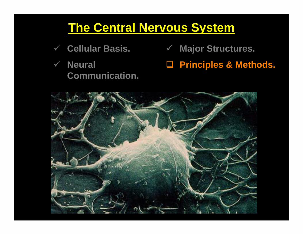
The Central Nervous System Cellular Basis.
Neural Communication.
Major Structures.
Principles & Methods.
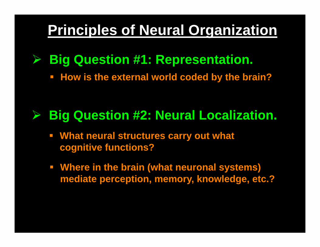
Principles of Neural Organization
Big Question #2: Neural Localization.
How is the external world coded by the brain? Big Question #1: Representation.
What neural structures carry out what cognitive functions?
Where in the brain (what neuronal systems) mediate perception, memory, knowledge, etc.?
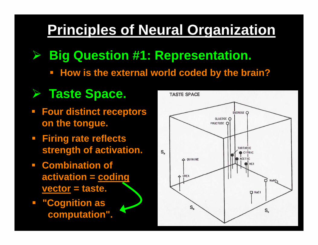
Principles of Neural Organization
How is the external world coded by the brain? Big Question #1: Representation.
Taste Space. Four distinct receptors
on the tongue. Firing rate reflects
strength of activation. Combination of
activation = coding vector = taste.
"Cognition as computation".
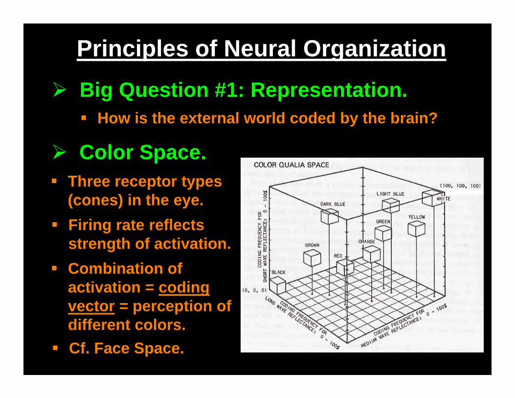
Principles of Neural Organization
How is the external world coded by the brain? Big Question #1: Representation.
Color Space. Three receptor types
(cones) in the eye. Firing rate reflects
strength of activation. Combination of
activation = coding vector = perception of different colors.
Cf. Face Space.
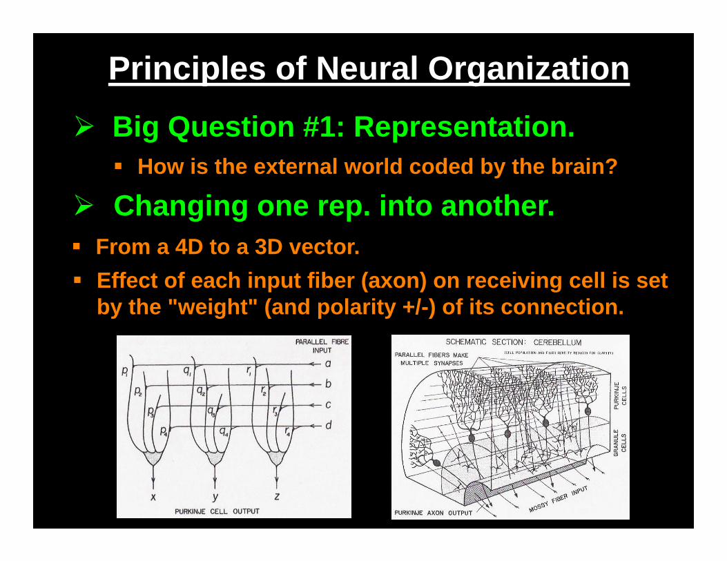
Principles of Neural Organization
How is the external world coded by the brain? Big Question #1: Representation.
Changing one rep. into another. From a 4D to a 3D vector. Effect of each input fiber (axon) on receiving cell is set
by the "weight" (and polarity +/-) of its connection.
Principles of Neural Organization
Cortical Maps. The Sensory and Motor Projection Areas. Retinotopic Mapping.
Flow of Information. Luria's Hierarchical Model. Felleman & van Essen's Parallel,
Distributed Hierarchies.
Cortical Areas. Brodmann's Cytoarchitectonics. Flechsig's Order of Myelination.
Big Question #2: Neural Localization.
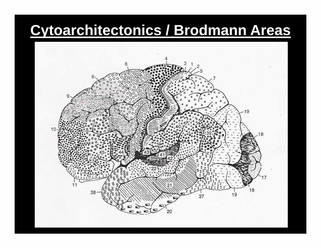
Cytoarchitectonics / Brodmann Areas
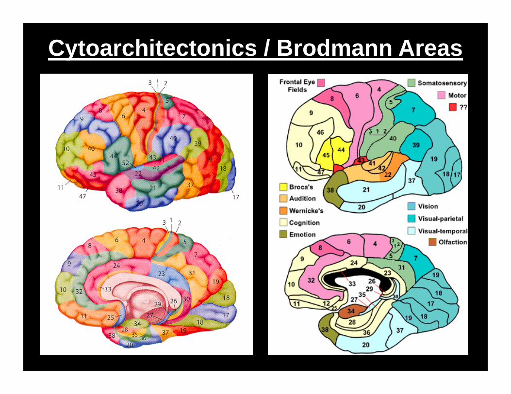
Cytoarchitectonics / Brodmann Areas
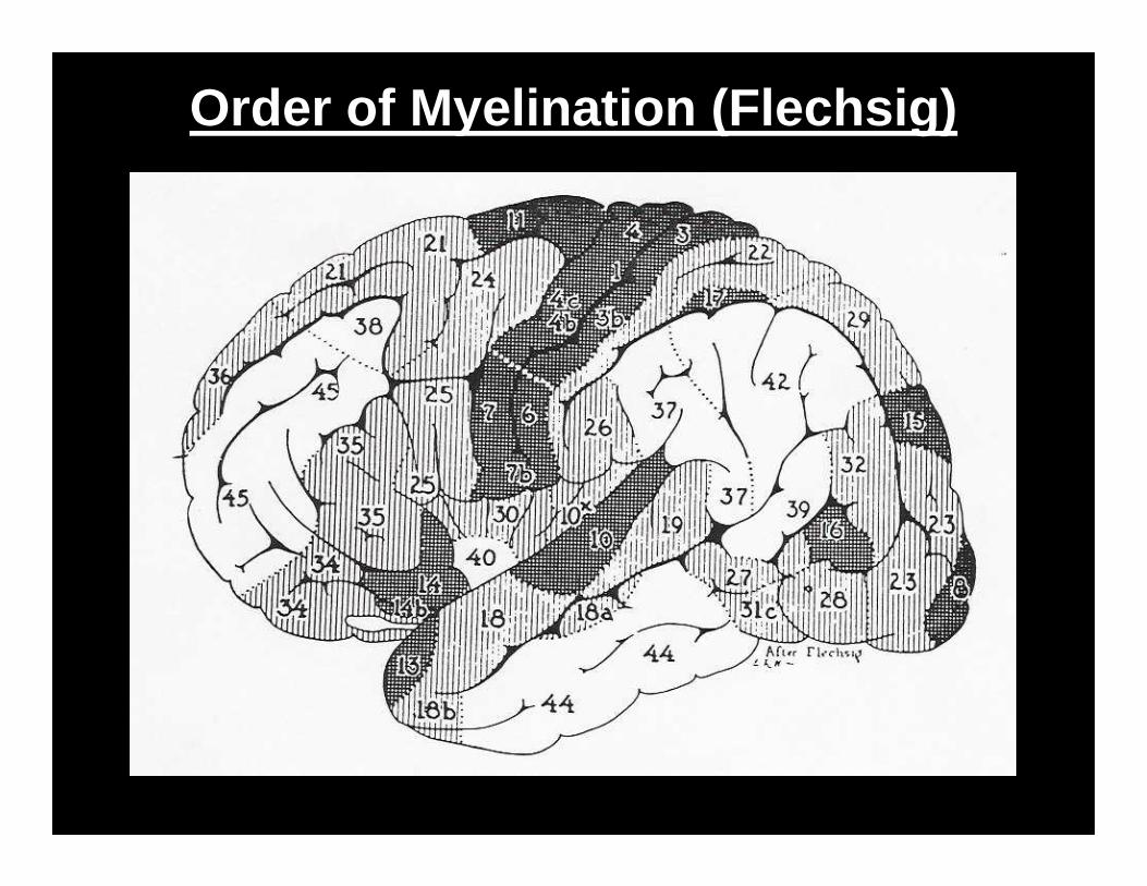
Order of Myelination (Flechsig)
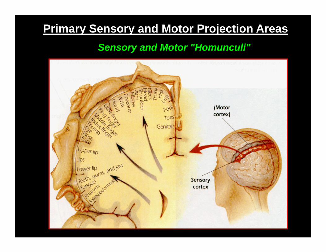
Primary Sensory and Motor Projection AreasSensory and Motor "Homunculi"
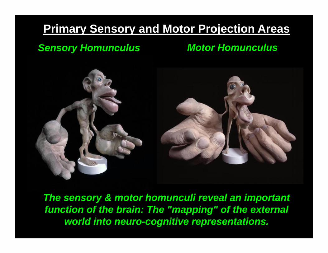
Primary Sensory and Motor Projection AreasSensory Homunculus Motor Homunculus
The sensory & motor homunculi reveal an important function of the brain: The "mapping" of the external
world into neuro-cognitive representations.
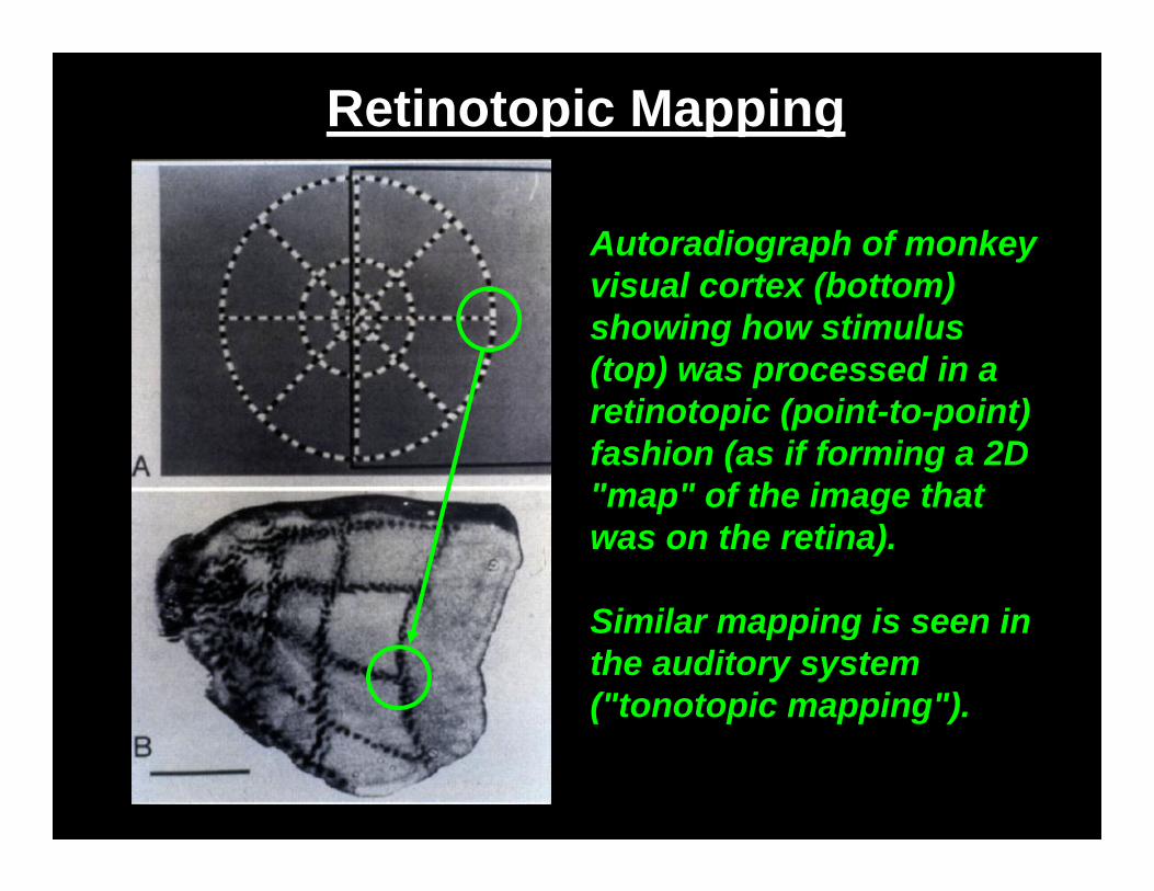
Retinotopic Mapping
Autoradiograph of monkey visual cortex (bottom) showing how stimulus (top) was processed in a retinotopic (point-to-point) fashion (as if forming a 2D "map" of the image that was on the retina).
Similar mapping is seen in the auditory system ("tonotopic mapping").
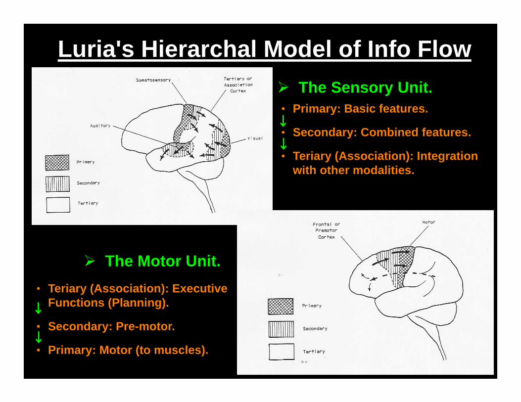
Luria's Hierarchal Model of Info Flow The Sensory Unit.• Primary: Basic features.
• Secondary: Combined features.
• Teriary (Association): Integration with other modalities.
The Motor Unit.• Teriary (Association): Executive
Functions (Planning).
• Secondary: Pre-motor.
• Primary: Motor (to muscles).
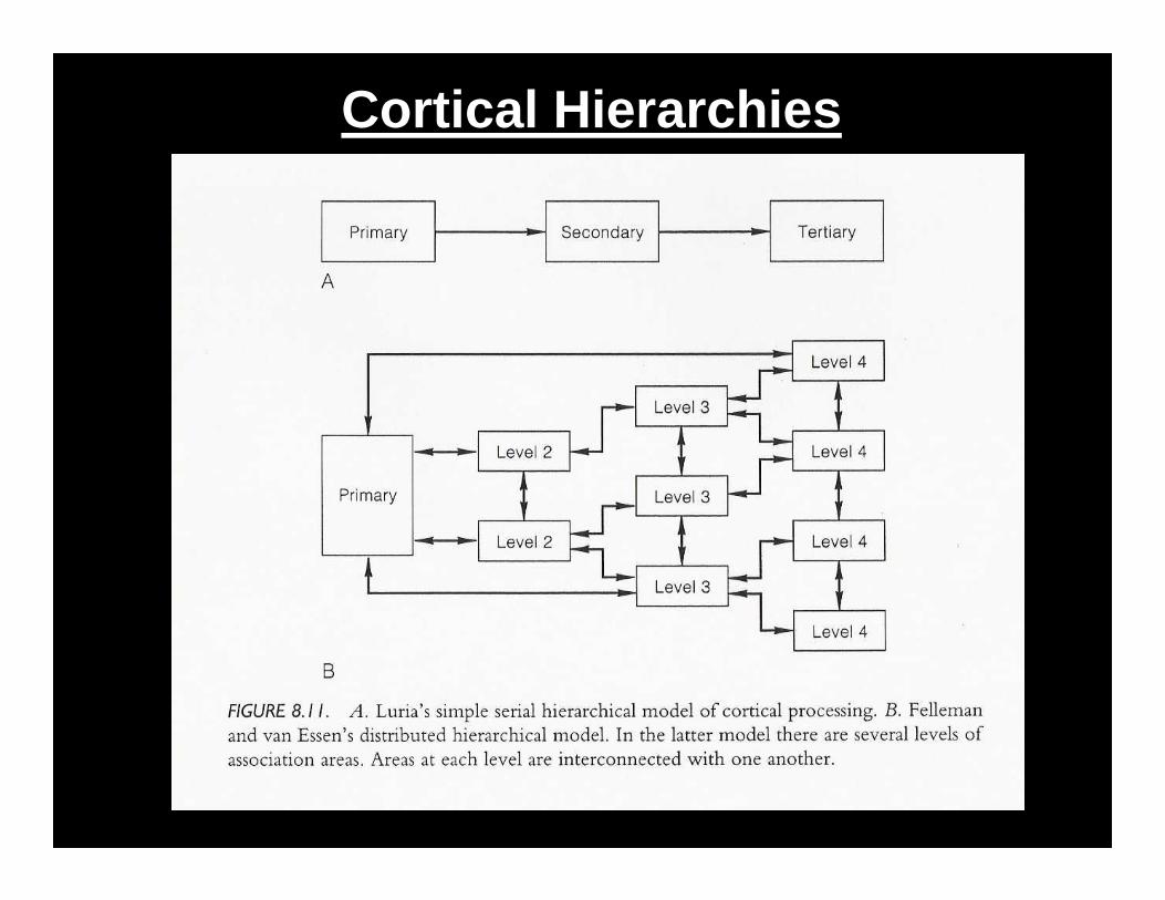
Cortical Hierarchies
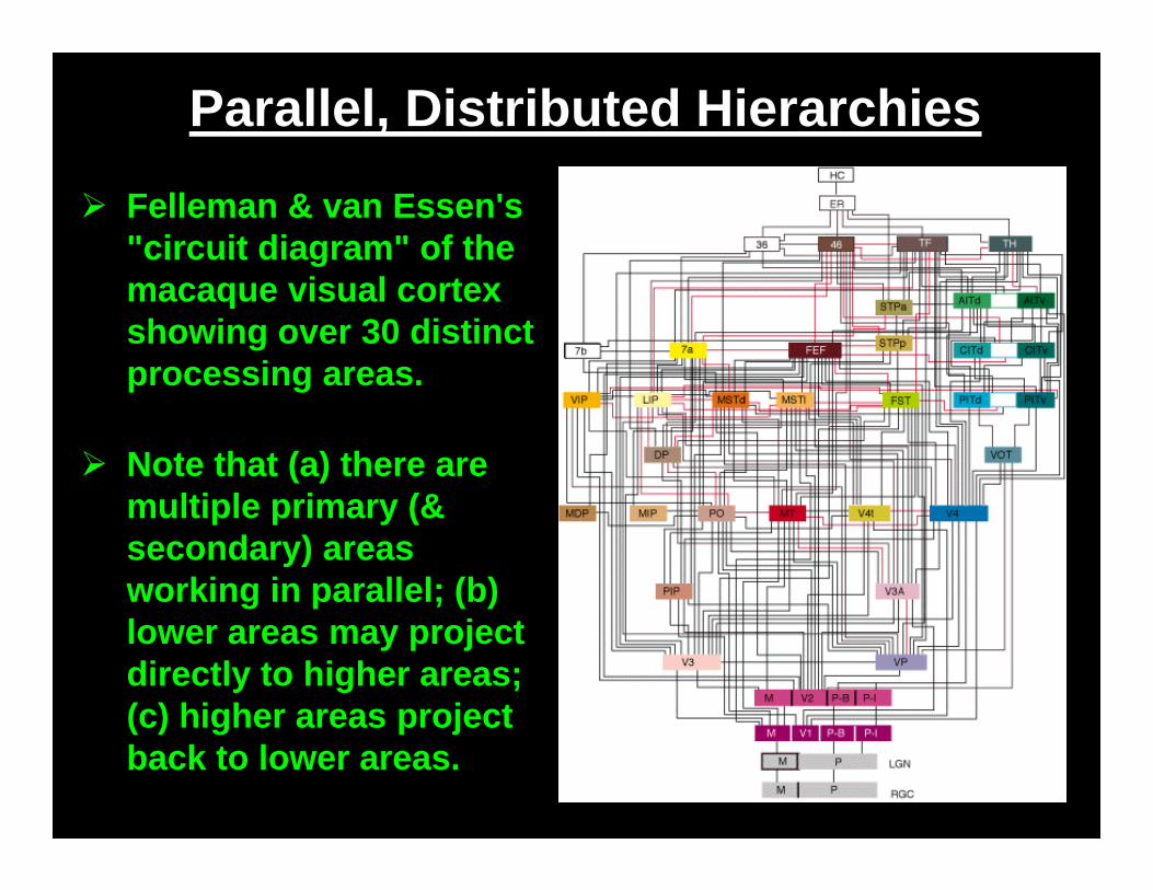
Parallel, Distributed Hierarchies
Felleman & van Essen's "circuit diagram" of the macaque visual cortex showing over 30 distinct processing areas.
Note that (a) there are multiple primary (& secondary) areas working in parallel; (b) lower areas may project directly to higher areas; (c) higher areas project back to lower areas.
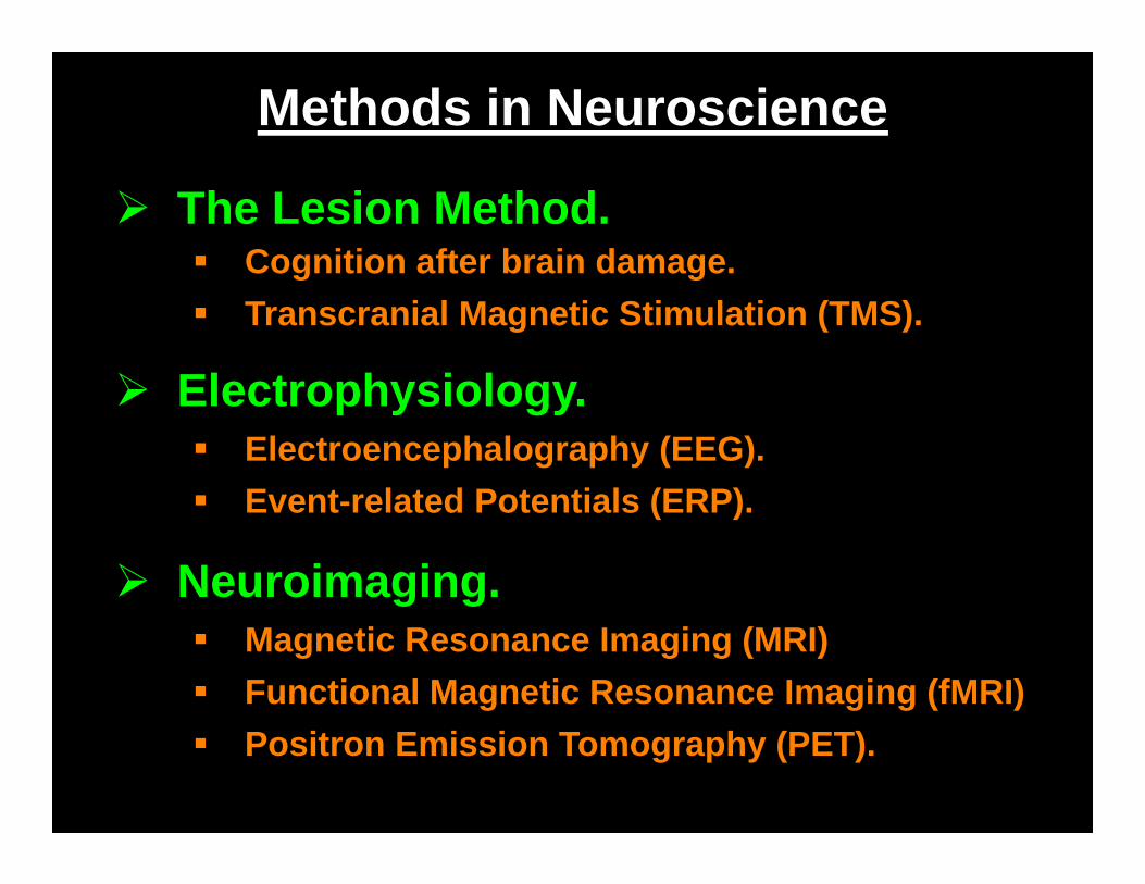
Methods in Neuroscience
The Lesion Method. Cognition after brain damage. Transcranial Magnetic Stimulation (TMS).
Electrophysiology. Electroencephalography (EEG). Event-related Potentials (ERP).
Neuroimaging. Magnetic Resonance Imaging (MRI) Functional Magnetic Resonance Imaging (fMRI) Positron Emission Tomography (PET).
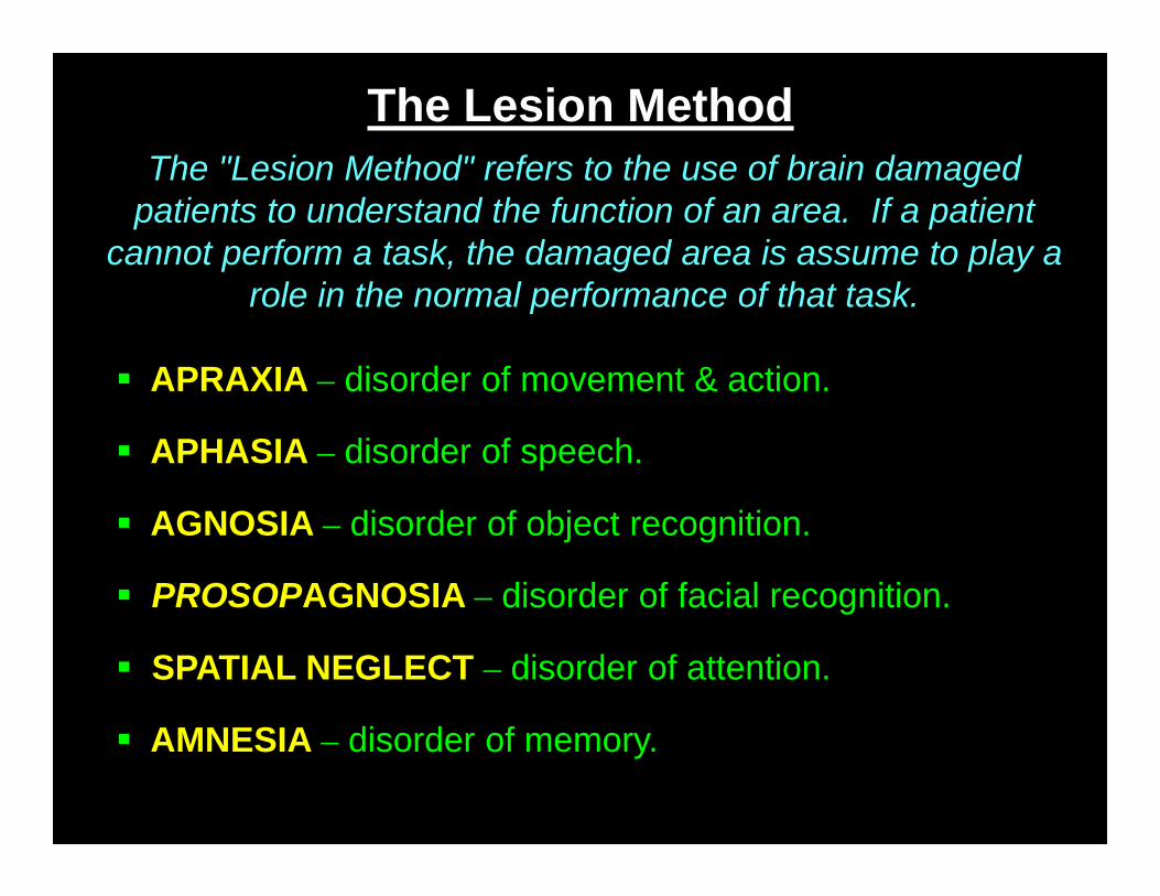
The Lesion Method
APRAXIA – disorder of movement & action.
AGNOSIA – disorder of object recognition.
PROSOPAGNOSIA – disorder of facial recognition.
APHASIA – disorder of speech.
AMNESIA – disorder of memory.
The "Lesion Method" refers to the use of brain damaged patients to understand the function of an area. If a patient
cannot perform a task, the damaged area is assume to play a role in the normal performance of that task.
SPATIAL NEGLECT – disorder of attention.
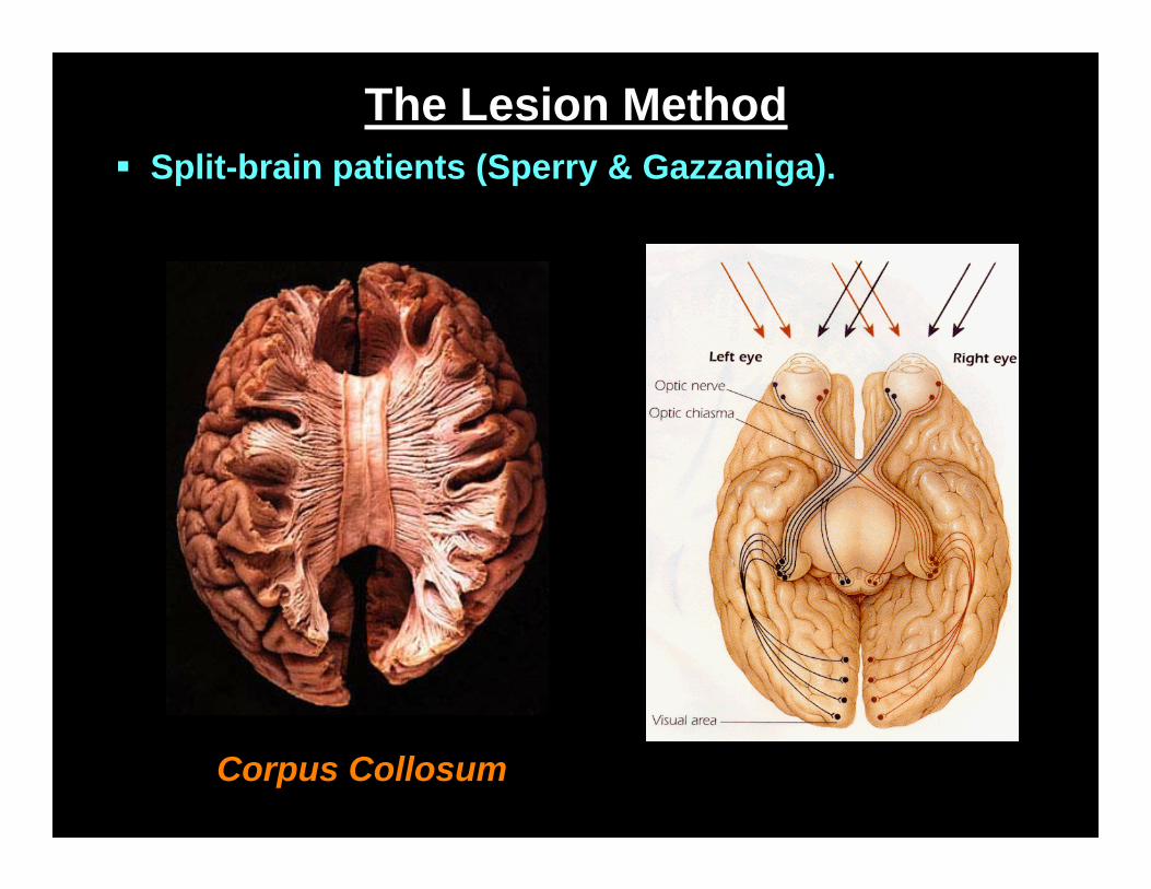
The Lesion Method
Corpus Collosum
Split-brain patients (Sperry & Gazzaniga).
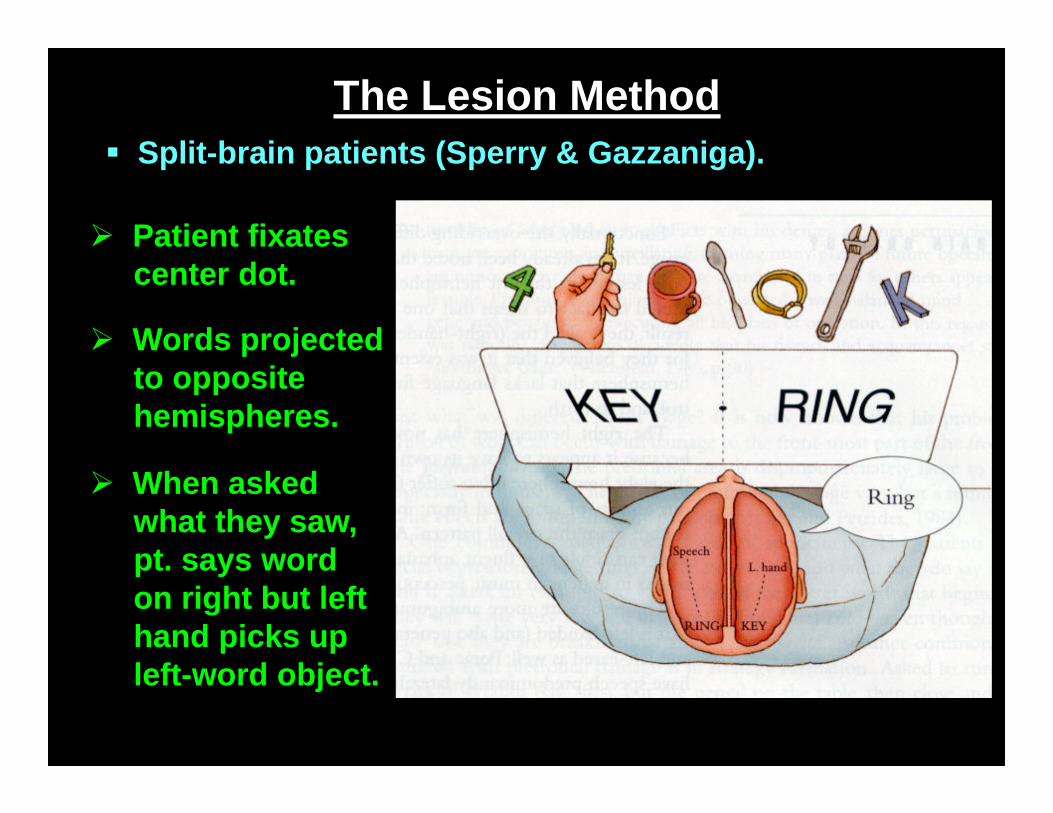
The Lesion Method Split-brain patients (Sperry & Gazzaniga).
Patient fixates center dot.
Words projected to opposite hemispheres.
When asked what they saw, pt. says word on right but left hand picks up left-word object.
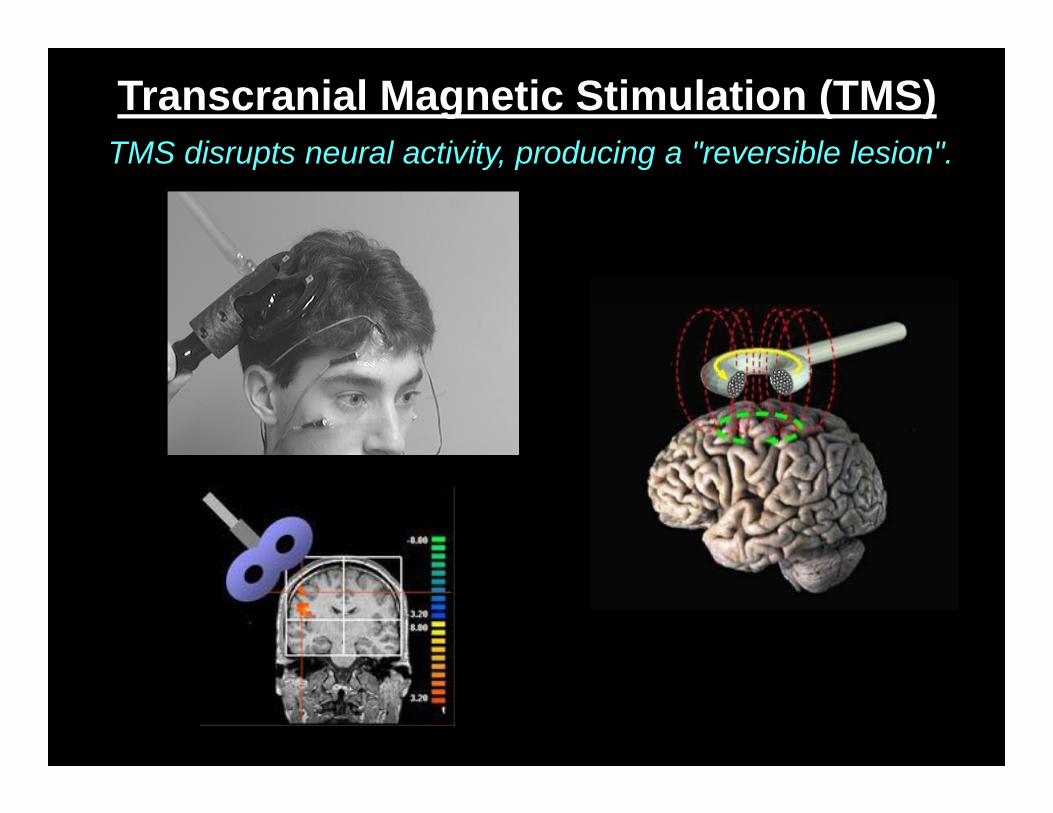
Transcranial Magnetic Stimulation (TMS)TMS disrupts neural activity, producing a "reversible lesion".
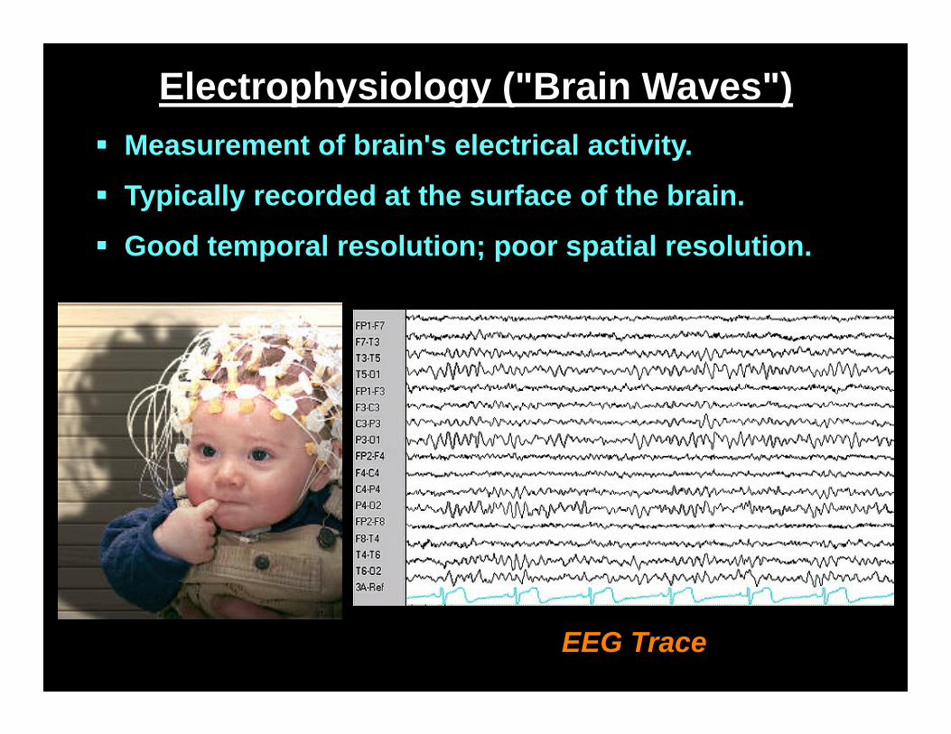
Electrophysiology ("Brain Waves") Measurement of brain's electrical activity.
Typically recorded at the surface of the brain.
Good temporal resolution; poor spatial resolution.
EEG Trace
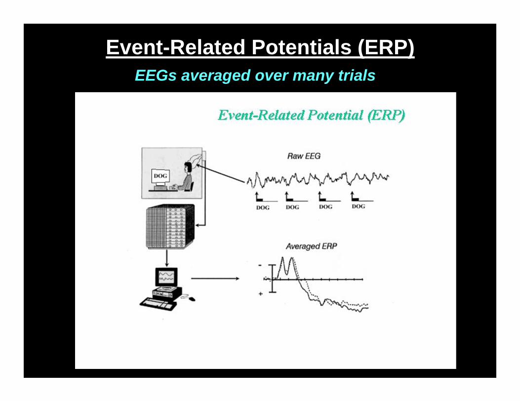
Event-Related Potentials (ERP)EEGs averaged over many trials
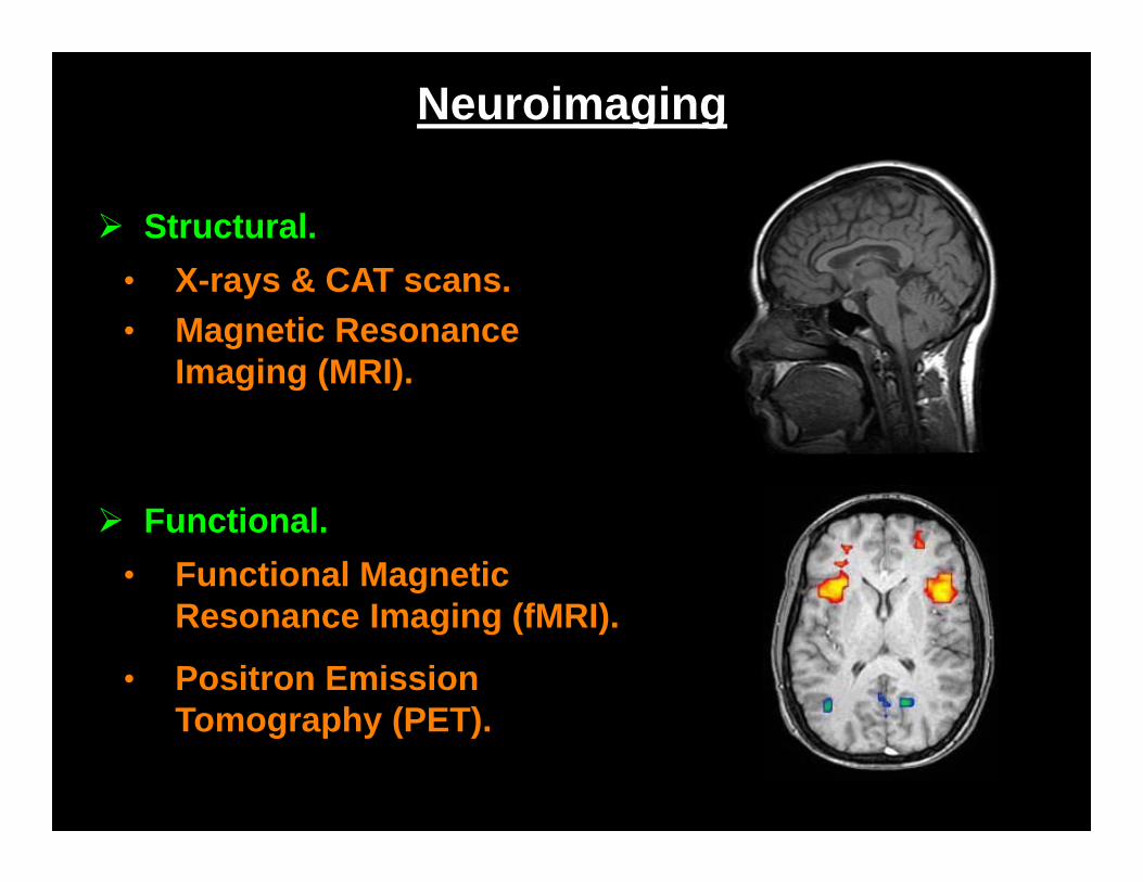
Neuroimaging
Structural.• X-rays & CAT scans.
Functional.• Functional Magnetic
Resonance Imaging (fMRI).
• Magnetic Resonance Imaging (MRI).
• Positron Emission Tomography (PET).
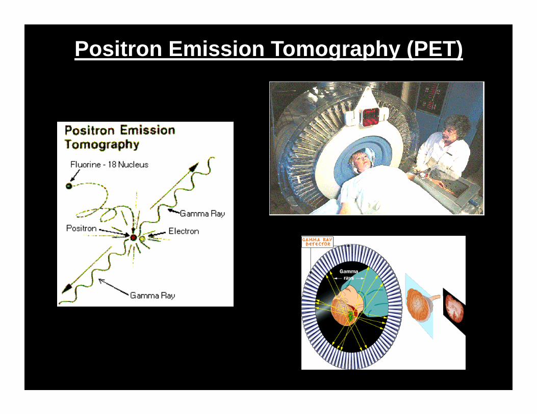
Positron Emission Tomography (PET)
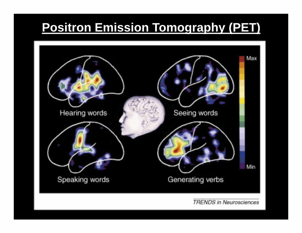
Positron Emission Tomography (PET)
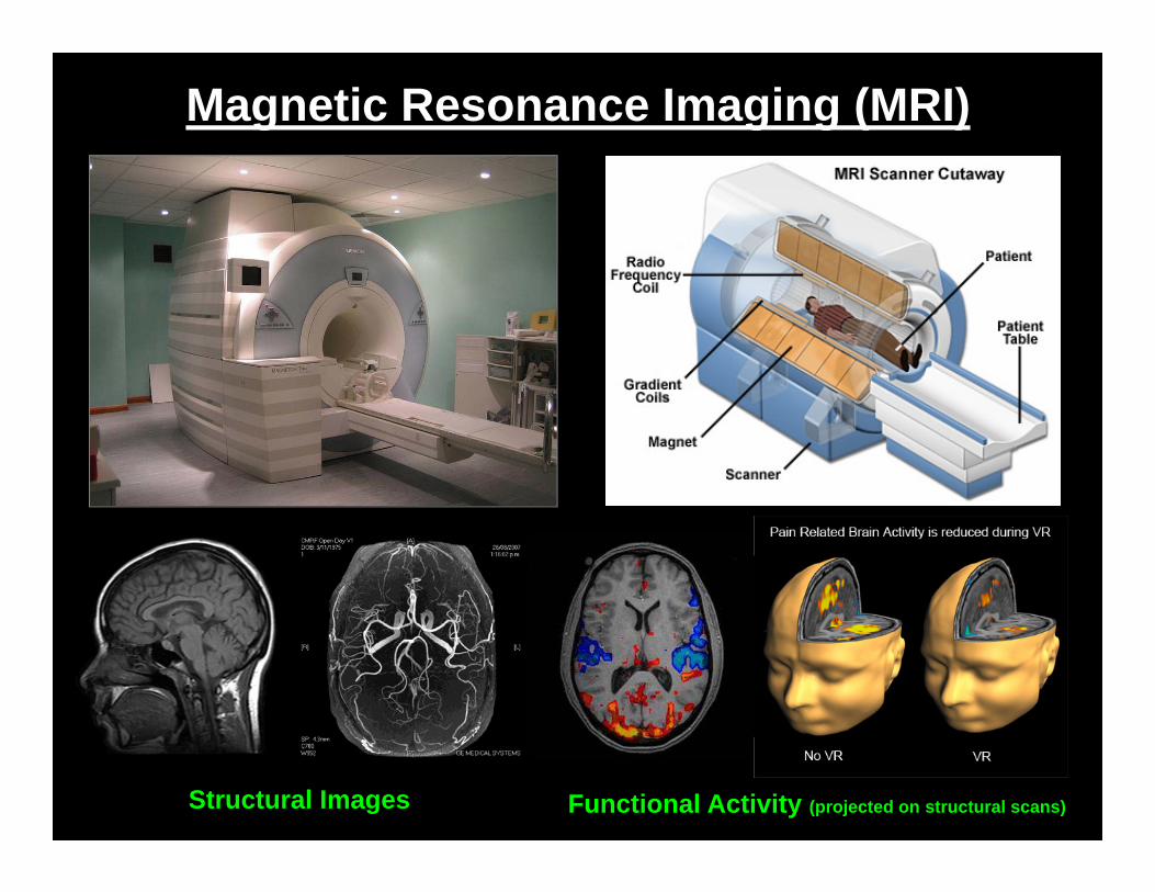
Magnetic Resonance Imaging (MRI)
Structural Images Functional Activity (projected on structural scans)
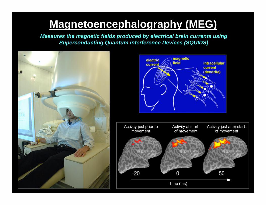
Magnetoencephalography (MEG)Measures the magnetic fields produced by electrical brain currents using
Superconducting Quantum Interference Devices (SQUIDS)
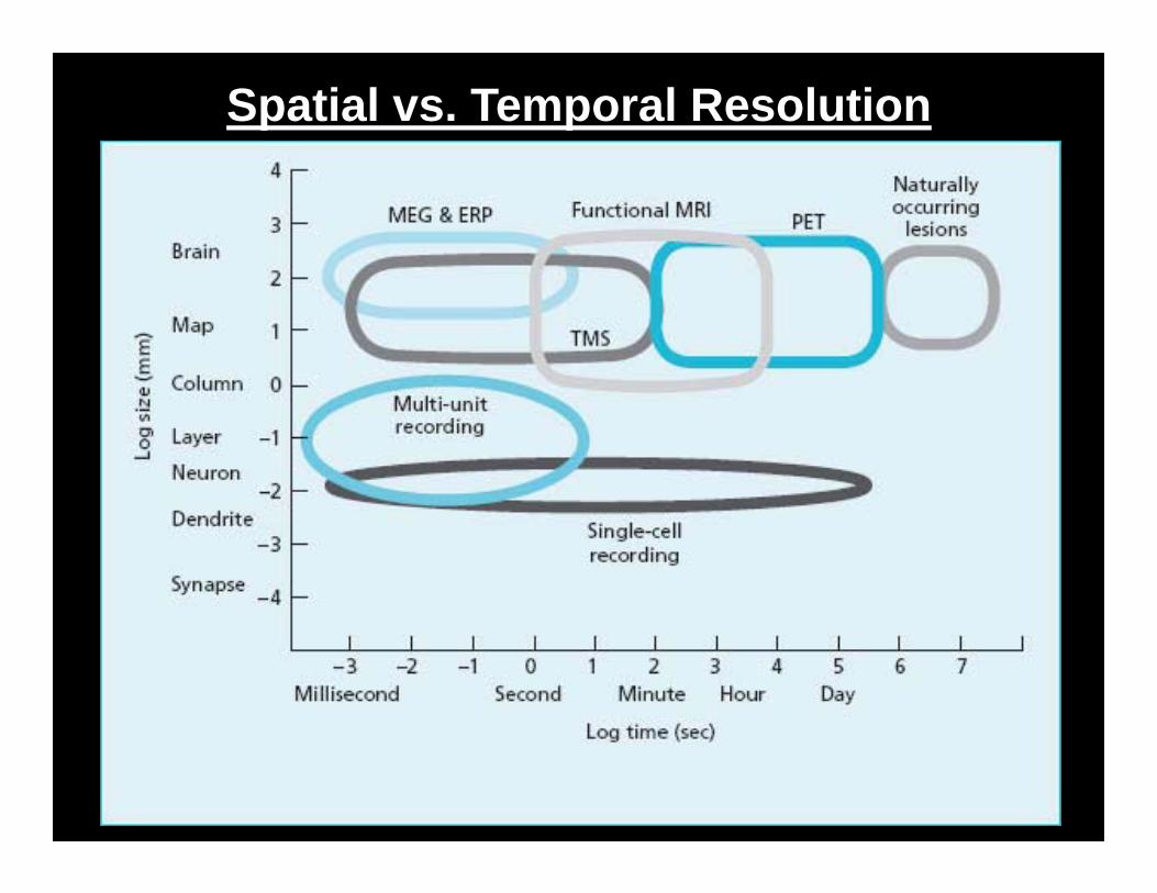
Spatial vs. Temporal Resolution
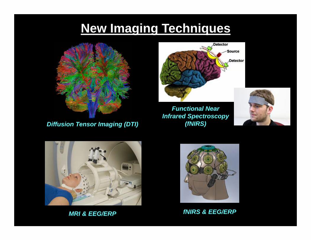
New Imaging Techniques
Diffusion Tensor Imaging (DTI)
Functional Near Infrared Spectroscopy
(fNIRS)
MRI & EEG/ERP fNIRS & EEG/ERP