Primary degeneration acromegaly case - bjo.bmj.com · tBlobnier (1937), MutchandMackay (1943),...
7
Brit. _J. Ophthal. (1972) 56, 25 Primary pigmentary degeneration of the retina and acromegaly in a case of * a0 pituitary acdenoma J. MI. SMAIL Sbhrewsbury Primary pigmentary degeneration of the retina has been linked with pituitary dysfunction since the complete clinical picture of primary dystrophy, hypogenitalism, obesity, poly- dactyly, and mental deficiency was fully documented several years ago (Laurence and Moon, i866; Biedl, I922). This syndrome, known as the Laurence-Mloon-Biedl syndrome, typifies the increasing tendency to associate retinal pigment dysplasia with endocrine dysfu-nction. The intermediate lobe of the pituitary gland secretes a hormone, melanophore-stimulat- ing hormone (MSH), which has been shown to have a marked effect on the skin colour of frogs (Hogben, I924) and to increase the skin pigmentation in human beings (Lerner and McGuire, I96I). While the above actions of MSH are accepted, this hormone has not been shown conclusively to have any effect on the pigment cells of the choroid or retinal pigment epithelium. In the hope that pituitary extracts containing MISH might be beneficial to the retina affected by primary pigmentary degeneration, several workers have used such extracts for therapeutic purposes. Many authors have claimed favourable results* while a considerable number of other workers have found no such improvement.t It is not within the scope of this paper to enter into this controversy. One would expect that, if MSH has any controlling effect whatever on the retinal pigment epithelium, cases of retinal pigmentary dysplasia would occur from time to time in patients suffering from pituitary tumours, especially those tumours associated with pituitary dysfunction. The commonest of these is the chromophobe adenoma. A thorough search through the neuro-ophthalmic literature has failed to reveal a single case. It seems worth while therefore to record a case in which primary pigmentary degeneration of the retina occurred in a man with a pituitary chromophobe adenoma in which the pars intermedia appeared to have been totally destroyed. Case report A man aged 44 years was admitted to the Midland Centre for Neurosurgery and Neurology, Smethwick, on July 2 I, i 968. About IO years previously his vision had started to deteriorate. Objects to each side became progressively more difficult to see and he could only be sure of his sight when observing objects directly in front. He had also become impotent over the past year. For about 6 months his Receised for publicationi June i6, 1971 Acddress for reprints: J. XI. Smail, FR.C.S., Ese Lat and 'Ihroat Hospital, Murisance, Slhrewsbury *Mussio-Fournier, Conti, Gonzales Vanirell and, Carriquiry (i949), Jones (193y). Scardaccionie (1938, I94), Basile (i939), Beauvieux, Ferron, anid Houssin (0950), 'Velter, D)esvignes, and Salmorn (1950), Diinitriu (1951), Stefrensen antii Kukora (1951), Rama (1952), Campbell anid Gebertt (955), Gundorova, Sclhekina, arnd Koitikova (1960), Nagdasaeva anid Savitskaya (1960), Buschke ('934) tBlobnier (1937), Mutch and Mackay (1943), Fran4ois (1950), Moreu Gonzalez Pola (1950), Gordon and Burke (1958), Rokitskaya ati(d Adishrin-Zade (ig6i1) on 9 June 2019 by guest. Protected by copyright. http://bjo.bmj.com/ Br J Ophthalmol: first published as 10.1136/bjo.56.1.25 on 1 January 1972. Downloaded from
Transcript of Primary degeneration acromegaly case - bjo.bmj.com · tBlobnier (1937), MutchandMackay (1943),...
Brit. _J. Ophthal. (1972) 56, 25
Primary pigmentary degeneration of theretina and acromegaly in a case of
* a0pituitary acdenoma
J. MI. SMAIL
Sbhrewsbury
Primary pigmentary degeneration of the retina has been linked with pituitary dysfunctionsince the complete clinical picture of primary dystrophy, hypogenitalism, obesity, poly-dactyly, and mental deficiency was fully documented several years ago (Laurence andMoon, i866; Biedl, I922). This syndrome, known as the Laurence-Mloon-Biedl syndrome,typifies the increasing tendency to associate retinal pigment dysplasia with endocrinedysfu-nction.The intermediate lobe of the pituitary gland secretes a hormone, melanophore-stimulat-
ing hormone (MSH), which has been shown to have a marked effect on the skin colour offrogs (Hogben, I924) and to increase the skin pigmentation in human beings (Lerner andMcGuire, I96I). While the above actions of MSH are accepted, this hormone has notbeen shown conclusively to have any effect on the pigment cells of the choroid or retinalpigment epithelium. In the hope that pituitary extracts containing MISH might bebeneficial to the retina affected by primary pigmentary degeneration, several workershave used such extracts for therapeutic purposes. Many authors have claimed favourableresults* while a considerable number of other workers have found no such improvement.tIt is not within the scope of this paper to enter into this controversy.One would expect that, if MSH has any controlling effect whatever on the retinal
pigment epithelium, cases of retinal pigmentary dysplasia would occur from time to timein patients suffering from pituitary tumours, especially those tumours associated withpituitary dysfunction. The commonest of these is the chromophobe adenoma. A thoroughsearch through the neuro-ophthalmic literature has failed to reveal a single case. Itseems worth while therefore to record a case in which primary pigmentary degenerationof the retina occurred in a man with a pituitary chromophobe adenoma in which the parsintermedia appeared to have been totally destroyed.
Case report
A man aged 44 years was admitted to the Midland Centre for Neurosurgery and Neurology,Smethwick, on July 2 I, i 968.About IO years previously his vision had started to deteriorate. Objects to each side became
progressively more difficult to see and he could only be sure of his sight when observing objectsdirectly in front. He had also become impotent over the past year. For about 6 months his
Receised for publicationi June i6, 1971Acddress for reprints: J. XI. Smail, FR.C.S., Ese Lat and 'Ihroat Hospital, Murisance, Slhrewsbury
*Mussio-Fournier, Conti, Gonzales Vanirell and, Carriquiry (i949), Jones (193y). Scardaccionie (1938, I94), Basile (i939),Beauvieux, Ferron, anid Houssin (0950), 'Velter, D)esvignes, and Salmorn (1950), Diinitriu (1951), Stefrensen antii Kukora (1951), Rama(1952), Campbell anid Gebertt (955), Gundorova, Sclhekina, arnd Koitikova (1960), Nagdasaeva anid Savitskaya (1960), Buschke('934)
tBlobnier (1937), Mutch and Mackay (1943), Fran4ois (1950), Moreu Gonzalez Pola (1950), Gordon and Burke (1958), Rokitskayaati(d Adishrin-Zade (ig6i1)
on 9 June 2019 by guest. Protected by copyright.
http://bjo.bmj.com
/B
r J Ophthalm
ol: first published as 10.1136/bjo.56.1.25 on 1 January 1972. Dow
nloaded from
6. M. Smail
fingers and hands had increased in size, and his feet were also larger. He began to have headaches,mainly in the frontal region, about 2 months before admission, and he started to perspire freelyfrom about the same time.
PREVIOUS MEDICAL HISTORY
Apart from bilateral inguinal hernias which had been repaired surgically, he had never been illbefore.
FAMILY HISTORY
He was married and had two sons aged 25 and I9 years. His wife and children were well and therewas no relevant family history.
PHYSICAL EXAMINATION
He was a heavily built middle-aged man. His face was long with coarse features and his tonguelarge. His speech was laboured and not clearly understandable. He had large hands and feetand there was clubbing of the fingers. There was generalized weakness in all four limbs and hismovements were sluggish.The corrected visual acuity was right eye-counting fingers, left eye-6/6o. Both fundi showed
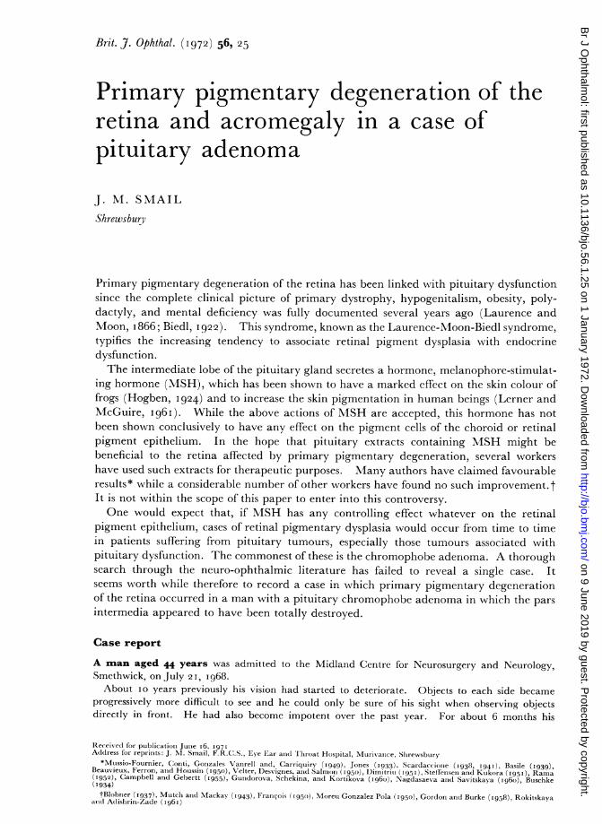
the typical changes of primary retinal pigmentary dysplasia of retinitis pigmentosa type. Thesechanges involved the whole of both fundi apart from the posterior poles. The optic discs wereof good colour, considering the extensive pigment changes and the arterial narrowing.The visual fields were plotted on the Goldmann perimeter and revealed tubular fields in both
eyes reduced to 30 from fixation with a white IV 4 target (Fig. I).
FIG. I Goldmann perimetrv on uly23, 968. White IV 4. Visualacuity: left counting fingers at 3 ft.,right 6/6o
LABORATORY INVESTIGATIONS
I7 oxosteroids 14 mg./24 hrs; protein-bound iodine 3.4 mg./ 100 ml.; blood sugar (random) IOO mg./IOO ml.; urea 20 mg./ioo ml.; haemoglobin I4-4 g./ioo ml.; erythrocyte sedimentation rate (Wester-gren) 26 mm./ist hr; white blood cells 5000/cubic mm. (neutrophils 52 per cent., lymphocytes41 per cent.; monocytes 6 per cent.; basophils I per cent.).
Wassermann reaction and Kahn test negative.Cerebrospinal fluid protein 78 mg./Ioo ml.
26
on 9 June 2019 by guest. Protected by copyright.
http://bjo.bmj.com
/B
r J Ophthalm
ol: first published as 10.1136/bjo.56.1.25 on 1 January 1972. Dow
nloaded from
Pituitary adenoma
X rays Chest: Showed some scarring of right apex due probably to healed tuberculosis.
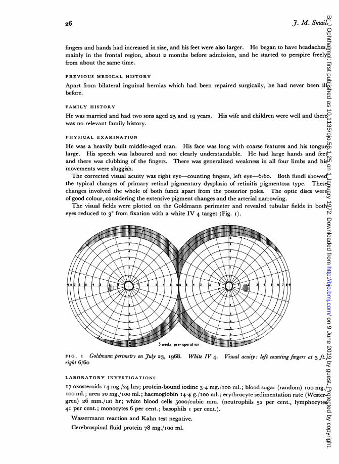
Skull: Showed moderate ballooning of the sella turcica with thinning of the dorsumsellae and erosion of the posterior clinnid processes (Fig. 2).l | l | , ~~~~~~~~~~~~~~~~~~~~~~~~~~~~~~~~~~~~~.E'.'.:..o...:.. ...: .:... .:..
FIG. 2 Lateral x ray.......... .Of skull, showing ba/-
loaning of the sella~turcica, thinness of the
dorstm sellae, and eosionof the posterior clinoidprocesses
,- ........... k ~~~~~~~~~~~~~~~~~~~~~~~~~~~~~~~~~~~........
C6arotid angiogramil: Revealed a tumour having a small suprasellai extension with no evidenceof further extension laterally or in any other direction.
Electroencephalographic findings were a few theta waves at 5 or 6 cycles per second of slightly higheramplitude than the background activity in the fronto-temporal regions, L> R, in the transverseposition after overbreathing.
OPERATION
On August 14, I968, a large irregular dumb-bell shaped pituitary tumour was completely removedby frontal craniotomy. The upper part of the tumour lay above the diaphragma sellae and was notencapsulated. This was possibly compressing the left optic nerve only very slightly and the rightone not at all. The lower part of the tumour was successfully excised from below the diaphragmasellae and the hypophyseal remains also removed.
PATHOLOGICAL REPORT
The specimen consisted of several fragments which were removed at the operation. All the materialwas examined.
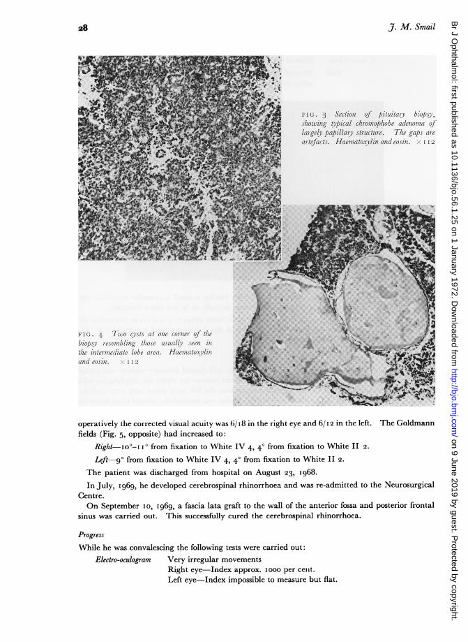
HistologyThe major part of the specimen consisted of a chromophobe adenoma, mostly of papillary pattern,but with occasional more solid areas (Fig. 3, overleaf). The usual amount of nuclear pleomorphismwas evident. Compressed but recognizable anterior lobe tissue appeared in one or two sections,together with degenerate foci containing cholesterol crystals and giant cells (Fig. 4, overleaf). Therewas also one single small fragment of posterior lobe, together with a collection of tiny colloid-filledcysts, but no recognizable intermediate lobe tissue.
POST-OPERATIVE PERIOD
The patient made a good recovery from the operation. It was necessary to prescribe pituitary snuffeach night since he had signs of diabetes insipidus; and cortisone 25 mg. twice daily and pheno-barbitone 30 mg. twice daily were also prescribed in an attempt to avert epilepsy. One week post-
27
on 9 June 2019 by guest. Protected by copyright.
http://bjo.bmj.com
/B
r J Ophthalm
ol: first published as 10.1136/bjo.56.1.25 on 1 January 1972. Dow
nloaded from
J. M. Smail
in IC;.'( Sectiojn u /ti/oitriy bOPS/ J. I
ShowlZing tlc/al c/th nomoPhobe adenomna o?flarge) pa/pi/lary' structure. 'The gal/s tio-artefacAs. Haentettoxylin and eosinP.1. I 12
i (,. -i 'T'nfo cys.ts aIt otnte torner u] /he,bin/isv relsembling thf/tse tisual/ seen initle itt/eltrmediate lobe area. HIematoax li/tand11 eott.sl. I I2
operatively the corrected visual acuity was 6/ I 8 in the right eye and 6/i 2 in the left. The Goldmannfields (Fig. 5, opposite) had increased to:
Right IO°-I I 0 from fixation to White IV 4, 40 from fixation to White II 2.
Left 90 from fixation to White IV 4, 40 from fixation to White II 2.
The patient was discharged from hospital on August 23, I968.In July, I969, he developed cerebrospinal rhinorrhoea and was re-admitted to the Neurosurgical
Centre.On September IO, I969, a fascia lata graft to the wall of the anterior fossa and posterior frontal
sinus was carried out. This successfully cured the cerebrospinal rhinorrhoea.
ProgressWhile he was convalescing the following tests were carried out:
Electro-oculogram Very irregular movementsRight eye Index approx. iooo per cent.Left eye-Index impossible to measure but flat.
28
on 9 June 2019 by guest. Protected by copyright.
http://bjo.bmj.com
/B
r J Ophthalm
ol: first published as 10.1136/bjo.56.1.25 on 1 January 1972. Dow
nloaded from
Pituitary adenoma
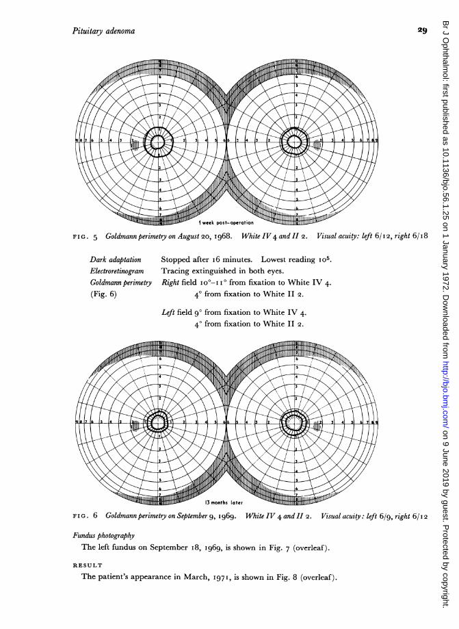
FIG. 5 Goldmannperimetry on August 20, I968. White IV4 and II 2. Visual acuity: left 6/12, right 6/i8
Dark adaptation Stopped after i6 minutes. Lowest reading Io5.Electroretinogram Tracing extinguished in both eyes.Goldmann perimetry Right field I0o-I I 0 from fixation to White IV 4.(Fig. 6) 40 from fixation to White II 2.
Left field 90 from fixation to White IV 4.40 from fixation to White II 2.
~' 13 months later "
F I G . 6 Goldmann perimetry on September 9, I 969. White IV 4 and II 2. Visual acuity: left 6/9, right 6/I 2
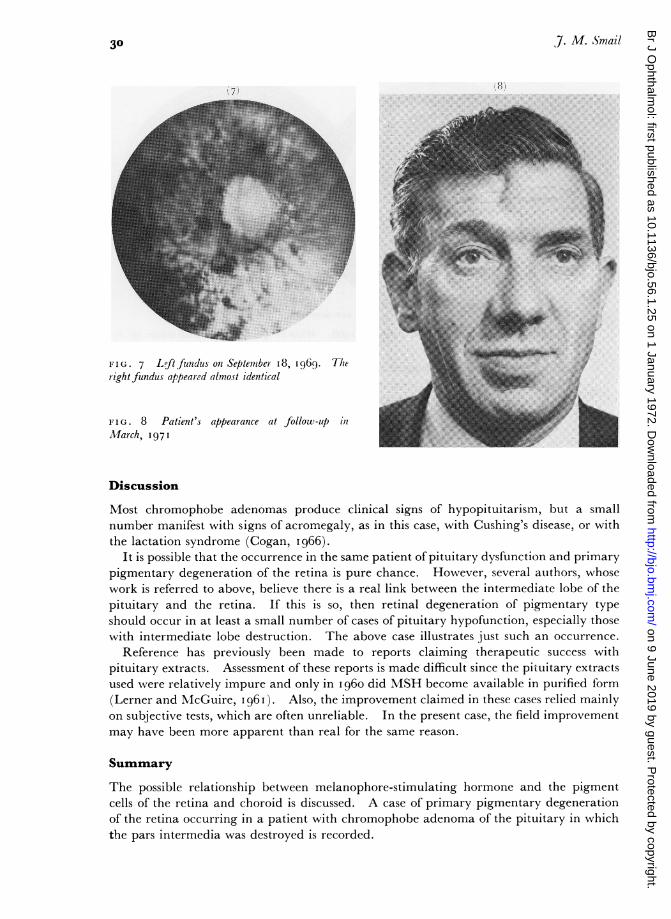
Fundus photographyThe left fundus on September i8, I969, is shown in Fig. 7 (overleaf).
RESULT
The patient's appearance in March, 1971, is shown in Fig. 8 (overleaf).
29
on 9 June 2019 by guest. Protected by copyright.
http://bjo.bmj.com
/B
r J Ophthalm
ol: first published as 10.1136/bjo.56.1.25 on 1 January 1972. Dow
nloaded from
07. M. Smail
FIG.7 L~ftfinds onl Septesbe I8, I969. Theright fundus appeared almost identical
FTIG. 8 Patient's appearance at follow-up in h........
Mach 19111
Discussion
Most chromophobe adenomas produce clinical signs of hypopituitarism, but a smallnumber manifest with signs of acromegaly, as in this case, with Cushing's disease, or withthe lactation syndrome (Cogan, i966).
It is possible that the occurrence in the same patient of pituitary dysfunction and primarypigmentary, degeneration of the retina is pure chance. However, several authors, whosework is referred to above, believe there is a real link between the intermediate lobe of thepituitary and the retina. If this is so, then retinal degeneration of pigmentary typeshould occur in at least a small number of cases of pituitary hypofunction, especially thosewith intermediate lobe destruction. The above case illustrates just such an occurrence.
Reference has previously been made to reports claiming therapeutic success withpituitary extracts. Assessment of these reports is made difficult since the pituitary extractsused were relatively impure and only in i1960 did MSH become available in purified f'orm(Lerner and MlcGuire, I1961I). Also, the improvement claimed in these cases relied mainlyon subjective tests, which are often unreliable. In the present case, the field improvementmay have been more apparent than real for the same reason.
SummaryThe possible relationship between melanophore-stimulating hormone and the pigmentcells of the retina and choroid is discussed. A case of primary pigmentary degenerationof the retina occurring in a patient with chromophobe adenoma of the pituitary in whichthe pars intermedia was destroyed is recorded.
30
on 9 June 2019 by guest. Protected by copyright.
http://bjo.bmj.com
/B
r J Ophthalm
ol: first published as 10.1136/bjo.56.1.25 on 1 January 1972. Dow
nloaded from
Pituitary adenoma 3'
I should like to thank Mr. J. S.nall and Mr. M. Rope:--Htll for pir nitting ml- to record this case, and toDr. Salmon for his help with the pathology.
References
BASILE, G. B. (I939) Ann. Ottal. clin. Ocul., 67, 4I2BEAUVIEUX, FERRON, and HOUSSIN (1950) Bull. Soc. Ophtal. Fr., p. 223BIEDL, A. (I922) Dtsch. med. Wschr., 48, I630BLOBNER, F. (I937) Klin. Mbl. Augenheilk., 98, 289BUSCHKE, W. (I934) Klin. Wschr., 13, 1785CAMPBELL, D. R.,and GEBERTT, S. (I955) Trans. ophthal. Soc. U.K., 75, 667COGAN, D. G. (I966) "Neurology of the Visual System", p. 226. Thomas, Springfield, Ill.DIMITRIU, T. (I 95 I) Bull. Soc. hillen. Ophtal., I9' 386FRAN9OIS, J. (1950) Bull. Soc. belge Ophtal., no. 95, p. 426GORDON, D. M., and BURKE, G. E. (1958) Amer. J. Ophthal., 45, 666GUNDAROVA, R. A., SCHEKINA, A. N., and KORTIKOVA, E. A. (I960) Vestn. Oftal., 73, no. 4, p. 37HOGBEN, L. T. (I924) "The Pigmentary Effector System". Oliver and Boyd, LondonJORES, L. (I933) Klin. Wschr., 12, I559LAURENCE, j. z., and MOON, R. C. (i866) Ophthal. Rev., 2, 32LERNER, A. B., and McGUIRE, j. S. (I96I) Nature (Lond.), 189, 176MOREU GONZALEZ POLA, A. (I950) Arch. Soc. oftal. hisp.-amer., 10, 458MUSSIO-FOURNIER, J. C., CONTI, O., GONZALES VANRELL, F., and CARRIQUIRY, P. (1949) Bull. Acad.
nat. Mid. Paris, 133, I03MUTCH, J. R., and MACKAY, D. (I943) Brit. J. Ophthal., 27, 434NAGDASAEVA, A. L., and SAVITSKAYA, N. F. (I960) Vestn. Oftal., 73, no. 4, p. 35RAMA, G. (1952) Rass. ital. Ottal., 21, 143ROKITSKAYA, L. V., and ADISHRNI-ZADE, R. F. (I96I) Oftal. Zh., I6, no. 5, p. 28ISCARDACCIONE, M. (I938) Boll. Oculist., 17, 755
(I94i) Ibid., 20, Io8STEFFENSEN, E. H., and KUKORA, J. (1951) Amer. J. Ophthal., 34, i665VELTER, E., DESVIGNES, P., and SALMON (1950) Bull. Soc. Ophtal. Fr., p. 9
on 9 June 2019 by guest. Protected by copyright.
http://bjo.bmj.com
/B
r J Ophthalm
ol: first published as 10.1136/bjo.56.1.25 on 1 January 1972. Dow
nloaded from