Preparation and characterization of PLGA loaded ...
10
International Journal of Green Pharmacy • Jul-Sep 2017 (Suppl) • 11 (3) | S438 Preparation and characterization of PLGA loaded nanoparticles obtained from D. melanoxylon Roxb. leaves for their antiproliferative and antidiabetic activity Md. Harun Al Rashid 1 , P. V. P. Deepak Bharadwaj 2 , Vivekananda Mandal 3 , Mahadeb Pal 2 , Subhash C. Mandal 1 , Rajarajan Amirthalingam Thandavarayan 4 1 Department of Pharmaceutical Technology, Pharmacognosy and Phytotherapy Research Laboratory, Division of Pharmacognosy, Jadavpur University, Kolkata, West Bengal, India, 2 Department of molecular medicine, Bose Institute, Kolkata, West Bengal, India, 3 Department of Pharmacy, Guru Ghasidas Central University, Bilaspur, Chhattisgarh, India, 4 Department of Cardiovascular Sciences, Center for Cardiovascular Regeneration, Houston Methodist Research Institute, TX 77030, USA Abstract Introduction: Diospyros melanoxylon Roxb. is traditionally used medicinal plant. Previously, we confirmed the better antiproliferative and antidiabetic activity of ethyl acetate fractions (EAF) obtained from D. Melanoxylon. To assessment the hypothesis if nanoparticle-encapsulated fraction could progress bioactivity. Materials and Methods: We prepared nanoparticle encapsulation based on poly (lactide-co-glycolide) (PLGA) and confirmed encapsulation by scanning electron microscopy. Sub EAF (SEAF) and nanoparticles of sub ethyl acetate (NSEAF) were characterized for their antiproliferative and antidiabetic activity. Antiproliferative activity was performed using the methylene blue assay. Antidiabetic activity was performed on the basis of α-amylase and α-glucosidase assay. Results and Discussion: Comparatively, NSEAF showed better activity on different cancerous cell lines HCT116 (inhibitory concentration 50% [IC 50 ] 32.89 ± 1.71 µg/ml), MCF-7 (IC 50 36.13 ± 0.96 µg/ml) and PC-3 (IC 50 32.39 ± 1.91 µg/ml), and antidiabetic activity on the basis of α-amylase (IC 50 35.62 ± 2.56 µg/ml), and α- glucosidase (IC 50 73.52 ± 1.13 µg/ml) action at a dose-dependent manner. It is also demonstrated that NSEAF exerted very low toxic effect against normal cell lines. Conclusion: For this reason, NSEAF studied in the present research may be considered as possible future drug candidates for the treatment and management of cancer and diabetes. Key words: Antidiabetes, antiproliferative, Diospyros melanoxylon, nanoparticles, poly (lactide-co-glycolide) Address for Correspondence: Md. Harun Al Rashid, Department of Pharmaceutical Technology, Pharmacognosy and Phytotherapy Research Laboratory, Division of Pharmacognosy, Jadavpur University, Kolkata, West Bengal, India. E-mail: [email protected] Received: 23-06-2017 Revised: 26-07-2017 Accepted: 04-08-2017 INTRODUCTION C ancer is the most leading causes of death throughout the Worldwide because patients diagnosed with it have a poor prognosis. [1,2] Chemotherapy is the essential module of cancer management. The common restrictions related with chemotherapy are enhanced drug toxicity and unwanted side effects. [3,4] Furthermore, tumors are highly aggressive and development of multidrug resistance to chemotherapy is a challenging aspect for cancer therapy. In this regard, the natural herbal extracts curcumin (food chemical present in turmeric), which is less toxic than other chemotherapeutic agents and can sensitize the cancer cells to chemotherapy has fascinated much awareness nowadays. [2,4] Lung cancer is a common type of cancer, but still the patients continue to have a poor prognosis. Cancer is a dangerous disease with the property of uncontrolled proliferation and growth of cells, look for agents that can cause apoptosis or programmed cell death in tumor cells has become a main objective in anticancer drug detection. [5] In response to apoptotic stimuli caused by ORIGINAL ARTICLE
Transcript of Preparation and characterization of PLGA loaded ...
International Journal of Green Pharmacy • Jul-Sep 2017 (Suppl) • 11 (3) | S438
Preparation and characterization of PLGA loaded nanoparticles obtained from
D. melanoxylon Roxb. leaves for their antiproliferative and antidiabetic activity
Md. Harun Al Rashid1, P. V. P. Deepak Bharadwaj2, Vivekananda Mandal3, Mahadeb Pal2, Subhash C. Mandal1, Rajarajan Amirthalingam Thandavarayan4
1Department of Pharmaceutical Technology, Pharmacognosy and Phytotherapy Research Laboratory, Division of Pharmacognosy, Jadavpur University, Kolkata, West Bengal, India, 2Department of molecular medicine, Bose Institute, Kolkata, West Bengal, India, 3Department of Pharmacy, Guru Ghasidas Central University, Bilaspur, Chhattisgarh, India, 4Department of Cardiovascular Sciences, Center for Cardiovascular Regeneration, Houston Methodist Research Institute, TX 77030, USA
Abstract
Introduction: Diospyros melanoxylon Roxb. is traditionally used medicinal plant. Previously, we confirmed the better antiproliferative and antidiabetic activity of ethyl acetate fractions (EAF) obtained from D. Melanoxylon. To assessment the hypothesis if nanoparticle-encapsulated fraction could progress bioactivity. Materials and Methods: We prepared nanoparticle encapsulation based on poly (lactide-co-glycolide) (PLGA) and confirmed encapsulation by scanning electron microscopy. Sub EAF (SEAF) and nanoparticles of sub ethyl acetate (NSEAF) were characterized for their antiproliferative and antidiabetic activity. Antiproliferative activity was performed using the methylene blue assay. Antidiabetic activity was performed on the basis of α-amylase and α-glucosidase assay. Results and Discussion: Comparatively, NSEAF showed better activity on different cancerous cell lines HCT116 (inhibitory concentration 50% [IC50] 32.89 ± 1.71 µg/ml), MCF-7 (IC50 36.13 ± 0.96 µg/ml) and PC-3 (IC50 32.39 ± 1.91 µg/ml), and antidiabetic activity on the basis of α-amylase (IC50 35.62 ± 2.56 µg/ml), and α- glucosidase (IC50 73.52 ± 1.13 µg/ml) action at a dose-dependent manner. It is also demonstrated that NSEAF exerted very low toxic effect against normal cell lines. Conclusion: For this reason, NSEAF studied in the present research may be considered as possible future drug candidates for the treatment and management of cancer and diabetes.
Key words: Antidiabetes, antiproliferative, Diospyros melanoxylon, nanoparticles, poly (lactide-co-glycolide)
Address for Correspondence: Md. Harun Al Rashid, Department of Pharmaceutical Technology, Pharmacognosy and Phytotherapy Research Laboratory, Division of Pharmacognosy, Jadavpur University, Kolkata, West Bengal, India. E-mail: [email protected]
Received: 23-06-2017 Revised: 26-07-2017 Accepted: 04-08-2017
INTRODUCTION
Cancer is the most leading causes of death throughout the Worldwide because patients diagnosed with it have a poor
prognosis.[1,2] Chemotherapy is the essential module of cancer management. The common restrictions related with chemotherapy are enhanced drug toxicity and unwanted side effects.[3,4] Furthermore, tumors are highly aggressive and development of multidrug resistance to chemotherapy is a challenging aspect for cancer therapy. In this regard, the natural herbal extracts curcumin (food chemical present in turmeric), which is less toxic than other chemotherapeutic agents and can sensitize the cancer cells to chemotherapy has fascinated much awareness nowadays.[2,4] Lung cancer is
a common type of cancer, but still the patients continue to have a poor prognosis. Cancer is a dangerous disease with the property of uncontrolled proliferation and growth of cells, look for agents that can cause apoptosis or programmed cell death in tumor cells has become a main objective in anticancer drug detection.[5] In response to apoptotic stimuli caused by
OR
IGIN
AL
AR
TIC
LE
Al Rashid, et al.: Antiproliferative and antidiabetic activity of PLGA loaded nanoparticles
International Journal of Green Pharmacy • Jul-Sep 2017 (Suppl) • 11 (3) | S439
anticancer drugs, some proteins (such as cytochrome C) translocate to the mitochondrial intermembrane space into the cytoplasm that mediates the activation of caspase cascades, which in turn eventually encourages apoptosis. Key regulator calcium act as a cell survival because cytosolic free calcium concentration is a chief regulatory factor for huge number of cellular processes such as muscle contraction, secretion, cell differentiation, and apoptosis. Although sustained elevation of intracellular calcium plays a responsibility in cell death as well.[6] The proapoptotic properties of calcium are mediated by a various series of calcium-sensitive issues that are compartmentalized in different intracellular organelles, together with endoplasmic reticulum and mitochondria.[7] Extreme calcium fill to the mitochondria could encourage apoptosis by damaging mitochondrial function, for example, generation of reactive oxygen species (ROS) by damaged respiratory chain.[8] Calcium accumulation has also been reported to stimulate mitochondrial ROS production in cells.[9,10] Mitochondrial calcium uptake also activates important membrane depolarization, ensuing in a reduce in the mitochondrial membrane potential.[7]
Diabetes mellitus is a complex metabolic disease of the endocrine system, characterized by hyperglycemia due to deficiency in insulin secretion, insulin action, or both.[11] This metabolic disease leads to multiple macro- and microvascular difficulties which affect some organs, including the kidneys, heart, skin, and brain.[12] The incidence of the disease is on rise worldwide, is likely to hit 300 million by 2025 with India anticipated to have the biggest number of diabetic cases.[13]
Type 2 diabetes is one of the major intimidations to human health due to rising incidence, chronic course and disabling complications.[14] The conformist obtainable approaches for diabetes consist of stimulation of endogenous insulin secretion, enhancement of the action of insulin at the target tissues, oral hypoglycemic agents, such as biguanids and sulfonylureas and the inhibition or degradation of dietary starch by glycosidases such as α-amylase and α-glucosidase.[15]
Pancreatic α-amylase is a key enzyme in the digestive system and catalyzes the preliminary step in hydrolysis of starch to a combination of lesser oligosaccharides consisting of maltose, maltotriose, and a number of a-(l-6) and a-(1-4) oligoglucans. These are then acted on by α-glucosidases and additionally degraded glucose which on absorbtion enters the bloodstream. Degradation of this dietary starch proceeds quickly and leads to eminent PPHG (post-prandial hyperglycemia). The activity of human pancreatic a-amylase has been revealed in the small intestine associates to an increase in post-prandial glucose levels, the control of which is a significant feature in the treatment of type 2 diabetes.[16] Therefore, retardation of starch digestion by inhibition of enzymes such as α-amylase acting a key function in the management of diabetes. Inhibitors of pancreatic α-amylase delay
carbohydrate digestion causing a decrease in the rate of glucose absorption and lowering the post-prandial serum glucose levels.[17] Several inhibitors presently in clinical utilize are acarbose and miglitol which reduce glycosidases such as α-glucosidase and α-amylase while others such as voglibose reduce α-glucosidase. On the other hand, several of these fail to reduce diabetic complications. The major side effects of these inhibitors are gastrointestinal, viz., bloating, abdominal discomfort, diarrhea, and flatulence.[18]
Presently, nanotechnology offered diverse proficient and safe drug delivery system where a constant dose of therapeutic agents is delivered directly to the cells, tissues and organs over an extended period resulting in an alternative therapeutic approach. By verifying the performance of the drugs within the tissues and cells such delivery systems are predicted as potential therapy candidates leading to determined obliteration of the target area by minimizing arbitrary drug distribution.[19] Modern nanoparticulate dosage forms such as polymeric nanoparticles, nanocapsules, liposomes, solid lipid nanoparticles, and nanoemulsions, all of which can improve drug solubility. Broadly, nanoparticulate drug delivery improves solubility and bioavailability, pharmacological activity and tissue macrophage distribution, while preventing physical and chemical degradation.[20,21]
Application of nanotechnology to plant extracts has exposed an beneficial approach for herbal drugs bearing the abundant features that nanostructured systems have to offer including solubility, bioavailability, pharmacological activity improvement, protection from toxicity, sustained delivery, and protection from physical, and chemical degradation.[9,10] Presently, nanotechnological procedures concerning medicinal plants have given numerous novel delivery systems, including polymeric nanoparticles. These are made from biodegradable and biocompatible polymers such as chitosan (CS), symbolize an alternative for controlled drug delivery.[10] Hereby, we select D. melanoxylon which has a potent antiproliferative and antidiabetic potential apart from other medicinal significances such as anti-inflammatory, antiatherosclerotic, antimutagenic, anticarcinogenic, antibacterial, and antiviral activities.[22,23]
Many plant derived compounds are known to have curative potentials. World Health Organization estimated that approximately 80% of world’s inhabitants rely mainly on traditional herbal medicines for their primary health care and at least 119 chemical substances derived from 90 plant species can be considered as important drugs.[24] Traditional herbal medicines are getting more importance in the treatment of various diseases as they are free from side effects and less expensive when compared to synthetic agents.[25-27]
In sight of the above conditions, it is of enormous consequence to investigate the preparation of polymeric nanoparticles by Diospyros melanoxylon and check its antiproliferative and antidiabetic activities. In our previous

Al Rashid, et al.: Antiproliferative and antidiabetic activity of PLGA loaded nanoparticles
International Journal of Green Pharmacy • Jul-Sep 2017 (Suppl) • 11 (3) | S440
work, we confirmed the antiproliferative activity against different cancerous cell lines and antidiabetic activity on the basis of α-amylase and α-glucosidase enzyme of different fractions from D. melanoxylon. It is also seen that ethyl acetate fraction (EAF) is more potent for their antiproliferative and antidiabetic activity. In this circumstance, we have prepared poly (lactide-co-glycolide) (PLGA) loaded nanoparticles of sub EAF (SEAF) and characterized them. In this research work, we also report the antiproliferative and antidiabetic activity of SEAF as well as NSEAF obtained from D. melanoxylon.
MATERIALS AND METHODS
Collection of Plant Materials
The leaves of D. melanoxylon known as kendu were collected from Dhenkanal District, Bhubaneswar, Odisha. The plant was authenticated using scientific literature, Botanical Survey of India, Central National Herbarium, PO- Botanic Garden, Howrah-711103. The specimen voucher No. CNH/28/2014/Tech.II was deposited in the Department of Pharmaceutical Technology, Jadavpur University, Kolkata, India.
Extraction and Fractionation Methods
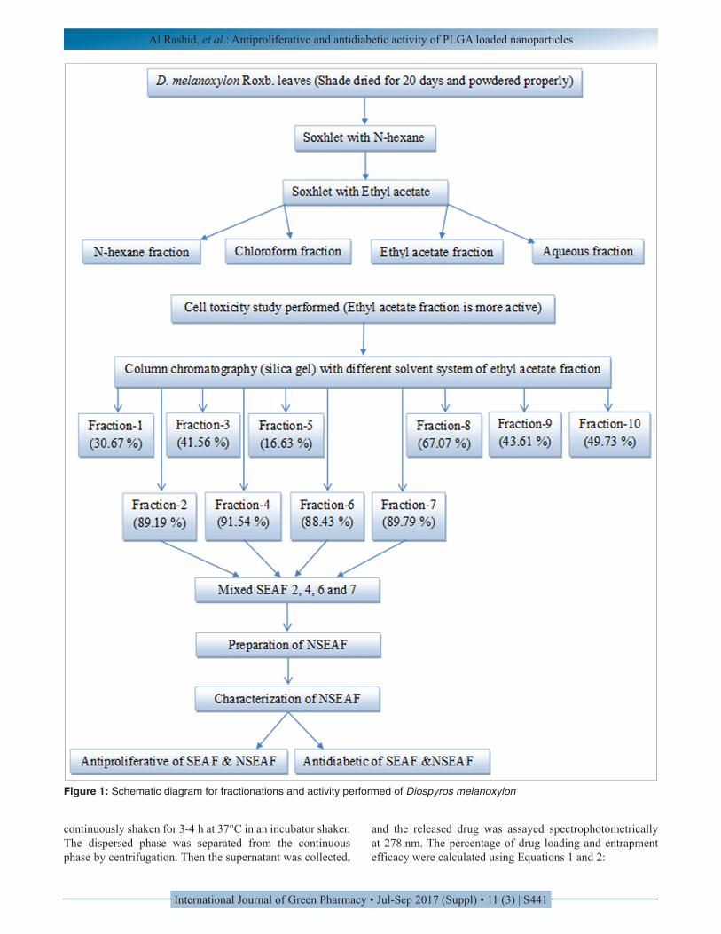
The leaves were washed properly, shade dried for 20 d, cut into small pieces and powdered by mechanical grinder. The powdered material was stored in an airtight container before extraction. Dried 500 g D. melanoxylon leaves were extracted with N-hexane (800 ml) by Soxhlet extraction method for 24 h for removing waxy and fatty materials. The residue was dried at room temperature and again extracted with ethyl acetate solvent for 36 h using Soxhlet apparatus. The extracts collected by filtering through Whatman filter paper and dried using rotary vacuum evaporator. With this extract four fractions such as N-hexane fraction, chloroform fraction (CF), EAF, and aqueous fraction was prepared using liquid–liquid partitioning system with the help of a separating funnel. EAF was collected and loaded in silica gel column chromatography (mesh size 230-400, bed volume ~130 ml, L = 25 cm, D = 4 cm) as our previous work has been proved that EAF having more activity. Mobile phase was selected by checking movement of the compounds on a thin layer chromatography (TLC) plate. For 40 g of EAF, 350 ml each of the following solvents mixture was applied: N-hexane:Ethyl acetate (NH:EA) 95:5, 90:10, 85:15, 80:20, 75:25, 70:30, 65:35, 60:40, 55:45, 50:50, 45:55, 40:60, 35:65, 30:70, 25:75, and 20:80. N-hexane:Ethyl acetate:Dichloromethane (NH:EA:DCM) 15:80:05, 10:80:10, and 05:85:10. Ethyl acetate: Dichloromethane:Methanol (EA:DCM:MEOH) 90:05:05. Ethyl acetate:Methanol (EA:MEOH) 90:10. Those fractions showed the same spots in TLC plate were collected in conical flask, dried in a vacuum evaporator
and tested for cell toxicity study against MCF-7. SEAF 2, 4, 6, and 7 showed maximum cell toxicity. Mixed these active SEAF properly and prepared nanoparticles for antiproliferative and antidiabetic activity [Figure 1].
Chemicals and Reagents
PLGA obtained as gift sample from Evonik India Pvt. Ltd., Mumbai - 400 072. (MW 50,000-75,000; poly D, L-lactide-co-glycolide ratio 75:25), polyvinyl alcohol (PVA, MW 125,000; S.D. Fine Chem. Pvt. Ltd., Mumbai, India), hydroxypropyl-β-cyclodextrin; HiMedia Laboratories, Mumbai. Dulbecco’s Modified Eagle’s Medium (DMEM), and fetal bovine serum (FBS) were obtained from Gibco, Grand Island, NY 14072, USA 1-716-774-6700. Roswell Park Media Institute (RPMI) 1640, non-essential amino acids, penicillin-streptomycin (PS), L-glutamine, and gentamicin were obtained from HiMedia Laboratories Pvt. Ltd., Mumbai, India. All other chemicals used were of reagent grade and used as purchased without further purification.
Preparation of Nanoparticles
NSEAF were prepared by emulsion solvent evaporation method according to Maji et al. with some modifications.[28] SEAF and subsequently PLGA (75:25) (~250 mg) were dissolved in 1.5 ml of methanol and 1.5 ml of dichloromethane mixture (phase 1). PVA was dissolved in water (2.0 % w/v) (phase 2), and 0.5 mL of phase 2 was added dropwise into Phase 1 with homogenization at speed (8,000 rpm) using a high-speed homogenizer (IKA Laboratory Equipment, Model T10B Ultras-Turrax, Staufen, Germany). The prepared primary emulsion was then added slowly into 75 ml of 1.5% (w/v) PVA solution (phase 3) with a continuous homogenization (8,000 rpm), which produced a secondary emulsion and then sonicated for 2 min. The secondary emulsion was then placed on a magnetic stirrer and stirred overnight for evaporation to remove organic solvent and solidification of the particles. The NPs were first separated by centrifugation at 5,000 rpm for 4 min to separate larger particles, and then the supernatant was collected and recentrifuged at 18,000 rpm for 1 h. The solid particles, thus separated, were resuspended in Milli-Q water, and centrifuged to wash the particles to remove the excess PVA attached on the surface of the NPs and to remove the free drug. The washing was repeated 2 times. The separated NPs were frozen at −20°C and lyophilized (Laboratory Freeze Dryer, Instrumentation India Ltd., Kolkata, India) for 8-9 h to obtain a solid product.
Determination of Drug Loading
For the determination of drug loading, an accurately weighed amount of NSEAF (2 mg) was placed into a centrifuge tube, and 2 ml of 5% SLS-NaOH solution was added. It was
Al Rashid, et al.: Antiproliferative and antidiabetic activity of PLGA loaded nanoparticles
International Journal of Green Pharmacy • Jul-Sep 2017 (Suppl) • 11 (3) | S441
Figure 1: Schematic diagram for fractionations and activity performed of Diospyros melanoxylon
continuously shaken for 3-4 h at 37°C in an incubator shaker. The dispersed phase was separated from the continuous phase by centrifugation. Then the supernatant was collected,
and the released drug was assayed spectrophotometrically at 278 nm. The percentage of drug loading and entrapment efficacy were calculated using Equations 1 and 2:
Al Rashid, et al.: Antiproliferative and antidiabetic activity of PLGA loaded nanoparticles
International Journal of Green Pharmacy • Jul-Sep 2017 (Suppl) • 11 (3) | S442
Amount of drug loadingActual drug loading in nanoparticles 100%
Weight of nanoparticles sample
=
×
( ) Amount of drug loadingEntrapment efficiency % 100%Theoretical drug loading
= ×
Determiation of Particle Size and Zeta (ζ) Potential
Average particle size, polydispersity index, and zeta potential of different formulations were determined based on quasi-elastic light scattering method using Data Transfer Assistance software by Zetasizer Nano ZS 90 (Malvern Instruments, Malvern, UK) at 25°C. Briefly, 1 mg of sample was dissolved in 2 ml Milli-Q water and sonicated for 30 s in an ice bath. All measurements were performed in triplicate.
Surface Morphology and Particle Size Measurement Using Scanning Electron Microscopy (SEM)
Particle size and external morphology of the nanoparticles were studied by SEM (Model-JSM-6360; JEOL, Tokyo, Japan).
Cell Lines and Culture Conditions
The cell lines were ordered from the American Type Culture Collection, and those used in this study were Human colon cancer cells (HCT116), breast cancer cell (MCF-7) and prostate cancer cells (PC-3), normal human kidney epithelium cells (NKE), and normal lung tissue cells (WI-38). HCT116, MCF-7, and WI-38 cells were grown in DMEM medium with 10% FBS and 1% PS. PC-3 and NKE cells were grown in RPMI 1640 medium with 10% FBS and 1% PS. All cells were supplemented in a humidified atmosphere at 37°C in 5% CO2. Afterward, the exhausted medium was removed, replaced with fresh medium, and incubated again for 24 h. The cell cultures were then washed with PBS and were suspended using trypsin. Fresh medium was added to the cells.
Antiproliferative Assay
The methylene blue cytotoxicity assay was carried out according to the method previously described by Rathore et al. with slight modification.[22] Cells were seeded at 1.5 × 10⁴ cells in each well of 96-well plate using 100 µl of fresh culture medium and were allowed to attach for overnight. Cell counts were made to determine cell viability using the Trypan Blue exclusion method.[29] For screening, the cells (60-70% confluence) were treated with the fractions at different concentrations. Afterward, to obtain a curve, the
most active fractions were tested for cytotoxicity at different concentrations (6.25-100 µg/ml). After 48 h of treatment, the medium was removed from each well; methylene blue was added and incubated for 90 min on a shaker followed by decanting and washing with water to remove the extracellular dye. A solution (0.1 N HCl) was then added for detaching the dead cells. The colorimetric assay at 580 nm was performed on 96 wells micro plate. The cytotoxicity effects of different samples were calculated as a percentage of cell growth inhibition with respect to control and inhibitory concentration 50% (IC50) value calculated. The assay was performed in triplicates.
Inhibition of α-amylase Activity
α-amylase inhibition assay was carried out using a microtiter plate according to Sudha et al. based on the starch-iodine test with slight modification.[30] 75 µl of different concentrations of NPs (6.25-100 mg/ml) were added to 150 µl 0.02 M sodium phosphate buffer (pH 6.9 containing 6 mM sodium chloride) and 75 µl α-amylase solution, and incubated at 37°C for 15 min. Take 100 µl from each sample reaction solution, 750 µl soluble starch (1%, w/v), and 500 µl phosphate buffer solution was added and incubated at 37°C for 45 min. Take 25 µl of above mixture and added 2.5 ml of iodine reagent (5 mM I2 and 5 mM KI) and mixed properly for a uniform mixture. The color change was observed, and the absorbance was taken at 565 nm on a microplate reader. Absence of sample is the control reaction representing 100% enzymatic activity. To eliminate the absorbance produced by NSEAF, appropriate NSEAF controls without the enzyme were also included. The standard drug acarbose (α-amylase inhibitor) was used as a positive control. It is observed the dark blue color which indicates the presence of starch; a brownish color indicates partially degraded starch and a yellow color indicates the absence of starch in the reaction mixture. In the presence of inhibitors from the fraction, the starch added to the enzyme assay mixture is not degraded and gives a dark blue color complex whereas no color complex is developed in the absence of the inhibitor, indicating that starch is completely hydrolyzed by α-amylase. The assay was performed in triplicates.
Inhibition of α-glucosidase Activity
The α-glucosidase enzyme inhibition activity was carried out according to Kuppusamy et al. with slight modification.[31] It was determined by incubating100 µl of α-glucosidase enzyme (1 U/ml) solution with100 µl of phosphate buffer (pH 7.0) which contains 100 µl of enzyme inhibitor such as acarbose (6.25-100 µg/mL) at 37°C for 60 min in maltose solution. To stop the α-glucosidase action on maltose, the above reaction mixture was kept in boiling water for 3 min and cooled. To this, 2 mL of glucose reagent was added and its absorbance was measured at 540 nm to measure the amount of liberated glucose by the action of α-glucosidase enzyme.
Al Rashid, et al.: Antiproliferative and antidiabetic activity of PLGA loaded nanoparticles
International Journal of Green Pharmacy • Jul-Sep 2017 (Suppl) • 11 (3) | S443
The percentage inhibition and IC50 value was calculated. The assay was performed in triplicates.
Statistical Analysis
All tests were performed as three independent experiments; each carried out in triplicate. Results were expressed as mean values with standard deviations (mean ± SD) and were subjected to two-way analysis of variance. All the data were calculated using GraphPad Prism version 7.02 software. The results were considered significant when P ˂ 0.001.
RESULTS
Particle Size and Surface Morphology Using SEM
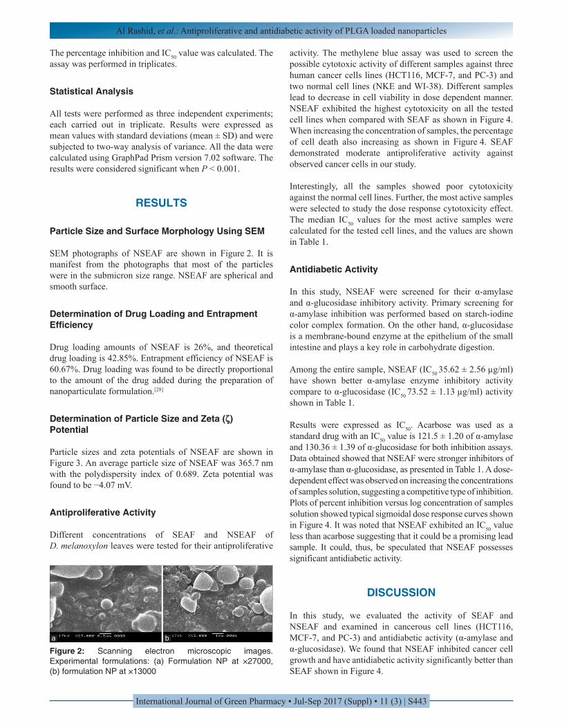
SEM photographs of NSEAF are shown in Figure 2. It is manifest from the photographs that most of the particles were in the submicron size range. NSEAF are spherical and smooth surface.
Determination of Drug Loading and Entrapment Efficiency
Drug loading amounts of NSEAF is 26%, and theoretical drug loading is 42.85%. Entrapment efficiency of NSEAF is 60.67%. Drug loading was found to be directly proportional to the amount of the drug added during the preparation of nanoparticulate formulation.[28]
Determination of Particle Size and Zeta (ζ) Potential
Particle sizes and zeta potentials of NSEAF are shown in Figure 3. An average particle size of NSEAF was 365.7 nm with the polydispersity index of 0.689. Zeta potential was found to be −4.07 mV.
Antiproliferative Activity
Different concentrations of SEAF and NSEAF of D. melanoxylon leaves were tested for their antiproliferative
activity. The methylene blue assay was used to screen the possible cytotoxic activity of different samples against three human cancer cells lines (HCT116, MCF-7, and PC-3) and two normal cell lines (NKE and WI-38). Different samples lead to decrease in cell viability in dose dependent manner. NSEAF exhibited the highest cytotoxicity on all the tested cell lines when compared with SEAF as shown in Figure 4. When increasing the concentration of samples, the percentage of cell death also increasing as shown in Figure 4. SEAF demonstrated moderate antiproliferative activity against observed cancer cells in our study.
Interestingly, all the samples showed poor cytotoxicity against the normal cell lines. Further, the most active samples were selected to study the dose response cytotoxicity effect. The median IC50 values for the most active samples were calculated for the tested cell lines, and the values are shown in Table 1.
Antidiabetic Activity
In this study, NSEAF were screened for their α-amylase and α-glucosidase inhibitory activity. Primary screening for α-amylase inhibition was performed based on starch-iodine color complex formation. On the other hand, α-glucosidase is a membrane-bound enzyme at the epithelium of the small intestine and plays a key role in carbohydrate digestion.
Among the entire sample, NSEAF (IC50 35.62 ± 2.56 µg/ml) have shown better α-amylase enzyme inhibitory activity compare to α-glucosidase (IC50 73.52 ± 1.13 µg/ml) activity shown in Table 1.
Results were expressed as IC50. Acarbose was used as a standard drug with an IC50 value is 121.5 ± 1.20 of α-amylase and 130.36 ± 1.39 of α-glucosidase for both inhibition assays. Data obtained showed that NSEAF were stronger inhibitors of α-amylase than α-glucosidase, as presented in Table 1. A dose-dependent effect was observed on increasing the concentrations of samples solution, suggesting a competitive type of inhibition. Plots of percent inhibition versus log concentration of samples solution showed typical sigmoidal dose response curves shown in Figure 4. It was noted that NSEAF exhibited an IC50 value less than acarbose suggesting that it could be a promising lead sample. It could, thus, be speculated that NSEAF possesses significant antidiabetic activity.
DISCUSSION
In this study, we evaluated the activity of SEAF and NSEAF and examined in cancerous cell lines (HCT116, MCF-7, and PC-3) and antidiabetic activity (α-amylase and α-glucosidase). We found that NSEAF inhibited cancer cell growth and have antidiabetic activity significantly better than SEAF shown in Figure 4.
Figure 2: Scanning electron microscopic images. Experimental formulations: (a) Formulation NP at ×27000, (b) formulation NP at ×13000
a b

Al Rashid, et al.: Antiproliferative and antidiabetic activity of PLGA loaded nanoparticles
International Journal of Green Pharmacy • Jul-Sep 2017 (Suppl) • 11 (3) | S444
Table 1: IC50 (µg/ml) of SEAF and NSEAF of D. melanoxylon for various antiproliferative and antidiabetic systems
Treatment Cancerous cell lines Normal cell lines Antidiabetic activity
HCT116 MCF-7 PC-3 NKE WI-38 α-amylase α-glucosidaseSEAF 47.71±3.09 49.57±1.25 47.75±3.25 319.95±3.36 464.14±1.62 47.60±2.76 89.21±1.08
NSEAF 32.89±1.71 36.13±0.96 32.39±1.91 453.88±5.37 564.99±1.67 35.62±2.56 73.52±1.13Each value in the table is represented as mean±SD (n=3) Means not sharing the same letter are significantly different (LSD) at P<0.0001 probability level in each column
Figure 3: (a) Average particle size: 365.7 nm, (b) zeta potential: −4.07 mV of NSEAF
a b
Figure 4: Cell toxicity studies of different samples in (a) HCT116, (b) MCF-7, (c) PC-3, (d) WI-38, and (e) NKE, antidiabetic activity of different samples in (f) α-amylase and (g) α-glucosidase. Standard curve of acarbose in (h) α-amylase and (i) α-glucosidase
a
d
g
b
e
h
c
f
i

Al Rashid, et al.: Antiproliferative and antidiabetic activity of PLGA loaded nanoparticles
International Journal of Green Pharmacy • Jul-Sep 2017 (Suppl) • 11 (3) | S445
Chemotherapy agents used for cancer therapy have limited efficacy mainly due to their low specificity toward cancer cells and poor pharmaco bioavailability in vivo, resulting in incidence of normal tissue toxicity and major side effects.[32-34] To address the problem, there is a high demand to explore therapeutic modalities with no or minimal side effects to normal cells. In this case, the study of using targeting molecules for cancer-specific therapy, linking to nanosized drug carriers, has been a promising strategy for targeted drug delivery and controlled drug release in the battle against cancer.[35,36]
On the other hand, drugs that inhibit hyperglycemia by suppressing hydrolysis of starch for α-amylase and carbohydrate for α-glucosidase have been found in the control of diabetes mellitus.[37,38] Many herbal extracts have been reported for their antidiabetic activities and are currently being used in Ayurveda for the treatment of diabetes.
More than 800 plants with antidiabetic properties according to the ancient Indian literature intelligences and that more than 1200 plants according to ethnopharmacological assessments are used hypoglycemic action.[39] Two carbohydrate hydrolyzing enzymes such as α-amylase and α-glucosidase are mainly responsible for post-prandial hyperglycemia. α-amylase begins the process of carbohydrate digestion by hydrolysis of 1, 4 glycosidic linkages of polysaccharides, viz., starch and glycogen to disaccharides and α-glucosidase catalyzes the disaccharides to monosaccharides, which leads to post-prandial hyperglycemia.[40,41] Therefore, inhibitors of α-amylase and α-glucosidase are helpful in the management of hyperglycemia as they hinder carbohydrate digestion, which subsequently decreases the post-prandial plasma glucose level.
In this study, we wanted to check if the efficacy of the drug could be improved by nanoparticle encapsulation. This would be of further benefit because the encapsulated drug could be directly carried into the cells, making it more bioactive. Here, in this circumstance, we tested our hypothesis on different cancerous cell lines, α-amylase and α-glucosidase of SEAF as well as NSEAF.
Our present study showed that NSEAF having high drug loading efficacy and entrapment efficacy. SEM images indicate that the NSEAF were spherical in shape and had a smooth surface. Average particle size of NSEAF was 365.7nm with a polydispersity index (PDI) 0.689 mV. Hence, the majority of the particles were found to be in submicron size with a relatively narrow variety of distribution as maintained by the PDI significance. The zeta potential of NSEAF was found to be around −4.07 mV. The electrostatic potential at the surface of a particle was determined by the zeta potential. Zeta potential value of −30-+30 mV recommends that the particles would stay in a suspended state for a longer time and are not disposed to rapid agglomeration in the liquid circumstances.[42,43] Therefore, zeta potential value of our
NSEAF proposes that should be stored in a lyophilized state and should be reconstituted only before experimental use.
Our previous study confirmed that EAF has better antiproliferative and antidiabetic potentials obtained from D. melanoxylon in in vitro model. furthermore, the role of some nanoparticles was also predicted in making slight changes in molecular orientation of the drug molecules and thereby biological activities. Therefore, the results of this study assume further significance. In this study, we confirm the antiproliferative and antidiabetic potentials of PLGA loaded NPs in five dose-dependent manner. The outcome recommended that the higher dose was more competent as compared with the lower dose. In this study, the cytotoxicity and antidiabetic activity of NSEAF were more than that of SEAF. The possible mechanism underlying the enhanced efficacy of NSEAF against different cancerous cell lines may include the enhanced intracellular drug accumulation by nanoparticle uptake.[44-46]
By the way to encapsulate orthodox drugs, PLGA had formally been used including coenzyme Q[47] Taxol[48] and camptothecin.[49] Although in the field of herbal medicine has also been reported to have enhanced cellular uptake and increased bioavailability of nanoparticle encapsulated the yellow curcumin present in turmeric known as Curcumin longma.[50,51] Hence, in this consequence, it is seen that NSEAF was established to be still more powerful and dynamic as compared with SEAF may be due to enhanced cellular uptake and increased bioavailability of nanoparticle encapsulated.
To our best information, this is the first effort to encapsulate SEAF of D. melanoxylon that is notorious to have antiproliferative and antidiabetic potentials of NSEAF. The results of exposure to cancerous cell lines, viz., HCT116, MCF-7, and PC-3 in vitro are encouraging for their possible therapeutic use as an anticancer agent. Interestingly, all the samples showed either mild or negligible cytotoxicity against both the normal cell lines (NKE and WI-38) which were used as the model normal cell lines.
Advance research will also be essential to investigate if the value of the principal pharmacologically active compound(s) can also be improved by PLGA nanoparticles encapsulation by carry out an experiment in both in vivo and in vitro methods. In this field, this work is rapidly gaining and should prove satisfying in the areas of toxicology and therapeutics and also in drug development for frightful diseases such as cancer and diabetes.
CONCLUSION
Preparation of PLGA loaded nanoparticle was effectively proficient by D. melanoxylon fraction. This one step, eco-friendly efficient process involves plant based bioresource

Al Rashid, et al.: Antiproliferative and antidiabetic activity of PLGA loaded nanoparticles
International Journal of Green Pharmacy • Jul-Sep 2017 (Suppl) • 11 (3) | S446
serving as both reducing as well as stabilizing agents. We report here the admirable inhibitory potential of SEAF as well as NSEAF against different cancerous cell lines, α-amylase and α-glucosidase activity. These activities are considered to be significant pharmacological targets for their antiproliferative properties. Similarly, it exhibited superior activityagainst α-amylase and α-glucosidase toward the exploration of its antidiabetic potential. This is the first ever report on different cancerous cell lines and antidiabetic activity on the basis of α-amylase and α-glucosidase inhibitory action of PLGA loaded nanoparticles of SEAF obtained from D. melanoxylon. The high potency of these biogenic NSEAF against cancerous cell lines and glycosidase inhibitory activities in vitro provided strong scientific evidence for antiproliferative and antidiabetic potential which intensely rationalize its use in the therapy and management of cancer and diabetes.
ACKNOWLEDGMENTS
Authors are grateful to “Innovation in Science Pursuit for Inspire Research” (INSPIRE) implemented by the Department of Science and Technology (DST, Government of India, New Delhi, India) for financial support. We are also thankful to the authority of Jadavpur University and Bose Institute, Kolkata, for affording necessary facilities.
REFERENCES
1. Erkan M, Kleeff J, Esposito I, Giese T, Ketterer K, Büchler MW, et al. Loss of BNIP3 expression is a late event in pancreatic cancer contributing to chemoresistance and worsened prognosis. Oncogene 2005;24:4421-32.
2. Fryer RA, Galustian C, Dalgleish AG. Recent advances and developments in treatment strategies against pancreatic cancer. Curr Clin Pharmacol 2009;4:102-2.
3. Anand P, Kunnumakkara AB, Newman RA, Aggarwal BB. Bioavailability of curcumin: Problems and promises. Mol Pharm 2007;4:807-18.
4. Somasundaram S, Edmund NA, Moore DT, Small GW, Shi YY, Orlowski RZ. Dietary curcumin inhibits chemotherapy-induced apoptosis in models of human breast cancer. Cancer Res 2002;62:3868-75.
5. Reed JC. Apoptosis-targeted therapies for cancer. Cancer Cell 2003;3:17-22.
6. Demaurex N, Distelhorst C. Cell biology. Apoptosis-the calcium connection. Science 2003;300:65-7.
7. Hajnóczky G, Davies E, Madesh M. Calcium signaling and apoptosis. Biochem Biophys Res Commun 2003;304:445-54.
8. Wang X. The expanding role of mitochondria in apoptosis. Genes Dev 2001;15:2922-33.
9. Ajazuddin, Saraf S. Applications of novel drug delivery system for herbal formulations. Fitoterapia 2010;81:680-9.
10. Bonifácio BV, Silva PB, Ramos MA, Negri KM, Bauab TM, Chorilli M. Nanotechnology-based drug delivery systems and herbal medicines: A review. Int J Nanomedicine 2014;9:1-15.
11. Arunachalam K, Parimelazhagan T. Antidiabetic activity of Ficus amplissima Smith. Bark extract in streptozotocin induced diabetic rats. J Ethnopharmacol 2013;147:302-10.
12. Sarma AD, Mallick AR, Ghosh A. Free radicals and their role in different clinical conditions: An overview. Int J Pharm Sci Res 2010;1:185-92.
13. Gupta OP, Phatak S. Pandemic trends in prevalence of diabetes mellitus and associated coronary heart disease in India-their causes and prevention. Int J Diabetes Dev Ctries 2003;23:37-50.
14. Leena AA, Jill PC. Type 2 diabetes prevention: A review. Clin Diabetes 2003;28:53-9.
15. Rang HP, Dale MM, Ritter JM, Moore PK. Pharmacology. 5th ed. New York: Churchill Livingstone; 2003. p. 382.
16. Eichler HG, Korn A, Gasic S, Pirson W, Businger J. The effect of a new specific alpha-amylase inhibitor on post-prandial glucose and insulin excursions in normal subjects and Type 2 (non-insulin-dependent) diabetic patients. Diabetologia 1984;26:278-81.
17. Tarling CA, Woods K, Zhang R, Brastianos HC, Brayer GD, Andersen RJ, et al. The search for novel human pancreatic alpha-amylase inhibitors: High-throughput screening of terrestrial and marine natural product extracts. Chembiochem 2008;9:433-8.
18. Cheng AY, Fantus IG. Oral antihyperglycemic therapy for Type 2 diabetes mellitus. CMAJ 2005;172:213-6.
19. Sahoo SK, Labhasetwar V. Nanotech approaches to drug delivery and imaging. Drug Discov Today 2003;8:1112-20.
20. Kumar CSSR. Nanotechnology tools in pharmaceutical R and D. Mater Today 2010;12:24-30.
21. Raffa V, Vittorio O, Riggio C, Cuschieri A. Progress in nanotechnology for healthcare. Minim Invasive Ther Allied Technol 2010;19:127-35.
22. Rathore K, Singh VK, Jain P, Rao SP, Ahmed Z, Singh VD. In vitro and in vivo antiadipogenic, hypolipidemic and antidiabetic activity of Diospyros melanoxylon (Roxb). J Ethnopharmacol 2014;155:1171-6.
23. Aiyegoro OA, Okoh AI. Preliminary phytochemical screening and in vitro antioxidant activities of the aqueous extract of Helichrysum longifolium DC. BMC Complement Altern Med 2010;10:21.
24. Farnsworth NR, Akerele O, Bingel AS, Soejarto DD, Guo Z. Medicinal plants in therapy. Bull World Health Organ 1985;63:965-81.
25. Rashid HA, Mandal V, Choudhury SR, Mandal SC. In: Choudhury H, editor. Herbal Nanotechnology: An Emerging Tool in Cancer Therapy. Biology, biotechnology and sustainable development. New Delhi: Research India Publications (RIP); 2015. p. 86-108.
26. Grover JK, Yadav S, Vats V. Medicinal plants of
Al Rashid, et al.: Antiproliferative and antidiabetic activity of PLGA loaded nanoparticles
International Journal of Green Pharmacy • Jul-Sep 2017 (Suppl) • 11 (3) | S447
India with anti-diabetic potential. J Ethnopharmacol 2002;81:81-100.
27. Mukherjee PK, Maiti K, Mukherjee K, Houghton PJ. Leads from Indian medicinal plants with hypoglycemic potentials. J Ethnopharmacol 2006;106:1-28.
28. Maji R, Dey NS, Satapathy BS, Mukherjee B, Mondal S. Preparation and characterization of Tamoxifen citrate loaded nanoparticles for breast cancer therapy. Int J Nanomedicine 2014;9:3107-8.
29. Kedar P, Chakrabarti CH. Effects of bittergourd (Momordica charantia) seed and glibenclamide in streptozotocin induced diabetes mellitus. Indian J Exp Biol 1982;20:232-5.
30. Sudha P, Zinjarde SS, Bhargava SY, Kumar AR. Potent a-amylase inhibitory activity of Indian Ayurvedic medicinal plants. BMC Complement Altern Med 2011;11:5.
31. Kuppusamy A, Muthusamy U, Thirumalaisamy SA, Varadharajan S, Ramasamy K, Ramanathan S. In vitro (a-glucosidase and a-amylase inhibition) and in vivo antidiabetic property of phytic acid (IP6) in streptozotocin-nicotinamide-induced Type 2 diabetes mellitus (NIDDM) in rats. J Complement Integr Med 2011;8. Doi: 10.2202/1553-3840.1483.
32. Yallapu MM, Gupta BK, Jaggi M, Chauhan SC. Fabrication of curcumin encapsulated PLGA nanoparticles for improved therapeutic effects in metastatic cancer cells. J Colloid Interface Sci 2010;351:19-29.
33. Markman M. Pharmaceutical management of ovarian cancer: Current status. Drugs 2008;68:771-89.
34. Herzog TJ, Pothuri B. Ovarian cancer: A focus on management of recurrent disease. Nat Clin Pract Oncol 2006;3:604-11.
35. Farokhzad OC, Jon S, Khademhosseini A, Tran TN, Lavan DA, Langer R. Nanoparticle-aptamer bioconjugates: A new approach for targeting prostate cancer cells. Cancer Res 2004;64:7668-2.
36. Langer R. Drug delivery. Drugs on target. Science 2001;293:58-9.
37. Mandal V, Mohan Y, Hemalatha S. Microwave assisted extraction of curcumin by sample-solvent dual heating mechanism using Taguchi L9 orthogonal design. J Pharm Biomed Anal 2008;46:322-7.
38. Martino E, Ramaiola I, Urbano M, Bracco F, Collina S. Microwave-assisted extraction of coumarin and related compounds from Melilotus officinalis (L.) Pallas as an alternative to Soxhlet and ultrasound-assisted extraction. J Chromatogr A 2006;1125:147-51.
39. Mishra SB, Rao CV, Ojha SK. An analytical review of plants for anti diabetic activity with their Phytoconstituents and mechanism of action. Int J Pharm Sci Res 2010;1:1647-52.
40. Hara Y, Honda M. The inhibition of alpha amylase by tea polyphenols. Agric Biol Chem 1990;54:1939-45.
41. Matsui T, Tanaka T, Tamura S, Toshima A, Tamaya K, Miyata Y, et al. Alpha-glucosidase inhibitory profile
of catechins and theaflavins. J Agric Food Chem 2007;55:99-105.
42. Meißner T, Potthoff A, Richter V. Suspension characterization as important key for toxicological investigations. J Phys Conf Ser 2009;170:012012.
43. Basu S, Mukherjee B, Chowdhury SR, Paul P, Choudhury R, Kumar A, et al. Colloidal gold-loaded, biodegradable, polymer-based stavudine nanoparticle uptake by macrophages: An in vitro study. Int J Nanomedicine 2012;7:6049-1.
44. Li X, Li R, Qian X, Ding Y, Tu Y, Guo R, et al. Superior antitumor efficiency of cisplatin-loaded nanoparticles by intratumoral delivery with decreased tumor metabolism rate. Eur J Pharm Biopharm 2008;70:726-34.
45. Zhang L, Yang M, Wang Q, Li Y, Guo R, Jiang X, et al. 10-Hydroxycamptothecin loaded nanoparticles: Preparation and antitumor activity in mice. J Control Release 2007;119:153-62.
46. Zhang L, Hu Y, Jiang X, Yang C, Lu W, Yang YH. Camptothecin derivative-loaded poly(caprolactone-co-lactide)-b-PEG-b-poly(caprolactone-co-lactide) nanoparticles and their biodistribution in mice. J Control Release 2004;96:135-48.
47. Ankola DD, Viswanad B, Bhardwaj V, Ramarao P, Kumar MN. Development of potent oral nanoparticulate formulation of coenzyme Q10 for treatment of hypertension: Can the simple nutritional supplements be used as first line therapeutic agents for prophylaxis/therapy? Eur J Pharm Biochem 2007;67:361-9.
48. Kim TY, Kim DW, Chung JY, Shin SG, Kim SC, Heo DS, et al. Phase I and pharmacokinetic study of Genexol-PM, a cremophor-free, polymeric micelle-formulated paclitaxel, in patients with advanced malignancies. Clin Cancer Res 2004;10:3708-16.
49. Vasey PA, Kaye SB, Morrison R, Twelves C, Wilson P, Duncan R, et al. Phase I clinical and pharmacokinetic study of PK1 [N-(2-hydroxypropyl) methacrylamide copolymer doxorubicin]: First member of a new class of chemotherapeutic agents-drug-polymer conjugates. Cancer Research Campaign Phase I/II Committee. Clin Cancer Res 1999;5:83-94.
50. Bisht S, Feldmann G, Soni S, Ravi R, Karikar C, Maitra A, et al. Polymeric nanoparticle-encapsulated curcumin (“nanocurcumin”): A novel strategy for human cancer therapy. J Nanobiotechnology 2007;5:3.
51. Anand P, Nair HS, Sung B, Kunnumakkara AB, Yadav VR, Tekmal RR, et al. Design of curcumin loaded PLGA nanoparticles formulation with enhanced cellular uptake, and increased bioactivity in vitro and superior bioavailability in vivo. Biochem Pharmacol 2010;79:330-8.
Source of Support: Funding for the present research provided by Department of Science and Technology (DST), Government of India, New Delhi. Conflict of Interest: None declared.