Open Access Full Text Article endostar-loaded Peg-PLgA ... · Fax +86 531 8216 9293 email...
10
© 2010 Hu and Zhang, publisher and licensee Dove Medical Press Ltd. This is an Open Access article which permits unrestricted noncommercial use, provided the original work is properly cited. International Journal of Nanomedicine 2010:5 1039–1048 International Journal of Nanomedicine Dovepress submit your manuscript | www.dovepress.com Dovepress 1039 ORIGINAL RESEARCH open access to scientific and medical research Open Access Full Text Article DOI: 10.2147/IJN.S14753 Endostar-loaded PEG-PLGA nanoparticles: in vitro and in vivo evaluation Sanyuan Hu 1 Yangde Zhang 2 1 Xiangya School of Medicine and 2 National Hepatobiliary and Enteric Surgery Research Center, Ministry of Health, Central South University, Changsha, Hunan Province, People’s Republic of China Correspondence: Yangde Zhang National Hepatobiliary and Enteric Surgery Research Center, Ministry of Health, Central South University, Changsha, Hunan Province 410008, People’s Republic of China Tel +86 531 8692 0598 Fax +86 531 8216 9293 Email [email protected] Abstract: Endostar, a novel recombinant human endostatin, which was approved by the Chinese State Food and Drug Administration in 2005, has a broad spectrum of activity against solid tumors. In this study, we aimed to determine whether the anticancer effect of Endostar is increased by using a nanocarrier system. It is expected that the prolonged circulation of endostar will improve its anticancer activity. Endostar-loaded nanoparticles were prepared to improve controlled release of the drug in mice and rabbits, as well as its anticancer effects in mice with colon cancer. A protein release system could be exploited to act as a drug carrier. Nanoparticles were formulated from poly (ethylene glycol) modified poly (DL-lactide-co-glycolide) (PEG- PLGA) by a double emulsion technique. Physical and release characteristics of endostar-loaded nanoparticles in vitro were evaluated by transmission electron microscopy (TEM), photon cor- relation spectroscopy (PCS), and micro bicinchoninic acid protein assay. The pharmacokinetic parameters of endostar nanoparticles in rabbit and mice plasma were measured by enzyme-linked immunosorbent assay. Western blot was used to detect endostatin in different tissues. To study the effects of endostar-loaded nanoparticles in vivo, nude mice in which tumor cells HT-29 were implanted, were subsequently treated with endostar or endostar-loaded PEG-PLGA nano- particles. Using TEM and PCS, endostar-loaded PEG-PLGA nanoparticles were found to have a spherical core-shell structure with a diameter of 169.56 ± 35.03 nm. Drug-loading capacity was 8.22% ± 2.35% and drug encapsulation was 80.17% ± 7.83%. Compared with endostar, endostar-loaded PEG-PLGA nanoparticles had a longer elimination half-life and lower peak concentration, caused slower growth of tumor cell xenografts, and prolonged tumor doubling times. The nanoparticles changed the pharmacokinetic characteristics of endostar in mice and rabbits, thereby reinforcing anticancer activity. In conclusion, PEG-PLGA nanoparticles are a feasible carrier for endostar. Endostar-loaded PEG-PLGA nanoparticles seem to have a better anticancer effect than conventional endostar. We believe that PEG-PLGA nanoparticles are an effective carrier for protein medicines. Keywords: medical physics, biologic physics, nanoparticles Background Cancer affects millions of men and women in all age groups. Colorectal cancer is one of the most common internal malignancies. However, many conventional chemotherapies are ineffective in colorectal cancer, and in many other cancers, because of short half- lives and inability to reach the tumor site in effective concentrations. 1 The emergence of nanotechnology has had a profound effect on chemotherapy for cancer. To date, many anticancer drugs have been incorporated into polymeric micelles, surface-modified particles, liposomes, or nanoparticles for delivery to the tumor. 2,3 There have been several problems with these approaches, including limited biodistribution, toxic side
Transcript of Open Access Full Text Article endostar-loaded Peg-PLgA ... · Fax +86 531 8216 9293 email...
© 2010 Hu and Zhang, publisher and licensee Dove Medical Press Ltd. This is an Open Access article which permits unrestricted noncommercial use, provided the original work is properly cited.
International Journal of Nanomedicine 2010:5 1039–1048
International Journal of Nanomedicine Dovepress
submit your manuscript | www.dovepress.com
Dovepress 1039
O r I g I N A L r e s e A r c H
open access to scientific and medical research
Open Access Full Text Article
DOI: 10.2147/IJN.S14753
endostar-loaded Peg-PLgA nanoparticles: in vitro and in vivo evaluation
sanyuan Hu1
Yangde Zhang2
1Xiangya school of Medicine and 2National Hepatobiliary and enteric surgery research center, Ministry of Health, central south University, changsha, Hunan Province, People’s republic of china
correspondence: Yangde Zhang National Hepatobiliary and enteric surgery research center, Ministry of Health, central south University, changsha, Hunan Province 410008, People’s republic of china Tel +86 531 8692 0598 Fax +86 531 8216 9293 email [email protected]
Abstract: Endostar, a novel recombinant human endostatin, which was approved by the
Chinese State Food and Drug Administration in 2005, has a broad spectrum of activity against
solid tumors. In this study, we aimed to determine whether the anticancer effect of Endostar is
increased by using a nanocarrier system. It is expected that the prolonged circulation of endostar
will improve its anticancer activity. Endostar-loaded nanoparticles were prepared to improve
controlled release of the drug in mice and rabbits, as well as its anticancer effects in mice with
colon cancer. A protein release system could be exploited to act as a drug carrier. Nanoparticles
were formulated from poly (ethylene glycol) modified poly (DL-lactide-co-glycolide) (PEG-
PLGA) by a double emulsion technique. Physical and release characteristics of endostar- loaded
nanoparticles in vitro were evaluated by transmission electron microscopy (TEM), photon cor-
relation spectroscopy (PCS), and micro bicinchoninic acid protein assay. The pharmacokinetic
parameters of endostar nanoparticles in rabbit and mice plasma were measured by enzyme-linked
immunosorbent assay. Western blot was used to detect endostatin in different tissues. To study
the effects of endostar-loaded nanoparticles in vivo, nude mice in which tumor cells HT-29
were implanted, were subsequently treated with endostar or endostar-loaded PEG-PLGA nano-
particles. Using TEM and PCS, endostar-loaded PEG-PLGA nanoparticles were found to have
a spherical core-shell structure with a diameter of 169.56 ± 35.03 nm. Drug-loading capacity
was 8.22% ± 2.35% and drug encapsulation was 80.17% ± 7.83%. Compared with endostar,
endostar-loaded PEG-PLGA nanoparticles had a longer elimination half-life and lower peak
concentration, caused slower growth of tumor cell xenografts, and prolonged tumor doubling
times. The nanoparticles changed the pharmacokinetic characteristics of endostar in mice and
rabbits, thereby reinforcing anticancer activity. In conclusion, PEG-PLGA nanoparticles are a
feasible carrier for endostar. Endostar-loaded PEG-PLGA nanoparticles seem to have a better
anticancer effect than conventional endostar. We believe that PEG-PLGA nanoparticles are an
effective carrier for protein medicines.
Keywords: medical physics, biologic physics, nanoparticles
BackgroundCancer affects millions of men and women in all age groups. Colorectal cancer is one of
the most common internal malignancies. However, many conventional chemotherapies
are ineffective in colorectal cancer, and in many other cancers, because of short half-
lives and inability to reach the tumor site in effective concentrations.1 The emergence of
nanotechnology has had a profound effect on chemotherapy for cancer. To date, many
anticancer drugs have been incorporated into polymeric micelles, surface-modified
particles, liposomes, or nanoparticles for delivery to the tumor.2,3 There have been
several problems with these approaches, including limited biodistribution, toxic side
International Journal of Nanomedicine 2010:5submit your manuscript | www.dovepress.com
Dovepress
Dovepress
1040
Hu and Zhang
effects, and rapid clearance by the reticuloendothelial sys-
tem. However, nanoparticles are different from other drug
carriers, with many potential chemotherapeutic advantages,
including convenient injection, deposition in target tis-
sues, an enhanced targeting effect in primary or metastatic
tumors, and reduction of toxicity to normal tissues.4–6 In
addition, poly(ethylene glycol) modified poly (DL-lactide-
co-glycolide) (PEG-PLGA) nanoparticles can incorporate
water-soluble anticancer drugs and reduce drug interaction
within the reticuloendothelial system. Moreover, poly(DL-
lactide-co-glycolide) (PLGA) and poly(ethylene glycol)
(PEG), which are hydrophilic-hydrophobic diblock copoly-
mers, are biodegradable, nontoxic,7 nonimmunogenic, and
often act as drug incorporation sites.
Endostatin, a 20 kDa internal fragment of the carboxyter-
minus of collagen XVIII, has been demonstrated to inhibit
the growth of a variety of human tumors by inhibiting
neovascularization.8,9 However, most available endostatins
are either unstable or expensive, which limits their clinical
application. Endostar, a novel recombinant human endosta-
tin, was expressed and purified in Escherichia coli. It was
approved by the Chinese State Food and Drug Administra-
tion for the treatment of nonsmall cell lung cancer in 2005
and has a broad spectrum of activity against solid tumors.
Endostar had been shown to inhibit endothelial cell prolif-
eration, migration, and vessel formation.9 Moreover, it is
more stable than conventional endostatin, because it has an
additional nine-amino acid sequence at the N terminus of
the protein.10,11
In this study, we hypothesized that PEG-PLGA nano-
carrier systems could increase the circulation half-life of
endostar by exploiting the enhanced permeation retention
phenomenon effectively, and thus increase the effect of
the drug.
Materials and methodsMaterialsEndostar (5 mg/mL) was provided by Shandong Simcere
Medgenn Bio-Pharmaceutical Co Ltd (Nanjing, China).
PEG-PLGA was purchased from the Shandong Institute of
Medical Instruments (Shandong, China). The molar ratio
of D, L-lactic to glycolic acid of PLGA (molecular weight
45 kDa) was 50:50. Every 10 g of PEG-PLGA contained 1 g
PEG (molecular weight 2 kDa). Polyvinyl alcohol (molecular
weight 13–23 kDa) was obtained from Sigma-Aldrich (St
Louis, MO). The primary antibodies for CD-31, endostatin,
vascular endothelial growth factor, and β-actin were pur-
chased from Santa Cruz Biotechnology (Santa Cruz, CA).
The S-P detection kit was purchased from Fuzhou Maixin
Co (Fujian, China). All other chemicals were analytic grade.
Double-distilled water was used throughout the study. The
apparatus, including a low-temperature ultracentrifuge
(Hitachi, Japan), transmission electron microscopy (TEM,
Philips, The Netherlands), photon correlation spectroscopy
(PCS, Malvern, UK), ultraviolet spectrophotometer (Spec-
trum China Ltd, Shanghai, China), and an enzyme-linked
immunosorbent assay (ELISA) reader (BioTek, Winooski,
VT) were used. New Zealand rabbits (2–3 kg) and BALB/c
nude mice (4–6 weeks of age, 10–25 g) were purchased from
the Animal Center of Shandong University, Shangdong,
China, and Hunan Slac Laboratory Animal Co Ltd, Hunan,
China, respectively. All work performed with the animals
was in accordance with and approved by the ethics committee
of Shandong University.
synthesis of Peg-PLgA nanoparticlesPEG-PLGA nanoparticles were prepared by a double emul-
sion (mixing solvent) method as described in previous
studies.12,13 Firstly, PEG-PLGA was dissolved in dichlo-
romethane. The first emulsion (o/w) was formed between a
dichloromethane solution of PEG-PLGA (1 mL) and endostar
solution (0.05 mL) by shearing (2800 rpm for 60 seconds).
Then a 2 mL aqueous solution containing 0.1% polyvi-
nyl alcohol was added into this primary w/o emulsion to
obtain the double emulsion (w/o/w) by high-speed shearing
(25000 rpm for 60 seconds). The solvent was evaporated in
aqueous solution 10 mL containing 0.1% polyvinyl alcohol
(w/v) either by gentle magnetic stirring at room temperature
or using a vacuum rotating evaporator. Nanoparticles were
recovered by centrifugation (40000 rpm for 40 minutes) and
washed three times in phosphate-buffered saline.
Entrapment efficiencyAfter dissolving the lyophilized nanospheres in 0.05 N NaOH
and 1% sodium dodecyl sulfate, the endostar content of the
nanoparticles was estimated using the micro bicinchoninic
acid protein assay.14 No interference with the PEG-PLGA
or stabilizers was observed. The assay was validated using
purified recombinant human endostatin with a detection
limit of 0.5–20 µg/mL. The concentration of endostar in the
supernatant (C) was calculated according to the standard
curve equation. The drug-loading capacity and entrapment
efficiency were expressed as follows:
Endostar loading capacity (%)
= Mendostar
/Mendostar–loaded PEG–PLGA nanoparticles
× 100
Endostar encapsulation (%) = Mendostar
/Mendostar devoted
× 100
International Journal of Nanomedicine 2010:5 submit your manuscript | www.dovepress.com
Dovepress
Dovepress
1041
endostar-loaded Peg-PLgA nanoparticles
Mendostar
was the drug amount in the nanoparticles
[Mendostar
= C × V, C: concentration in the supernatant,
V: volume]; Mendostar-loaded
PEG-PLGA nanoparticles
was the amount
of PEG-PLGA nanoparticles containing endostar; and
Mendostar
devoted
was the initial amount of Endostar.
Particle size analysisMorphologic examination of the nanoparticles was performed
using TEM. Particle size distribution, ie, mean diameter and
polydispersity index (PDI) was determined by PCS. The PDI
was calculated as follows:
PDI = Mw/M
n ⋅ (M
w : the weight average molecular
weight; Mn : the number average molecular weight)
The presence of residual polyvinyl alcohol on the surface
of the nanoparticles was determined by direct and indirect
methods.15 The nanoparticles were digested in 0.05 N NaOH
and 1% sodium dodecyl sulfate. The solution obtained was
then neutralized using HCl diluted with phosphate-buffered
saline (pH 7.4), and analyzed for polyvinyl alcohol content
using the colorimetric method. Absorbance of the formation
of polyvinyl alcohol-iodine complexes was determined in the
presence of boric acid at 620 nm. The residual polyvinyl alco-
hol was also calculated according to the difference between the
total amount used and the amount present in the supernatant of
the washing steps. Corrections were made for the PEG-PLGA
nanoparticles, because PEG resulting from polymer degrada-
tion would interfere with the polyvinyl alcohol dosage.
In vitro releaseEndostar-loaded nanoparticles were washed three times with
phosphate-buffered saline and added to a dialysis Eppendorf
tube with phosphate-buffered saline (pH 7.4, 0.01% sodium
azide, 0.02% Tween 80). The Eppendorf tube was stirred at
100 rpm and at 37°C. At indicated time intervals, the suspension
was centrifuged at 40000 rpm for 40 minutes.14 The supernatant
was removed and detected using the micro bicinchoninic acid
protein assay with an ultraviolet spectrophotometer at 562 nm,
and new phosphate-buffered saline dialysis media was added
to the Eppendorf tube. The endostar released into the replaced
phosphate-buffered saline was calculated at different time inter-
vals according to the standard curve, which was established by
purified recombinant human endostatin. The release curve of
endostar-loaded nanoparticles was then described.
Pharmacokinetic study of endostar-loaded nanoparticles in vivoForty male BALB/c mice (mean weight 0.021 kg) were admin-
istered a single intravenous bolus of endostar or endostar-loaded
nanoparticles via the tail vein at a dose of 90 mg/m2 (n = 20
per dose group). Blood samples were collected by retro-orbital
bleeding from the two groups at the indicated time intervals.
Because blood samples could not be collected from the same
mouse repeatedly over a short time period, several mice died
after collection of blood, and New Zealand rabbits were used
subsequently. Ten New Zealand rabbits were randomized
into two groups. After 12 hours of fasting, a bolus of the
sample equivalent to 90 mg/m2 endostar or endostar-loaded
nanoparticles, was administered intravenously to each rabbit.
Each rabbit was given an equal amount of endostar intrave-
nously. Blood samples were collected from the aural vein at
the indicated time intervals. After centrifugation, the plasma
supernatant was detected by ELISA. The assay was validated
using purified recombinant human endostatin with a detection
limit of 2–500 ng/mL. The concentrations of endostar were
calculated according to the standard curve, established using
a standard endostar solution. The pharmacokinetic parameters
for Endostar distribution were calculated by using the DAS
2.0 program.16
Amount of endostatin in different tissues in vivoIn order to determine the clearance efficacy in the body,
the amounts of endostar in different tissues were analyzed
by Western blot assay using a polyclonal antiendostatin
antibody.16 Male BALB/c mice were used in this step. Each
mouse was administered an intravenous dose once and then
sacrificed at predetermined times (10 minutes or three hours).
Tissue samples of liver, spleen, and lung were collected,
weighed, and homogenated. The supernatant protein were
then concentrated and subjected to sodium dodecyl sulfate–
polyacrylamide gel electrophoresis followed by Western blot
assay with a polyclonal antihuman endostatin antibody.
Tumor inhibition effect of endostar-loaded nanoparticles in vivoThe therapeutic efficacy and biocompatibility of endostar-
loaded nanoparticles was evaluated in vivo by using a cancer
model. HT-29 colon cancer cells suspended at a density of
108 cells/mL were inoculated subcutaneously in the right flank
of BALB/c nude mice.17 After xenografts reached about 5 mm
in diameter, the nude mice were randomly assigned to four
treatment groups (n = 8 each), ie, control, endostar, endostar-
loaded nanoparticles, and blank PEG-PLGA nanoparticles.
Then, based on clinical dosing in humans, endostar 7.5 mg/m2/
day was administered intravenously once a day during the
first two weeks of the treatment cycle. Endostar-loaded
International Journal of Nanomedicine 2010:5submit your manuscript | www.dovepress.com
Dovepress
Dovepress
1042
Hu and Zhang
nanoparticles containing the same amount of endostar were
injected every seven days to investigate the effect of con-
trolled release in the endostar-loaded nanoparticle group. In
the control group, the same volume of phosphate-buffered
saline or blank PEG-PLGA nanoparticles was injected. Mice
were sacrificed on day 21. During the program, tumor size
was measured by calipers (length and width) every three days.
The tumor volume (V = 1/2 × length × width2) was calculated
and the tumor growth curve was generated.17,18 The tumor
doubling time during the logarithmic phase of tumor growth
and inhibition rate on day 21 was calculated. The inhibition
rate was calculated as follows:
Inhibition rate (%) = (1−Volume of experiment group
/
Volume of control group
) × 100
In addition, the amount of endostar and vascular endothe-
lial growth factor in the tumors was also analyzed by Western
blot assay.
ImmunohistochemistryTumor tissue specimens were fixed in neutral formalin and
embedded in paraffin after collection from the sacrificed
mice. Tissue sections 5 µm thick were dewaxed and incu-
bated with 0.01 M natrium citricum for antigen retrieval. The
slides were rinsed in phosphate-buffered saline and incubated
overnight at 4°C with diluted anti-CD31 antibody. Steps were
then performed using the immunostain kit according to the
manufacturer’s instructions. The sections were examined at a
magnification of 100 (10 objective and 10 ocular lens) under
a light microscope to identify three regions with the highest
microvascular density. Microvessels were counted in these
areas at a magnification of 200× , and the average numbers
of microvessels were recorded. The average number was the
microvascular density of the tumor.19
statistical analysisIn all cases, experiments were done in triplicate and data
represented as mean ± standard deviation. The inhibitory
effect on tumor growth was analyzed by one-way analysis
of variance and Student’s t-test.18 P , 0.05 was considered
statistically significant in all cases.
Resultscharacteristics of endostar-loaded nanoparticlesIn this study, the standard calibration curve equation for the
concentration of endostar in the supernatant (C) was assayed
using the micro bicinchoninic acid protein assay:
OD = 0.004C + 0.0556 (n = 3, r = 0.9996) (OD, optical
density value of absorbance).
The lower limit of determination was 0.5 µg/mL. The
intraday relative standard deviation and interday relative
standard deviation were less than 5%, justifying use of this
method for measurement of endostar concentration. Drug
loading capacity was 8.22% ± 2.35% and drug encapsulation
of the endostar-loaded nanoparticles was 80.17% ± 7.83%.
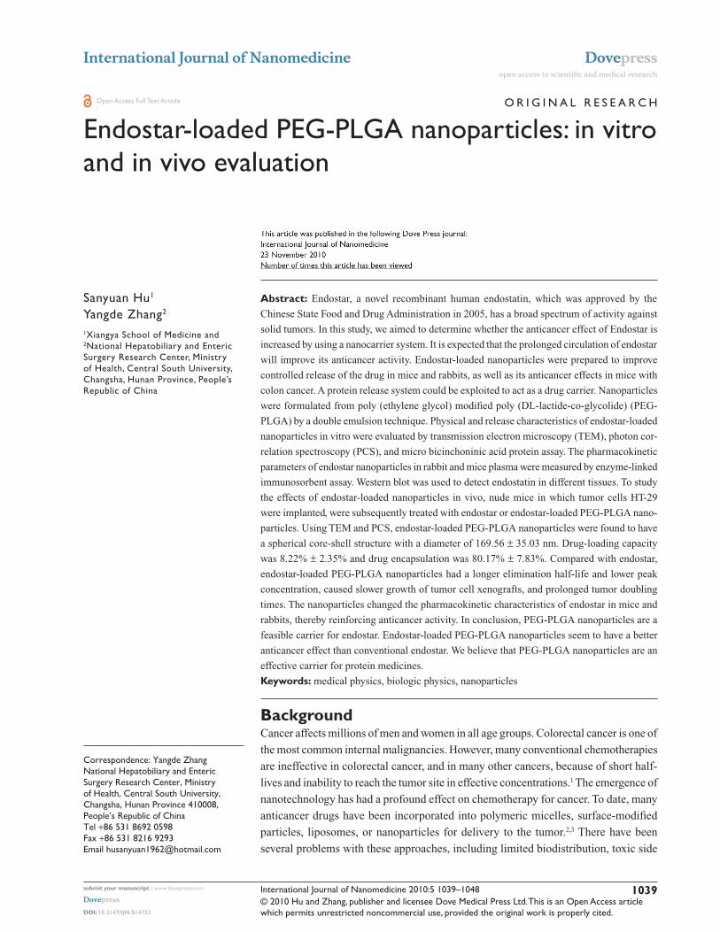
The morphology of endostar-loaded nanoparticles was found
to be a spherical core-shell structure with a relatively smooth
surface (Figure 1). It was approximately 169.56 ± 35.03 nm
in diameter (Figure 2). The PDI was 0.47 ± 0.18 by photon
correlation spectroscopy. Residual polyvinyl alcohol on the
surface was not detected by the two different methods.
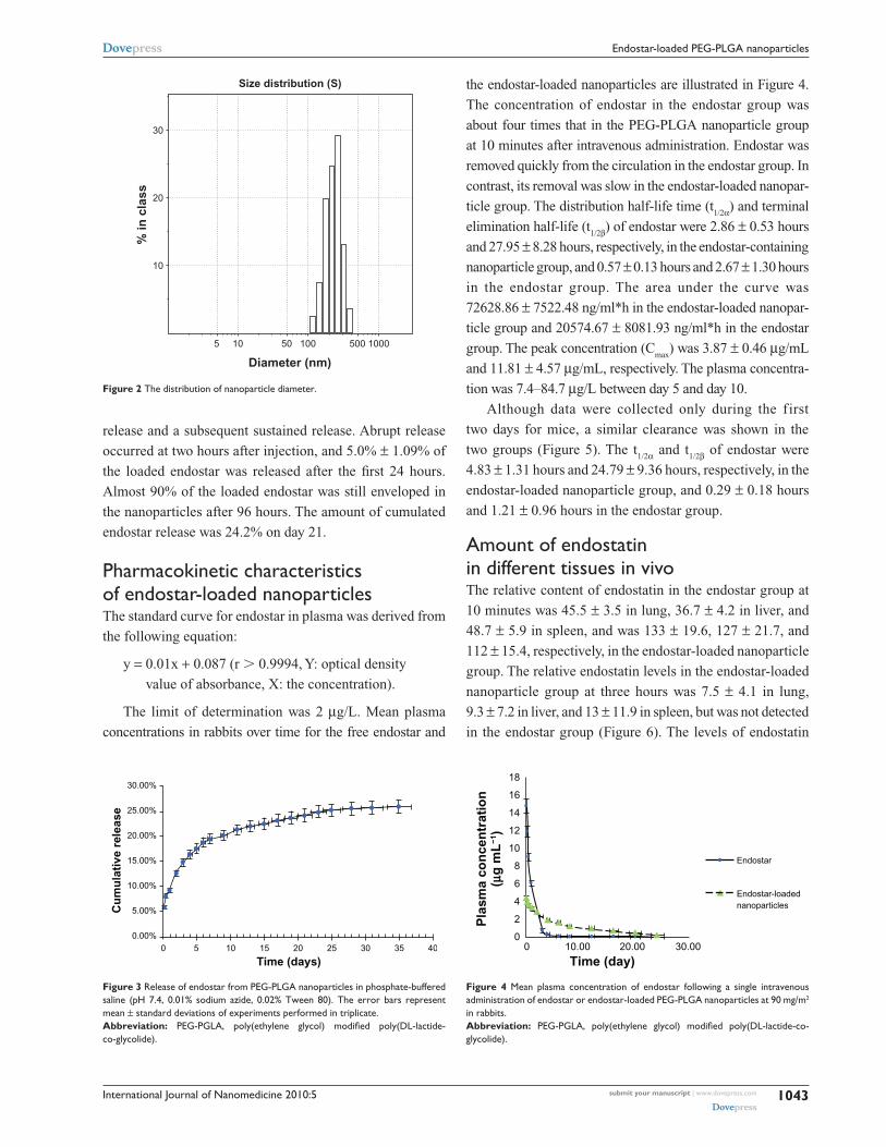
release of endostar-loaded nanoparticles in phosphate-buffered salineEndostar release from the endostar-loaded nanoparticles in
phosphate-buffered saline (pH 7.04) is shown as a standard
curve equation for the endostar solution in Figure 3. The
endostar release profile was biphasic, with an initial abrupt
Figure 1 The core-shell structure of endostar-loaded Peg-PLgA nanoparticles. Transmission electron microscopy showed that nanoparticles were round particles with relative smooth edges.Abbreviation: Peg-PgLA, poly(ethylene glycol) modified poly(DL-lactide-co-glycolide).
International Journal of Nanomedicine 2010:5 submit your manuscript | www.dovepress.com
Dovepress
Dovepress
1043
endostar-loaded Peg-PLgA nanoparticles
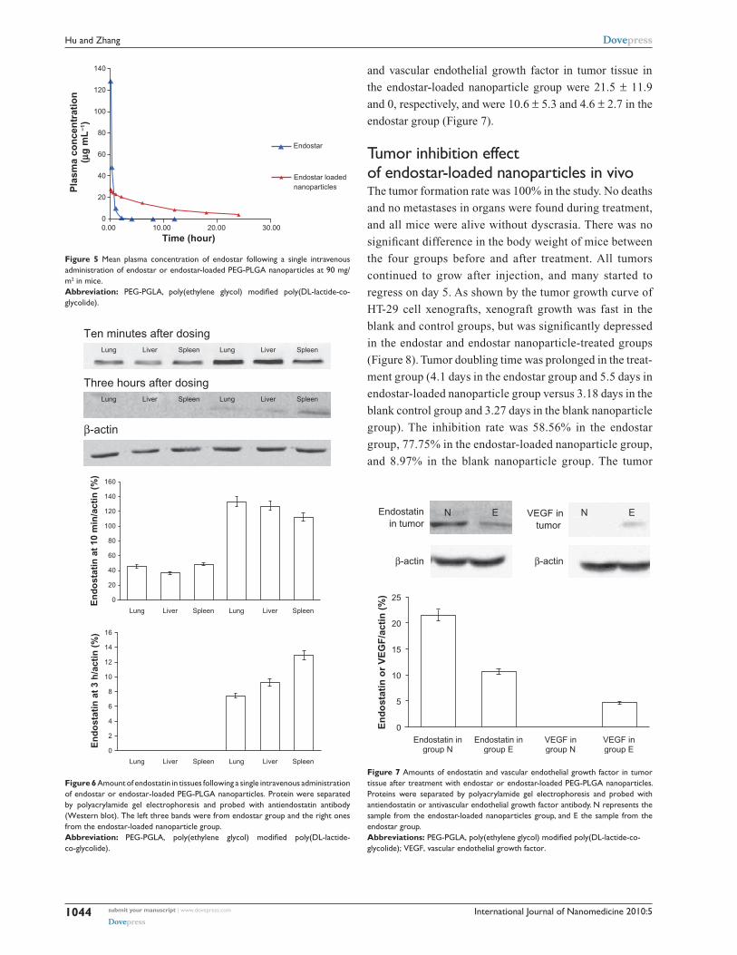
the endostar-loaded nanoparticles are illustrated in Figure 4.
The concentration of endostar in the endostar group was
about four times that in the PEG-PLGA nanoparticle group
at 10 minutes after intravenous administration. Endostar was
removed quickly from the circulation in the endostar group. In
contrast, its removal was slow in the endostar-loaded nanopar-
ticle group. The distribution half-life time (t1/2α) and terminal
elimination half-life (t1/2β) of endostar were 2.86 ± 0.53 hours
and 27.95 ± 8.28 hours, respectively, in the endostar-containing
nanoparticle group, and 0.57 ± 0.13 hours and 2.67 ± 1.30 hours
in the endostar group. The area under the curve was
72628.86 ± 7522.48 ng/ml*h in the endostar-loaded nanopar-
ticle group and 20574.67 ± 8081.93 ng/ml*h in the endostar
group. The peak concentration (Cmax
) was 3.87 ± 0.46 µg/mL
and 11.81 ± 4.57 µg/mL, respectively. The plasma concentra-
tion was 7.4–84.7 µg/L between day 5 and day 10.
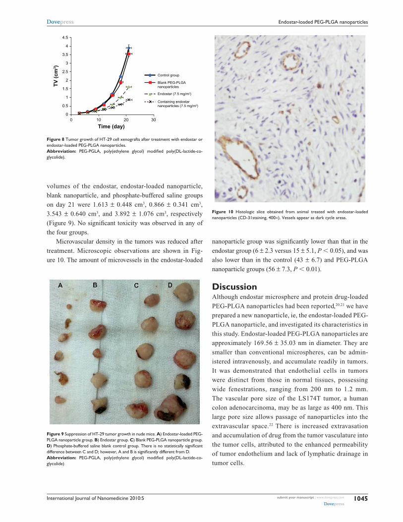
Although data were collected only during the first
two days for mice, a similar clearance was shown in the
two groups (Figure 5). The t1/2α and t
1/2β of endostar were
4.83 ± 1.31 hours and 24.79 ± 9.36 hours, respectively, in the
endostar-loaded nanoparticle group, and 0.29 ± 0.18 hours
and 1.21 ± 0.96 hours in the endostar group.
Amount of endostatin in different tissues in vivoThe relative content of endostatin in the endostar group at
10 minutes was 45.5 ± 3.5 in lung, 36.7 ± 4.2 in liver, and
48.7 ± 5.9 in spleen, and was 133 ± 19.6, 127 ± 21.7, and
112 ± 15.4, respectively, in the endostar-loaded nanoparticle
group. The relative endostatin levels in the endostar-loaded
nanoparticle group at three hours was 7.5 ± 4.1 in lung,
9.3 ± 7.2 in liver, and 13 ± 11.9 in spleen, but was not detected
in the endostar group (Figure 6). The levels of endostatin
30.00%
25.00%
20.00%
15.00%
10.00%
5.00%
0.00%0 5 10 15 20 25 30
Time (days)
Cu
mu
lati
ve r
elea
se
35 40
Figure 3 release of endostar from Peg-PLgA nanoparticles in phosphate-buffered saline (pH 7.4, 0.01% sodium azide, 0.02% Tween 80). The error bars represent mean ± standard deviations of experiments performed in triplicate.Abbreviation: Peg-PgLA, poly(ethylene glycol) modified poly(DL-lactide- co-glycolide).
release and a subsequent sustained release. Abrupt release
occurred at two hours after injection, and 5.0% ± 1.09% of
the loaded endostar was released after the first 24 hours.
Almost 90% of the loaded endostar was still enveloped in
the nanoparticles after 96 hours. The amount of cumulated
endostar release was 24.2% on day 21.
Pharmacokinetic characteristics of endostar-loaded nanoparticlesThe standard curve for endostar in plasma was derived from
the following equation:
y = 0.01x + 0.087 (r . 0.9994, Y: optical density
value of absorbance, X: the concentration).
The limit of determination was 2 µg/L. Mean plasma
concentrations in rabbits over time for the free endostar and
5 10
10
20
30%
in c
lass
Size distribution (S)
50 100 500 1000
Diameter (nm)
Figure 2 The distribution of nanoparticle diameter.
00 10.00
Time (day)
Pla
sma
con
cen
trat
ion
(µg
mL
−1)
20.00 30.00
Endostar
Endostar-loadednanoparticles
2
4
6
8
10
12
14
16
18
Figure 4 Mean plasma concentration of endostar following a single intravenous administration of endostar or endostar-loaded Peg-PLgA nanoparticles at 90 mg/m2 in rabbits.Abbreviation: Peg-PgLA, poly(ethylene glycol) modified poly(DL-lactide-co-glycolide).
International Journal of Nanomedicine 2010:5submit your manuscript | www.dovepress.com
Dovepress
Dovepress
1044
Hu and Zhang
and vascular endothelial growth factor in tumor tissue in
the endostar-loaded nanoparticle group were 21.5 ± 11.9
and 0, respectively, and were 10.6 ± 5.3 and 4.6 ± 2.7 in the
endostar group (Figure 7).
Tumor inhibition effect of endostar-loaded nanoparticles in vivoThe tumor formation rate was 100% in the study. No deaths
and no metastases in organs were found during treatment,
and all mice were alive without dyscrasia. There was no
significant difference in the body weight of mice between
the four groups before and after treatment. All tumors
continued to grow after injection, and many started to
regress on day 5. As shown by the tumor growth curve of
HT-29 cell xenografts, xenograft growth was fast in the
blank and control groups, but was significantly depressed
in the endostar and endostar nanoparticle-treated groups
(Figure 8). Tumor doubling time was prolonged in the treat-
ment group (4.1 days in the endostar group and 5.5 days in
endostar-loaded nanoparticle group versus 3.18 days in the
blank control group and 3.27 days in the blank nanoparticle
group). The inhibition rate was 58.56% in the endostar
group, 77.75% in the endostar-loaded nanoparticle group,
and 8.97% in the blank nanoparticle group. The tumor
0.00 10.00 20.00
Time (hour)30.00
Endostar
Endostar loadednanoparticles
0
140
20
40
60
80
100
120
Pla
sma
con
cen
trat
ion
(µg
mL
−1)
Figure 5 Mean plasma concentration of endostar following a single intravenous administration of endostar or endostar-loaded Peg-PLgA nanoparticles at 90 mg/m2 in mice.Abbreviation: PEG-PGLA, poly(ethylene glycol) modified poly(DL-lactide-co-glycolide).
Lung Liver Spleen Lung Liver Spleen
Lung Liver Spleen Lung Liver Spleen
Lung Liver Spleen Lung Liver Spleen
Lung Liver Spleen Lung Liver Spleen
0
140
20
40
60
80
100
120
160
0
14
2
4
6
8
10
12
16
En
do
stat
in a
t 10
min
/act
in (
%)
En
do
stat
in a
t 3
h/a
ctin
(%
)
Ten minutes after dosing
Three hours after dosing
β-actin
Figure 6 Amount of endostatin in tissues following a single intravenous administration of endostar or endostar-loaded Peg-PLgA nanoparticles. Protein were separated by polyacrylamide gel electrophoresis and probed with antiendostatin antibody (Western blot). The left three bands were from endostar group and the right ones from the endostar-loaded nanoparticle group.Abbreviation: Peg-PgLA, poly(ethylene glycol) modified poly(DL-lactide- co-glycolide).
0Endostatin in
group N
En
do
stat
in o
r V
EG
F/a
ctin
(%
)
Endostatin ingroup E
VEGF ingroup N
VEGF ingroup E
5
10
15
20
25
Endostatinin tumor
VEGF intumor
β-actinβ-actin
N E N E
Figure 7 Amounts of endostatin and vascular endothelial growth factor in tumor tissue after treatment with endostar or endostar-loaded Peg-PLgA nanoparticles. Proteins were separated by polyacrylamide gel electrophoresis and probed with antiendostatin or antivascular endothelial growth factor antibody. N represents the sample from the endostar-loaded nanoparticles group, and e the sample from the endostar group.Abbreviations: Peg-PgLA, poly(ethylene glycol) modified poly(DL-lactide-co-glycolide); VegF, vascular endothelial growth factor.
International Journal of Nanomedicine 2010:5 submit your manuscript | www.dovepress.com
Dovepress
Dovepress
1045
endostar-loaded Peg-PLgA nanoparticles
volumes of the endostar, endostar-loaded nanoparticle,
blank nanoparticle, and phosphate-buffered saline groups
on day 21 were 1.613 ± 0.448 cm3, 0.866 ± 0.341 cm3,
3.543 ± 0.640 cm3, and 3.892 ± 1.076 cm3, respectively
(Figure 9). No significant toxicity was observed in any of
the four groups.
Microvascular density in the tumors was reduced after
treatment. Microscopic observations are shown in Fig-
ure 10. The amount of microvessels in the endostar-loaded
nanoparticle group was significantly lower than that in the
endostar group (6 ± 2.3 versus 15 ± 5.1, P , 0.05), and was
also lower than in the control (43 ± 6.7) and PEG-PLGA
nanoparticle groups (56 ± 7.3, P , 0.01).
DiscussionAlthough endostar microsphere and protein drug-loaded
PEG-PLGA nanoparticles had been reported,20,21 we have
prepared a new nanoparticle, ie, the endostar-loaded PEG-
PLGA nanoparticle, and investigated its characteristics in
this study. Endostar-loaded PEG-PLGA nanoparticles are
approximately 169.56 ± 35.03 nm in diameter. They are
smaller than conventional microspheres, can be admin-
istered intravenously, and accumulate readily in tumors.
It was demonstrated that endothelial cells in tumors
were distinct from those in normal tissues, possessing
wide fenestrations, ranging from 200 nm to 1.2 mm.
The vascular pore size of the LS174T tumor, a human
colon adenocarcinoma, may be as large as 400 nm. This
large pore size allows passage of nanoparticles into the
extravascular space.22 There is increased extravasation
and accumulation of drug from the tumor vasculature into
the tumor cells, attributed to the enhanced permeability
of tumor endothelium and lack of lymphatic drainage in
tumor cells.
00 10
Time (day)
TV
(cm
3 )
20 30
Control group
Blank PEG-PLGAnanoparticles
Endostar (7.5 mg/m2)
Containing endostarnanoparticles (7.5 mg/m2)0.5
1
1.5
2
2.5
3
3.5
4
4.5
Figure 8 Tumor growth of HT-29 cell xenografts after treatment with endostar or endostar-loaded Peg-PLgA nanoparticles.Abbreviation: Peg-PgLA, poly(ethylene glycol) modified poly(DL-lactide-co-glycolide).
Figure 9 suppression of HT-29 tumor growth in nude mice. A) endostar-loaded Peg-PLgA nanoparticle group. B) endostar group. C) Blank Peg-PLgA nanoparticle group. D) Phosphate-buffered saline blank control group. There is no statistically significant difference between C and D; however, A and B is significantly different from D. Abbreviation: Peg-PgLA, poly(ethylene glycol) modified poly(DL-lactide-co-glycolide)
Figure 10 Histologic slice obtained from animal treated with endostar-loaded nanoparticles (cD-31staining, 400×). Vessels appear as dark cycle areas.
International Journal of Nanomedicine 2010:5submit your manuscript | www.dovepress.com
Dovepress
Dovepress
1046
Hu and Zhang
Endostar is a 20 kDa peptide and different from protein
drugs which are encapsulated within PLGA or PEG-PLGA
nanoparticles.21,23 It is smaller than a protein molecule and
more difficult to encapsulate. Thus, PEG and PLGA, which
are hydrophilic-hydrophobic diblock copolymers, were used
in this study. They have great potential as vehicles for the
delivery of anticancer drugs.24,25 PLGA, the hydrophobic
moiety, is biodegradable and acts as a drug incorporation
site. PEG, the hydrophilic moiety, is a nontoxic, nonimmu-
nogenic, and hydrophilic polymer which can prevent interac-
tions with cells and proteins.26,27 Studies have revealed that
nanoparticles of 100 nm in thickness with a PEG layer more
than 10 nm in thickness are not easily engulfed by phago-
cytes (Figure 11).28,29 Because of the hydrophilic moiety, the
encapsulation of endostar-loaded nanoparticles was high at
80.17% ± 7.83%.
Moreover, PEG-PLGA nanoparticles hydrolyze in
an aqueous environment (hydrolytic degradation or
biodegradation).30 The biodegradation rates of PLGA copo-
lymers are dependent on the molar ratio of the lactic and
glycolic acids in the polymer chain. Thus, PEG-PLGA nano-
particles have been used for controlling the release of drugs,
altering pharmacokinetics, enhancing anticancer effect, and
decreasing toxicity, especially for water-insoluble drugs.31–33
Similar to most proteins, endostar is administered by mul-
tiple injections at a high dose in order to maintain adequate
therapeutic levels.34,35 Clinically, endostar is administered
intravenously at a dose of 7.5 mg/m2 per day during the first
two weeks of a treatment cycle. However, the plasma concen-
tration of endostar would still fluctuate because of its short
biologic half-life and rapid metabolism. In addition, the need
for multiple injections causes poor patient compliance, which
limits its clinical use. However, PEG-PLGA nanoparticles
change the pharmacokinetic characteristics of endostar, with
the t1/2α and t
1/2β of endostar-containing nanoparticles being
longer than for endostar (P , 0.05) and the area under the
curve and maximum concentration being larger than for
endostar (P , 0.05).
As shown by Western blot assay, the amounts of endosta-
tin detected in the liver, spleen, and lung in the Endostar-
containing PEG-PLGA nanoparticle group were larger than
that in the endostar group after 10 minutes. There was no
statistically significant difference between amounts detected
in liver and spleen, although the amount of endostatin in
lung was higher than in liver or spleen. Endostatin was
detected only in the endostar-loaded PEG-PLGA group at
three hours after intravenous injection. Even when the mice
were sacrificed, the amount of endostatin in tumors for the
endostar-loaded nanoparticle group was larger than for the
endostar group.
The anticancer effect of endostar was improved when
incorporated into PEG-PLGA nanoparticles. As shown
in the colon cancer model, tumor doubling time was pro-
longed in the endostar-loaded nanoparticle group. The
inhibition rate for endostar-loaded nanoparticles was higher
than for endostar alone (P , 0.05). The tumor volumes
of the endostar and endostar-loaded nanoparticle-treated
groups were significantly smaller than those of the con-
trol and blank PEG-PLGA nanoparticle control groups
(P , 0.01). There was no significant difference in tumor
inhibition between the two control groups (P . 0.05).
Because endostar can inhibit neovascularization or induce
apoptosis of vascular endothelium to kill tumor cells,36
microvascular density was reduced in tumors after treat-
ment with endostar-loaded nanoparticles. The formation
of new capillaries in the existing vasculature is a process
fundamental to the development of a solid tumor, and it
was hard to elicit angiogenesis in the tumors treated with
endostar, and they did not grow fast. The antiangiogenic
effect of endostatin is related to vascular endothelial growth
factor.8 The amount of vascular endothelial growth factor
in tumors for the endostar-loaded nanoparticle group was
found to be less than for the endostar group when the mice
were sacrificed (P , 0.05).
PEG-PLGA nanoparticles could maintain adequate
concentrations of endostar in plasma and tumor, thereby
improving its antitumor effect. PEG-PLGA nanoparticles
have a great potential to be protein drug carriers. Although
more of their characteristics need to be investigated, and
PEG
PEG
PLGA
Endostar
Figure 11 endostar-loaded nanoparticles with a Peg layer.Abbreviation: Peg, poly(ethylene glycol); PgLA, poly(DL-lactide-co-glycolide).
International Journal of Nanomedicine 2010:5 submit your manuscript | www.dovepress.com
Dovepress
Dovepress
1047
endostar-loaded Peg-PLgA nanoparticles
some disadvantages need to be overcome, including passive
targeting, low drug-loading capacity, and sensitization, PEG-
PLGA nanoparticles could be modified to be specific for
cancer and applied in the clinical setting as a protein carrier
system in the future.
ConclusionIn this study, we prepared endostar-loaded PEG-PLGA
nanoparticles in an innovative way and found that they were
useful for sustained release of endostar. Although many other
characteristics need to be investigated, endostar-loaded PEG-
PLGA nanoparticles may improve the anticancer activity
of endostar by changing the pharmacokinetic behavior of
endostar in vivo.
AcknowledgmentThis work was supported by the National High Technology
Research and Development Program 863 Fund of China
(No.2007AA021802).
DisclosureThe authors report no conflicts of interest in this work.
References 1. Michor F, Iwasa Y, Lengauer C, Nowak MA. Dynamics of colorectal
cancer. Semin Cancer Biol. 2005;15(6):484–493. 2. Nishiyama N, Kataoka K. Current state, achievements, and future pros-
pects of polymeric micelles as nanocarriers for drug and gene delivery. Pharmacol Ther. 2006;112(3):630–648.
3. Torchilin VP. Targeted pharmaceutical nanocarriers for cancer therapy and imaging. AAPS J. 2007;9(2):E128–E147.
4. Jun YJ, Kim JI, Jun MJ, Sohn YS. Selective tumor targeting by enhanced permeability and retention effect. Synthesis and antitumor activity of polyphosphazene-platinum (II) conjugates. J Inorg Biochem. 2005;99(8):1593–1601.
5. Greish K. Enhanced permeability and retention of macromolecular drugs in solid tumors: A royal gate for targeted anticancer nanomedicines. J Drug Target. 2007;15(7–8):457–464.
6. Iyer AK, Khaled G, Fang J, Maeda H. Exploiting the enhanced perme-ability and retention effect for tumor targeting. Drug Discov Today. 2006;11(17–18):812–818.
7. Wang C, Pham PT. Polymers for viral gene delivery. Expert Opin Drug Deliv. 2008;5(4):385–401.
8. Ling Y, Yang Y, Lu N, et al. Endostar, a novel recombinant human endostatin, exerts antiangiogenic effect via blocking VEGF-induced tyrosine phosphorylation of KDR/Flk-1 of endothelial cells. Biochem Biophys Res Commun. 2007;361(1):79–84.
9. Zhuo W, Luo C, Wang X, Song X, Fu Y, Luo Y. Endostatin inhibits tumour lymphangiogenesis and lymphatic metastasis via cell surface nucleolin on lymphangiogenic endothelial cells. J Pathol. 2010;222(3):249–260.
10. Song HF, Liu XW, Zhang HN, et al. Pharmacokinetics of His-tag recombinant human endostatin in Rhesus monkeys. Acta Pharmacol Sin. 2005;26(1):124–128.
11. Jiang LP, Zou C, Yuan X, et al. N-terminal modification increases the stability of the recombinant human endostatin in vitro. Biotechnol Appl Biochem. 2009;54(2):113–120.
12. Cohen S, Yoshioka T, Lucarelli M, Hwang LH, Langer R. Controlled delivery systems for proteins based on poly(lactic/glycolic acid) micro-spheres. Pharm Res. 1991;8(6):713–720.
13. Roy A, Singh MS, Upadhyay PK, Bhaskar S. Combined chemo-immunotherapy as a prospective strategy to combat cancer: A nano-particle based approach. Mol Pharm. 2010 Sep 16. [Epub ahead of print].
14. Mukherjee B, Santra K, Pattnaik G, Ghosh S. Preparation, charac-terization and in-vitro evaluation of sustained release protein-loaded nanoparticles based on biodegradable polymers. Int J Nanomedicine. 2008;3(4):487–496.
15. Gref R, Quellec P, Sanchez A, et al. Development and characterization of CyA-loaded poly(lactic acid)-poly(ethylene glycol)PEG micro- and nanoparticles. Comparison with conventional PLA particulate carriers. Eur J Pharm Biopharm. 2001;51(2):111–118.
16. Thomas JP, Arzoomanian RZ, Alberti D, et al. Phase I pharma-cokinetic and pharmacodynamic study of recombinant human endostatin in patients with advanced solid tumors. J Clin Oncol. 2003;21(2):223–231.
17. Tentler JJ, Bradshaw-Pierce EL, Serkova NJ, et al. Assessment of the in vivo antitumor effects of ENMD-2076, a novel multitargeted kinase inhibitor, against primary and cell line-derived human col-orectal cancer xenograft models. Clin Cancer Res. 2010;16(11): 2989–2998.
18. Jie JZ, Wang JW, Qu JG, Hung T. Suppression of human colon tumor growth by adenoviral vector-mediated NK4 expression in an athymic mouse model. World J Gastroenterol. 2007;13(13):1938–1946.
19. Gao J, Knutsen A, Arbman G, et al. Clinical and biological significance of angiogenesis and lymphangiogenesis in colorectal cancer. Dig Liver Dis. 2009;41(2):116–122.
20. Wu J, Wu L, Xu X, et al. Microspheres made by w/o/o emulsion method with reduced initial burst for long-term delivery of endostar, a novel recombinant human endostatin. J Pharm Sci. 2009;98(6):2051–2058.
21. Li Y, Pei Y, Zhang X, et al. PEGylated PLGA nanoparticles as protein carriers: Synthesis, preparation and biodistribution in rats. J Control Release. 2001;71(2):203–211.
22. Hobbs SK, Monsky WL, Yuan F, et al. Regulation of transport pathways in tumor vessels: Role of tumor type and microenvironment. Proc Natl Acad Sci U S A. 1998;95(8):4607–4612.
23. Son S, Lee WR, Joung YK, et al. Optimized stability retention of a monoclonal antibody in the PLGA nanoparticles. Int J Pharm. 2009;368(1–2):178–185.
24. Mandal D, Chatterjee U. Synthesis and spectroscopy of CdS nano-particles in amphiphilic diblock copolymer micelles. J Chem Phys. 2007;126(13):134507.
25. Arimura H, Ohya Y, Ouchi T. Formation of core-shell type bio-degradable polymeric micelles from amphiphilic poly(aspartic acid)-block-polylactide diblock copolymer. Biomacromolecules. 2005;6(2):720–725.
26. Wang M, Thanou M. Targeting nanoparticles to cancer. Pharmacol Res. 2010;62(2):90–99.
27. Li SD, Huang L. Nanoparticles evading the reticuloendothelial system: Role of the supported bilayer. Biochim Biophys Acta. 2009;1788(10):2259–2266.
28. Jeong YI, Kang MK, Sun HS, et al. All-trans-retinoic acid release from core-shell type nanoparticles of poly(epsilon- caprolactone)/poly(ethylene glycol) diblock copolymer. Int J Pharm. 2004; 273(1–2): 95–107.
29. Zhang X, Pan SR, Hu HM, et al. Poly(ethylene glycol)-block- polyethylenimine copolymers as carriers for gene delivery: Effects of PEG molecular weight and PEGylation degree. J Biomed Mater Res A. 2008;84(3):795–804.
30. Muthu M. Nanoparticles based on PLGA and its co-polymer: An overview. Asian J Pharm. 2009;3(4):266–273.
31. Peer D, Karp JM, Hong S, et al. Nanocarriers as an emerging platform for cancer therapy. Nat Nanotechnol. 2007;2(12):751–760.
International Journal of Nanomedicine
Publish your work in this journal
Submit your manuscript here: http://www.dovepress.com/international-journal-of-nanomedicine-journal
The International Journal of Nanomedicine is an international, peer-reviewed journal focusing on the application of nanotechnology in diagnostics, therapeutics, and drug delivery systems throughout the biomedical field. This journal is indexed on PubMed Central, MedLine, CAS, SciSearch®, Current Contents®/Clinical Medicine,
Journal Citation Reports/Science Edition, EMBase, Scopus and the Elsevier Bibliographic databases. The manuscript management system is completely online and includes a very quick and fair peer-review system, which is all easy to use. Visit http://www.dovepress.com/ testimonials.php to read real quotes from published authors.
International Journal of Nanomedicine 2010:5submit your manuscript | www.dovepress.com
Dovepress
Dovepress
Dovepress
1048
Hu and Zhang
32. Wang MD, Shin DM, Simons JW, Nie S. Nanotechnology for targeted cancer therapy. Expert Rev Anticancer Ther. 2007;7(6): 833–837.
33. Petros RA, de Simone JM. Strategies in the design of nanoparticles for therapeutic applications. Nat Rev Drug Discov. 2010;9(8):615–627.
34. Kisker O, Becker CM, Prox D, et al. Continuous administration of endostatin by intraperitoneally implanted osmotic pump improves the efficacy and potency of therapy in a mouse xenograft tumor model. Cancer Res. 2001;61(20):7669–7674.
35. Xu F, Ma Q, Sha H. Optimizing drug delivery for enhancing therapeutic efficacy of recombinant human endostatin in cancer treatment. Crit Rev Ther Drug Carrier Syst. 2007;24(5):445–492.
36. Ling Y, Lu N, Gao Y, et al. Endostar induces apoptotic effects in HUVECs through activation of caspase-3 and decrease of Bcl-2. Anticancer Res. 2009;29(1):411–417.


![IS 9293 (1991): Textiles - Canvas, Flax · IS 9293 (1991): Textiles - Canvas, Flax [TXD 33: Industrial Fabrics]. . IS 9293 :1991 * Pwvmw Indian Standard -TEXTILES - CANVAS, FLAX -](https://static.fdocuments.us/doc/165x107/6049a091d9c85e3b7c6d7cec/is-9293-1991-textiles-canvas-flax-is-9293-1991-textiles-canvas-flax.jpg)