pFLD and pFLD - Thermo Fisher Scientific
44
USER GUIDE For Research Use Only. Not intended for any animal or human therapeutic or diagnostic use. pFLD and pFLDα Pichia pastoris expression vectors for inducible expression with methylamine and selection on Zeocin ™ Catalog number V230-20 Revision date: 29 March 2012 Publication Part number 25-0547 MAN0000304
Transcript of pFLD and pFLD - Thermo Fisher Scientific

user guide
For Research Use Only. Not intended for any animal or human therapeutic or diagnostic use.
pFLD and pFLDα Pichia pastoris expression vectors for inducible expression with methylamine and selection on Zeocin™
Catalog number V230-20
Revision date: 29 March 2012 Publication Part number 25-0547
MAN0000304

ii

iii
Contents
Kit Contents and Storage ......................................................................................................... v
Introduction ................................................................................................................... 1
Product Overview .................................................................................................................... 1
Methods ......................................................................................................................... 2
General Cloning Considerations ............................................................................................ 2
Cloning into pFLD .................................................................................................................... 4
Cloning into pFLDα ................................................................................................................. 5
Transforming E. coli ................................................................................................................. 7
Pichia Transformation .............................................................................................................. 9
Expression in Pichia ................................................................................................................ 13
Purification .............................................................................................................................. 16
Appendix ...................................................................................................................... 18
Recipes ..................................................................................................................................... 18
Zeocin™ ..................................................................................................................................... 22
Map of pFLD ........................................................................................................................... 24
Map of pFLDα......................................................................................................................... 25
Vector Features ....................................................................................................................... 26
Map of pFLD/CAT ................................................................................................................ 27
Lithium Chloride Transformation Method ........................................................................ 28
Accessory Products ................................................................................................................ 29
Technical Support ................................................................................................................... 31
Purchaser Notification ........................................................................................................... 32
References ................................................................................................................................ 33

iv

v
Kit Contents and Storage
Shipping/Storage Vectors are shipped at room temperature. Upon receipt, store at -20°C.
Kit Contents Catalog number V230-20 includes the following vectors, supplied at a concentration of 0.5 µg/µL in 10 mM Tris-HCl, 1 mM EDTA, pH 8.0 in a total volume of 40 µL:
• pFLD
• pFLDα
• pFLD/CAT (control plasmid)
Reference Sources
The pFLD vectors may be used with the EasySelect™ Pichia Expression Kit (Catalog no. K1740-01) or the Original Pichia Expression Kit (Catalog no. K1710-01), available for purchase. Additional general information about recombinant protein expression in Pichia pastoris is provided in the manuals for the EasySelect™ Pichia Expression Kit and the Original Pichia Expression Kit. The manuals can be downloaded from www.lifetechnologies.com/support or obtained from Technical Support (see page 31). For more information about the EasySelect™ Pichia Expression Kit or the Original Pichia Expression Kit, refer to www.lifetechnologies.com/support or call Technical Support. More detailed information and protocols dealing with Pichia pastoris may also be found in the following general reference: Higgins, D. R., and Cregg, J. M. (1998) Pichia Protocols. In Methods in Molecular Biology, Vol. 103. (J. M. Walker, ed. Humana Press, Totowa, NJ)
Product Use For research use only. Not intended for any human or animal therapeutic or diagnostic use.
Continued on next page

vi
Kit Contents and Storage, Continued
Recommended Pichia Host Strain
We recommend using the X-33 Pichia strain as the host for expression of recombinant proteins from pFLD. Other Pichia strains including GS115, KM71H, and SMD1168H are suitable. The X-33 Pichia strain and other strains are available for purchase (see page 29 for ordering information). The X-33 Pichia strain has the following genotype and phenotype:
Genotype: Wild-type
Phenotype: Mut+
Materials Supplied by the User
Equipment
• Microbiological equipment • Electroporation device and 0.2 cm cuvettes or reagents for transformation • 16°C and 37°C water baths or temperature blocks
• 30°C and 37°C shaking and non-shaking incubators
• Hemacytometer
• Microtiter plates (optional)
Reagents
• Pichia host strain (e.g., X-33, GS115, SMD1168H, KM71H) • Electrocompetent or chemically competent E. coli (must be recA, endA) for
transformation • Restriction enzymes and appropriate buffers • Agarose and low-melt agarose • PureLink® HQ Mini Plasmid Purification Kit (see page 29 for ordering) or
glass milk • Sterile water • CIAP (calf intestinal alkaline phosphatase, 1 unit/ μL) • 10X CIAP Buffer • Phenol/chloroform • 3 M sodium acetate • 100% ethanol • 80% ethanol • T4 Ligase (2.5 units/ μL) • 10X Ligation Buffer (with ATP) • Zeocin™ selection agent (see page 29 for ordering information) • YPDS plates containing the appropriate concentration of Zeocin™ (see
page 18 for recipe) • 50 mL conical centrifuge tubes • 15 mL polypropylene tubes
• ProBond™ Purification System (optional, see page 30 for ordering)

1
Introduction
Product Overview
Introduction pFLD (4.4 kb) and pFLDα (4.7 kb) are vectors used to express recombinant proteins in Pichia pastoris. Recombinant proteins are expressed as fusions to a C-terminal peptide containing the V5 epitope and a polyhistidine (6xHis) tag. The vector allows high-level, methanol- and methylamine-inducible expression of the gene of interest in Pichia, and can be used in any Pichia strain, including X-33, GS115, SMD1168H, and KM71H. The pFLD vectors contain the following elements:
• 5′ fragment containing the FLD1 promoter for methanol- or methylamine-induced expression of the gene of interest (Shen et al, 1998)
• Zeocin™ resistance gene for selection in both E. coli and Pichia (Baron et al., 1992; Drocourt et al., 1990)
• Ampicillin resistance gene for selection in E. coli
• C-terminal peptide containing the V5 epitope and a polyhistidine (6xHis) tag for detection and purification of a recombinant fusion protein (if desired)
Experimental Overview
The following table describes the basic steps needed to clone and express your gene of interest in pFLD.
Step Action Page
1 Propagate pFLD by transformation into a recA, endA1 E. coli strain such as TOP10, DH5α™, or JM109.
2
2 Develop a cloning strategy and ligate your gene into one of the pFLD vectors in frame with the C-terminal tag.
3–6
3 Transform into E. coli and select transformants on select on LB with 50–100 µg/mL ampicillin (LB-Amp).
7
4 Analyze 10–20 transformants by restriction mapping or sequencing to confirm in-frame fusion of your gene with the C-terminal tag.
8
5 Purify and linearize the recombinant plasmid for transformation into Pichia pastoris.
7–10
6 Transform your Pichia strain and plate onto YPDS plates containing the appropriate concentration of Zeocin™.
9–12
7 Select for Zeocin™-resistant transformants. 9
8 Optimize expression of your gene. 13–14
9 Purify your fusion protein on metal-chelating resin (i.e. ProBond™).
16–17

2
Methods
General Cloning Considerations
Introduction The multiple cloning site for pFLD is shown on page 4 and the multiple cloning site for pFLDα is shown on page 6. Use these diagrams to design a strategy to clone your gene of interest in frame with the C-terminal peptide. General considerations for cloning and transformation are discussed in this section.
General Molecular Biology Techniques
For assistance with E. coli transformations, restriction enzyme analysis, DNA biochemistry, and plasmid preparation, refer to Molecular Cloning: A Laboratory Manual (Sambrook et al., 1989) or Current Protocols in Molecular Biology (Ausubel et al., 1994).
E. coli Strain Many E. coli strains are suitable for the propagation of the pFLD vectors, including TOP10, JM109, and DH5α™. We recommend that you propagate the pFLD vectors in E. coli strains that are recombination deficient (recA) and endonuclease A deficient (endA).
For your convenience, TOP10 E. coli are available as chemically competent or electrocompetent cells (see page 29 for ordering).
Transformation Method
You may use any method of choice for transformation. Chemical transformation is the most convenient for many researchers. Electroporation is the most efficient and the method of choice for large plasmids.
Maintaining Plasmids
The pFLD vectors contain the ampicillin resistance gene and the Zeocin™ resistance gene to allow selection of the plasmid in E. coli using either ampicillin or Zeocin™. The following procedure uses ampicillin to propagate and maintain pFLD plasmids:
1. Use the supplied stock solution to transform a recA, endA E. coli strain like TOP10, DH5α™, JM109, or equivalent.
2. Select transformants on LB agar with 50–100 µg/mL ampicillin (LB-Amp).
3. Prepare a glycerol stock from each transformant containing plasmid for long-term storage (see page 7).
Continued on next page

3
General Cloning Considerations, Continued
General Considerations
The following are some general guidelines when using pFLD or pFLDα to express your gene of interest in Pichia:
• The codon usage in Pichia is believed to be similar to Saccharomyces cerevisiae.
• Many Saccharomyces genes have proven to be functional in Pichia.
• The premature termination of transcripts because of "AT rich regions" has been observed in Pichia and other eukaryotic systems (Henikoff and Cohen, 1984; Irniger et al., 1991; Scorer et al., 1993; Zaret and Sherman, 1984). If you have problems expressing your gene, check for premature termination by northern analysis and check your sequence for AT rich regions. It may be necessary to change the sequence in order to express your gene (Scorer et al., 1993).
Special Considerations for pFLD
For pFLD only:
• Your insert should contain an initiation ATG codon as part of a yeast consensus sequence (Romanos et al., 1992). An example of a yeast consensus sequence is provided below. The ATG initiation codon is shown underlined.
(G/A)NNATGG
• To express your gene as a recombinant fusion protein, you must clone your gene in frame with the C-terminal peptide containing the V5 epitope and the polyhistidine tag. Refer to the diagram on page 4 to develop a cloning strategy.
• To express your gene of interest without the C-terminal peptide, make sure that your gene contains a stop codon.
Special Considerations for pFLDα
For pFLDα only:
• pFLDα is an N-terminal fusion vector. To express your gene, you must clone your gene in frame with the N-terminal α-factor secretion signal. Refer to the diagram on page 6 to develop a cloning strategy.
• To express your gene of interest without the C-terminal peptide, make sure that your gene contains a stop codon.
• The predicted protease cleavage sites for the α-factor signal sequence are indicated in the figure on page 6.

4
Cloning into pFLD
Multiple Cloning Site of pFLD
Below is the multiple cloning site for pFLD. Restriction sites are labeled to indicate the cleavage site. The multiple cloning site has been confirmed by sequencing and functional testing. The vector sequence of pFLD is available for downloading from www.lifetechnologies.com or from Technical Support (see page 31). For a map and a description of the features of pFLD, see page 24.

5
Cloning into pFLDα
Introduction pFLDα possesses the α-factor mating signal sequence for secretion of your protein. The information in this section is provided to assist you in designing a cloning strategy. Details of the multiple cloning site of the pFLDα can be found on page 6.
Signal Sequence Processing
The processing of the α-factor mating signal sequence in pFLDα occurs in three steps:
1. Signal peptidase cleavage between Ala and Ala in the 19 and 20 positions.
2. Kex2 cleavage between Arg and Glu in the sequence Glu-Lys-Arg * Glu-Ala-Glu-Ala, where * is the site of cleavage.
3. The Glu-Ala repeats are further cleaved by the STE13 gene product.
Optimization of Signal Cleavage
In Saccharomyces cerevisiae, the Glu-Ala repeats are not necessary for cleavage by Kex2, but cleavage after Glu-Lys-Arg may be more efficient when followed by Glu-Ala repeats. A number of amino acids are tolerated at site X instead of Glu in the sequence Glu-Lys-Arg-X. These amino acids include the aromatic amino acids, small amino acids, and histidine. Proline, however, will inhibit Kex2 cleavage. For more information on Kex2 cleavage, see (Brake et al., 1984).
There are some cases where Ste13 cleavage of Glu-Ala repeats is not efficient, and Glu-Ala repeats are left on the N-terminus of the expressed protein of interest. This is generally dependent on the protein of interest.
Expression of Recombinant Protein with Native N-terminus
To have your protein expressed with a native N-terminus, you can use the Xho I site at bp 865–870 to clone your gene flush with the Kex2 cleavage site. Use PCR to rebuild the sequence from the Xho I site to the arginine codon at nucleotides 744–746. Remember to include the first amino acid(s) of your protein, if necessary, for correct fusion to the Kex2 cleavage site.
Continued on next page

6
Cloning into pFLDα, Continued
Multiple Cloning Site of pFLDα
Below is the multiple cloning site for pFLDα. Restriction sites are labeled to indicate the cleavage site. The multiple cloning site has been confirmed by sequencing and functional testing. The vector sequence of pFLDα is available for downloading for www.lifetechnologies.com or from Technical Support (see page 31). For a map and a description of the features of pFLDα, see page 25–26.

7
Transforming E. coli
Transforming E. coli
Transform your ligation mixtures into a competent recA, endA E. coli strain (e.g. TOP10, DH5α™, JM109) and select on LB agar plates containing 50-100 µg/mL ampicillin. Once you have obtained ampicillin-resistant colonies, pick 10 transformants and screen for the presence and orientation of your insert. Note that there is no blue/white screening for the presence of insert with the pFLD vectors.
We recommend that you sequence your construct to confirm that your gene is in the correct orientation for expression and cloned in frame with the C-terminal peptide (if desired). Refer to the multiple cloning site diagrams on the previous pages for the sequences and location of the priming sites. To order custom-synthesized primers, visit www.lifetechnologies.com and select Custom Primers.
Preparing a Glycerol Stock
Once you have identified the correct clone, purify the colony and make a glycerol stock for long-term storage. We also recommend keeping a DNA stock of your plasmid at –20°C.
1. Streak the original colony out on an LB plate containing 50–100 µg/mL ampicillin. Incubate the plate at 37°C overnight.
2. Isolate a single colony and inoculate into 1–2 mL of LB containing 50-100 µg/mL ampicillin.
3. Grow the culture to mid-log phase (OD600 = 0.5–0.7).
4. Mix 0.85 mL of culture with 0.15 mL of sterile glycerol and transfer to a cryovial.
5. Store at –80°C.
Plasmid Preparation
Once you have cloned and sequenced your insert, generate enough plasmid DNA to transform Pichia (5–10 µg of each plasmid per transformation). We recommend isolating plasmid DNA using the We recommend isolating plasmid DNA using the PureLink® HiPure Miniprep Kit (up to 30 µg) or the PureLink® HiPure Midiprep Kit (up to 150 µg), or CsCl gradient centrifugation. Once you have purified plasmid DNA, proceed to Pichia Transformation on page 9.
Continued on next page

8
Transformation into E. coli, Continued
Sequencing Recombinant Clones
We recommend that you sequence your construct before transforming into Pichia to confirm that your gene is in frame with the α-factor secretion signal and/or the C-terminal tag. To sequence your construct in pFLD or pFLDα, we recommend using FLD1 Forward and 3´ AOX1 primers. The 3´ AOX1 primer is available separately for purchase (see page 29 for ordering information). The FLD1 Forward primer can be custom-ordered from us; visit www.lifetechnologies.com and select Custom Primers.
Sequencing Primer Sequence
α-Factor 5´-TACTATTGCCAGCATTGCTGC-3´
3´ AOX1 5´-GCAAATGGCATTCTGACATCC-3´ For sequencing protocols, refer to Unit 7 in Current Protocols in Molecular Biology
(Ausubel et al., 1994) or Chapter 13 in Molecular Cloning: A Laboratory Manual (Sambrook et al., 1989).

9
Pichia Transformation
Introduction After your gene has been correctly cloned into one of the pFLD vectors, you are ready to transform the vector into your Pichia strain. This section provides general guidelines for preparing plasmid DNA, transformation, and selecting for Zeocin™-resistant clones.
Zeocin™ Selection We typically use 100 µg/mL Zeocin™ to select for transformants when using the X-33 Pichia strain. If you are transforming your pFLD or pFLDα construct into another Pichia strain, note that selection conditions may vary. We recommend performing a dose response curve to determine the appropriate concentration of Zeocin™ to use for selection of transformants in your strain.
Method of Transformation
We do not recommend spheroplasting for transformation of Pichia with plasmids containing the Zeocin™ resistance marker. Spheroplasting involves removal of the cell wall to allow DNA to enter the cell. Cells must first regenerate the cell wall before they are able to express the Zeocin™ resistance gene. For this reason, plating spheroplasts directly onto selective medium containing Zeocin™ does not yield any transformants.
We recommend electroporation for transformation of Pichia with pFLD. Electroporation yields 103 to 104 transformants per µg of linearized DNA and does not destroy the cell wall of Pichia. If you do not have access to an electroporation device, use the LiCl protocol on page 27 or the Pichia EasyComp™ Transformation Kit available for purchase (see below).
Pichia EasyComp™ Transformation Kit
For chemical transformation of Pichia strains with pFLD vectors, the Pichia EasyComp™ Transformation Kit is available for purchase (see page 29 for ordering information). The Pichia EasyComp™ Transformation Kit provides reagents to prepare six preparations of competent cells. Each preparation will yield enough competent cells for 20 transformations. Competent cells may be used immediately or frozen and stored for future use. For more information, refer to www.lifetechnologies.com/support or call Technical Support (see page 31).
Since the pFLD vectors contain DNA sequences from both the FLD1 and AOX1 loci (promoter and polyA addition, respectively), homologous recombination in the host can occur at either of these locations. The restriction enzyme site used to linearize the vector will determine the predominant integration locus. Cutting the vector within the FLD1 promoter sequence (see Linearizing Your pFLD Construct on page 10) will target the integration event to the FLD1 locus. If the vector is completely linearized within the FLD1 promoter—with no uncut, circular vector remaining—homologous recombination at the AOX1 locus will not occur.
Continued on next page

10
Pichia Transformation, Continued
pFLD vectors do not contain a yeast origin of replication. Transformants can only be isolated if recombination occurs between the plasmid and the Pichia genome.
Before Starting You will need the following reagents for transforming Pichia and selecting transformants on Zeocin™. Note: Inclusion of sorbitol in YPD plates stabilizes electroporated cells, which can be osmotically sensitive.
• 5–10 µg pure pFLD or pFLDα containing your insert
• YPD Medium
• 50-mL conical polypropylene tubes
• 1 liter cold (4°C) sterile water (place on ice the day of the experiment)
• 25 mL cold (4°C) sterile 1 M sorbitol (place on ice the day of the experiment)
• 30°C incubator
• Electroporation device and 0.2-cm cuvettes
• YPDS plates containing the appropriate concentration of Zeocin™ (see page 18 for recipe)
Linearizing the pFLD Construct
To promote integration, we recommend that you linearize your pFLD construct within the 5′ FLD1 region. The table below lists unique sites that may be used to linearize pFLD prior to transformation. Other restriction sites are possible. Be sure that your insert does not contain the restriction site you wish to use to linearize your vector.
Enzyme Restriction Site (bp)
Nsi I 285
Nde I 406
Cla I 493
Restriction Digest 1. Digest ~5–10 µg of plasmid DNA with one of the enzymes listed above.
2. Check a small aliquot of your digest by agarose gel electrophoresis for complete linearization.
3. If the vector is completely linearized, heat inactivate or add EDTA to stop the reaction, phenol/chloroform extract once, and ethanol precipitate using 1/10 volume 3 M sodium acetate and 2.5 volumes of 100% ethanol.
4. Centrifuge the solution to pellet the DNA, wash the pellet with 80% ethanol, air-dry, and resuspend in 10 μL sterile, deionized water. Use immediately or store at –20°C.
Continued on next page

11
Pichia Transformation, Continued
Preparing Pichia for Electroporation
Follow the procedure below to prepare your Pichia pastoris strain for electroporation.
1. Grow 5 mL of your Pichia pastoris strain in YPD in a 50 mL conical tube at 30°C overnight.
2. Inoculate 500 mL of fresh medium in a 2 liter flask with 0.1–0.5 mL of the overnight culture. Grow overnight again to an OD600 = 1.3–1.5.
3. Centrifuge the cells at 1500 × g for 5 minutes at 4°C. Resuspend the pellet with 500 mL of ice-cold (0°C), sterile water.
4. Centrifuge the cells as in Step 3, then resuspend the pellet with 250 mL of ice-cold (0°C), sterile water.
5. Centrifuge the cells as in Step 3, then resuspend the pellet in 20 mL of ice-cold (0°C) 1 M sorbitol.
6. Centrifuge the cells as in Step 3, then resuspend the pellet in 1 mL of ice-cold (0°C) 1 M sorbitol for a final volume of approximately 1.5 mL. Keep the cells on ice and use that day. Do not store cells.
Transformation by Electroporation
1. Mix 80 μL of the cells from Step 6 (above) with 5–10 µg of linearized pFLD or pFLDα DNA (in 5–10 μL sterile water) and transfer them to an ice-cold (0°C) 0.2-cm electroporation cuvette.
2. Incubate the cuvette with the cells on ice for 5 minutes.
3. Pulse the cells according to the parameters for yeast (Saccharomyces cerevisiae) as suggested by the manufacturer of the specific electroporation device being used.
4. Immediately add 1 mL of ice-cold 1 M sorbitol to the cuvette. Transfer the cuvette contents to a sterile 15 mL tube.
5. Let the tube incubate at 30°C without shaking for 1 to 2 hours.
6. Spread 50–200 μL each on separate, labeled YPDS plates containing the appropriate concentration of Zeocin™.
7. Incubate plates for 2 to 3 days at 30°C until colonies form.
8. Pick 10–20 colonies and purify (streak for single colonies) on fresh YPD or YPDS plates containing the appropriate concentration of Zeocin™.
Continued on next page

12
Pichia Transformation, Continued
Generally, several hundred Zeocin™-resistant colonies are generated using the protocol on page 11. If more colonies are needed, the protocol may be modified as described below. Note that you will need ~20 150-mm plates with YPDS agar containing the appropriate concentration of Zeocin™.
1. Set up two transformations per construct and follow Steps 1 through 5 of the Transformation by Electroporation protocol, page 11.
2. After 1 hour in 1 M sorbitol at 30°C (Step 5, page 11), add 1 mL of YPD medium to each tube.
3. Shake (~200 rpm) the cultures at 30°C.
4. After 1 hour, take one of the tubes and plate out all of the cells by spreading 200 μL on 150-mm plates containing the appropriate concentration of Zeocin™.
5. (Optional) Continue incubating the other culture for three more hours (for a total of four hours) and then plate out all of the cells by spreading 200 μL on 150-mm plates containing the appropriate concentration of Zeocin™.
6. Incubate plates for 2 to 4 days at 30°C until colonies form.
Mut Phenotype If you used a Pichia strain containing a native AOX1 gene (e.g., X-33, GS115, SMD1168H) as the host for your pFLD construct, your Zeocin™-resistant transformants will be Mut+. If you used a strain containing a deletion in the AOX1 gene (e.g., KM71H), your transformants will be MutS.
To verify the Mut phenotype of your Zeocin™-resistant transformants, refer to the general guidelines provided in the EasySelect™ Pichia Expression Kit manual or the Original Pichia Expression Kit manual or to published reference sources (Higgins and Cregg, 1998).
You are now ready to test your transformants for expression of your gene of interest. See Expression in Pichia, page 13.

13
Expression in Pichia
Introduction The primary purpose of small-scale expression is to identify/confirm a recombinant Pichia clone that is expressing the correct protein. Small-scale expression conditions may not be optimal for your protein. For this reason, the method you choose for detection (e.g., SDS-PAGE, Western, or functional assay) may be an important factor in determining the success of expression. If your method of detection does not reveal any expression, you may want to consider using a more sensitive method.
Once a positive clone has been identified, large-scale expression can be carried out in shake flask or fermentation, and expression conditions can be optimized.
Note that once you have obtained Zeocin™-resistant transformants, it is not necessary to maintain your recombinant Pichia clone in medium containing Zeocin™ for expression studies. Zeocin™ is only required for initial screening and selection of recombinant clones.
Detecting Recombinant Proteins in Pichia
We recommend that you use the following techniques to assay expression of your protein. Be sure to account for any additional amino acids that are in between the end of your protein and the C-terminal tag.
Technique Method of Detection Sensitivity
SDS-PAGE (Coomassie-stained)
Visualization by eye Can detect as little as 100 ng in a single band
SDS-PAGE (Silver-stained)
Visualization by eye Can detect as little as 2 ng in a single band
Western Analysis Antibody to your particular protein Anti-myc antibodies (see the next page) Anti-His(C-term) antibodies (see the next page)
Can detect as little as 1-10 pg, depending on detection method (alkaline phosphatase, horseradish peroxidase, radiolabeled antibody)
Functional assay Varies depending on assay. Varies depending on assay Used to compare relative amounts of protein.
Reminder: Because the pFLD vector does not contain the HIS4 gene, his4 Pichia strains containing the integrated plasmid must be grown in medium containing 0.004% histidine. If histidine is not present in the medium the cells will not grow. If you use X-33, SMD1168H, or KM71H as the host strain, supplementation of the medium with histidine is not required.
Continued on next page

14
Expression in Pichia, Continued
Expression Guidelines
Information and guidelines for performing small-scale expression, optimizing expression, and performing scale-up of expression are provided in the EasySelect™ Pichia Expression Kit manual and the Original Pichia Expression Kit manual. See below for information on inducing expression of the FLD1 gene with methanol, methylamine, or methanol plus methylamine.
Inducing with Methanol, Methylamine, or Methanol Plus Methylamine
Expression of the FLD1 gene may be induced with methanol, methylamine, or methanol plus methylamine. The following guidelines for induction have been developed using the wild-type X-33 strain; other strains may be suitable. For media recipes, see pages 19–21.
1. For all types of induction, grow cultures in MGAs medium at 30°C in a shaking incubator (250–300 rpm) until the culture reaches an OD600 of 4.
2. Harvest the cells by centrifuging at 1500–3000 × g for 5 minutes at room temperature. Then:
• For induction with methanol, decant supernatant and resuspend cell pellet in 1/5 of the original culture volume of MMAs medium.
• For induction with methylamine, decant supernatant and resuspend cell pellet in 1/5 of the original culture volume of MGMa medium.
• For induction with methanol plus methylamine, decant supernatant and resuspend cell pellet in 1/5 of the original culture volume of MMMa medium.
3. Incubate the resulting culture at 30°C for five days. For media containing methanol or methanol plus methylamine, add 100% methanol to a final concentration of 0.5% methanol every 24 hours to maintain induction.
Time points: At each of the times indicated below, transfer 1 mL of the expression culture to a 1.5-mL microcentrifuge tube, and centrifuge at maximum speed in a tabletop microcentrifuge for 2–3 minutes at room temperature. These samples will be used to analyze expression levels and determine the optimal time post-induction to harvest. Time points (hours): 0, 24 (1 day), 48 (2 days), 72 (3 days), 96 (4 days), and 120 (5 days).
Polyacrylamide Gel Electrophoresis
To facilitate separation and visualization of your recombinant protein by polyacrylamide gel electrophoresis, a wide range of pre-cast NuPAGE® and Tris-Glycine polyacrylamide gels are available for purchase. The NuPAGE® Gel System avoids the protein modifications associated with Laemmli-type SDS-PAGE, ensuring optimal separation for protein analysis. In addition, we also carry a large selection of molecular weight protein standards and staining kits. For more information about the appropriate gels, standards, and stains to use to visualize your recombinant protein, refer to www.lifetechnologies.com or call Technical Support (see page 31).
Continued on next page

15
Expression in Pichia, Continued
Western Analysis To detect expression of your recombinant fusion protein by Western blot analysis, you may use the Anti-V5 antibodies or the Anti-His(C-term) antibodies (see page 30 for ordering information) or an antibody to your protein of interest. In addition, the Positope™ Control Protein available for use as a positive control for detection of fusion proteins containing a V5 epitope or a polyhistidine (6xHis) tag (see page Error! Bookmark not defined. for ordering). WesternBreeze® Chromogenic Kits and WesternBreeze® Chemiluminescent Kits are available for detection of antibodies by colorimetric or chemiluminescent methods. For more information, refer to www.lifetechnologies.com or call Technical Support (see page 31).

16
Purification
Introduction In this section, you will grow and induce a 10–200 mL culture of your Pichia transformant for trial purification on a metal-chelating resin such as ProBond™. You can harvest the cells and store them at –80°C until you are ready to purify your fusion protein, or you can proceed directly with protein purification. Note that this section only describes preparation of cell lysates and sample application onto ProBond™. For instructions on how to prepare and use ProBond™ resin, refer to the ProBond™ Purification System manual.
ProBond™ Resin We recommend the ProBond™ Purification System to purify fusion proteins
expressed from pFLD or pFLDα (see page Error! Bookmark not defined.). Note that instructions for equilibration of and chromatography on ProBond™ resin are contained in the ProBond™ Purification Kit.
If you are using a metal-chelating resin other than ProBond™, follow the manufacturer's recommendations to purify fusion proteins expressed in bacteria or yeast.
Binding Capacity of ProBond™
One milliliter of ProBond™ resin binds at least 1 mg of recombinant protein. This amount can vary depending on the protein.
Throughout the following protocol, be sure to keep the cell lysate and fractions on ice. Small-scale purifications using the 2-mL ProBond™ columns and buffers can be performed at room temperature on the bench top. For large scale purifications, all reagents must be kept at 4°C.
Preparing Cell Lysates
Express your protein using a small-scale culture (10–20 mL for MutS strains; 100-200 mL for Mut+) and the optimal conditions for expression (if determined). Refer to the Pichia Expression Kit manual for details. Once your protein is expressed, follow the protocol below to prepare a cell lysate for chromatography on ProBond™.
Prepare Breaking Buffer (BB) as described in the Recipes, page 21.
1. Wash cells once in BB by resuspending them and centrifuging 5–10 minutes at 3000 × g at 4°C.
2. Resuspend the cells to an OD600 of 50–100 in BB.
3. Add an equal volume of acid-washed glass beads (0.5 mm). Estimate volume by displacement.
4. Vortex the mixture for 30 seconds, then incubate on ice for 30 seconds. Repeat 7 more times. Alternating vortexing with cooling keeps the cell extracts cold and reduces denaturation of your protein.
5. Centrifuge the sample at 4°C for 5–10 minutes at 12,000 × g.
6. Transfer the clear supernatant to a fresh container and analyze for your protein. The total protein concentration should be around 2–3 mg/mL.
7. Save the pellet and extract with 6 M urea or 1% Triton X-100 to check for insoluble protein.
Continued on next page

17
Purification, Continued
Sample Application (Native Conditions)
For sample application onto ProBond™, you will need Native Binding Buffer, pH 7.8 and a 2-mL ProBond™ column, pre-equilibrated using native conditions.
1. Combine 1 mL (2–3 mg/mL total protein) of Pichia lysate with 7 mL Native Binding Buffer.
2. Prepare a ProBond™ column with resin as described in the ProBond™ manual, and resuspend the resin in the column in 4 mL of the diluted lysate from Step 1.
3. Seal the column and batch-bind by rocking gently at room temperature for 10 minutes.
4. Let the resin settle by gravity or low speed centrifugation (800 × g) and carefully remove the supernatant. Save the supernatant to check for unbound protein.
5. Repeat Steps 2 through 4 with the remaining 4 mL of diluted lysate. Proceed with column washing and elution under native conditions as described in the ProBond™ manual. Use the recommendations noted for bacterial cell lysates.
Sample Applica-tion (Denaturing Conditions)
Use the protocol above except pre-equilibrate the ProBond™ column using Denaturing Binding Buffer and combine 1 mL of the Pichia cell lysate with 7 mL of the Denaturing Binding Buffer.
We have observed that some Pichia proteins may be retained on the ProBond™ column using native purification conditions. Optimization of the purification (see the ProBond™ manual) or using denaturing purification may remove these non-specific Pichia proteins.
Analysis of Purification
Save all fractions, washes, and flow-through for analysis by SDS-PAGE. You may need to use western blot analysis to detect your protein if expression is low or not enough protein was loaded onto the column. Refer to the ProBond™ System manual for a guide to troubleshoot chromatography.
Scale-up You may find it necessary to scale-up your purification to obtain sufficient amounts of purified protein. Adjust the pH and NaCl concentration of your lysate as indicated in the ProBond™ manual before adding it to the column. The pH should be greater than or equal to 7.5 and the NaCl concentration should be ~500 mM.

18
Appendix
Recipes
YPD (+ Zeocin™) Yeast Extract Peptone Dextrose Medium (1 liter)
1% yeast extract
2% peptone
2% dextrose (glucose)
+2% agar
+ appropriate concentration of Zeocin™
1. Dissolve 10 g yeast extract and 20 g of peptone in 900 mL of water.
2. Include 20 g of agar if making YPD slants or plates.
3. Autoclave for 20 minutes on liquid cycle.
4. Add 100 mL of 20% dextrose (filter-sterilize dextrose before use).
5. Cool solution to ~60°C and add the appropriate amount of Zeocin™ from a 100 mg/mL stock solution. Note: It is necessary to include Zeocin™ in the medium for selection of Pichia transformants only. Zeocin™ may be omitted from the medium when performing expression studies.
Store YPD slants or plates containing Zeocin™ at 4°C. The shelf life is 1–2 weeks.
YPDS (+ Zeocin™) Yeast Extract Peptone Dextrose Medium with Sorbitol (1 liter)
1% yeast extract
2% peptone
2% dextrose (glucose)
1 M sorbitol + 2% agar
+ appropriate concentration of Zeocin™
1. Dissolve: 10 g yeast extract
182.2 g sorbitol
20 g of peptone
in 900 mL of water.
2. Add 20 g of agar.
3. Autoclave for 20 minutes on liquid cycle.
4. Add 100 mL of 20% dextrose (filter-sterilize dextrose before use).
5. Cool solution to ~60°C and add the appropriate amount of Zeocin™ from a 100 mg/mL stock solution. Note: It is necessary to include Zeocin™ in the medium for selection of Pichia transformants only. Zeocin™ may be omitted from the medium when performing expression studies.
Store YPDS slants or plates containing Zeocin™ at 4°C. The shelf life is 1–2 weeks.
Continued on next page

19
Recipes, Continued
Stock Solutions 10X YNB (1.7% Yeast Nitrogen Base without Ammonium Sulfate)
Dissolve 17 g of yeast nitrogen base (YNB) without ammonium sulfate and without amino acids in 1000 mL of water and filter sterilize. Heat the solution to dissolve YNB completely in water. Store at 4°C. The shelf life of this solution is approximately one year.
10X As (5% Ammonium Sulfate)
Dissolve 50 g of ammonium sulfate in 1000 mL of water and filter sterilize.
500X B (0.02% Biotin)
Dissolve 20 mg biotin in 100 mL of water and filter sterilize. Store at 4°C. The shelf life of this solution is approximately one year.
10X M (5% Methanol)
Mix 5 mL of methanol with 95 mL of water. Filter sterilize and store at 4°C. The shelf life of this solution is approximately two months.
10X G (10% Glycerol)
Mix 100 mL of glycerol with 900 mL of water. Sterilize either by filtering or autoclaving. Store at room temperature. The shelf life of this solution is greater than one year.
10X Ma (2.5% Methylamine)
Mix 2.5 mL of methylamine HCl with 97.5 mL of water. Filter sterilize and store at 4°C. The shelf life of this solution is approximately two months.
MGAs Minimal Glycerol with Ammonium sulfate (1 liter)
0.17% YNB 0.5% ammonium sulfate 1% glycerol 4 × 10⁻⁵% biotin Combine aseptically 700 mL autoclaved water with 100 mL of 10X YNB, 100 mL of As, 2 mL of 500X B, and 100 mL of 10X G. Store at 4°C. The shelf life of this solution is approximately two months.
Continued on next page

20
Recipes, Continued
MMAs Minimal Methanol with Ammonium sulfate Medium (1 liter)
0.17% YNB 0.5% ammonium sulfate 4 × 10⁻⁵% biotin 0.5% methanol 1. For medium, autoclave 700 mL of water for 20 minutes on liquid cycle 2. Cool autoclaved water to 60°C and add:
100 mL of 10X YNB 100 mL of As 2 mL of 500X B 100 mL of 10X M
3. Mix and store at 4°C. 4. For plates, add 20 g agar to the water in Step 1 and proceed. 5. After mixing, pour the plates immediately. MMAs stores for several months
at 4°C.
MGMa Minimal Glycerol and Methylamine Medium (1 liter)
0.17% YNB 4 × 10⁻⁵% biotin 1% glycerol 0.25% methylamine 1. For medium, autoclave 700 mL of water for 20 minutes on liquid cycle 2. Cool autoclaved water to room temperature and add:
100 mL of 10X YNB 2 mL of 500X B 100 mL 10X G 100 mL of 10X Ma
3. Mix and store at 4°C. 4. For plates, add 20 g agar to the water in Step 1 and proceed. 5. After mixing, pour the plates immediately. MGMa stores for several months
at 4°C.
Continued on next page

21
Recipes, Continued
MMMa Minimal Methanol and Methylamine Medium (1 liter)
0.17% YNB 4 × 10⁻⁵% biotin 0.5% methanol 0.25% methylamine 1. For medium, autoclave 700 mL of water for 20 minutes on liquid cycle 2. Cool autoclaved water to 60°C and add:
100 mL of 10X YNB 2 mL of 500X B 100 mL of 10X M 100 mL of 10X Ma
3. Mix and store at 4°C. 4. For plates, add 20 g agar to the water in Step 1 and proceed. 5. After mixing, pour the plates immediately. MMMa stores for several months
at 4°C.
Breaking Buffer 50 mM sodium phosphate, pH 7.4 1 mM PMSF (phenylmethylsulfonyl fluoride. You may use other protease inhibitors) 1 mM EDTA 5% glycerol
1. Prepare a stock solution of your desired protease inhibitors and store appropriately. Follow manufacturer’s recommendations.
2. For 1 liter, dissolve: 6 g sodium phosphate (monobasic) 372 mg EDTA 50 mL glycerol
in 900 mL deionized water.
3. Use NaOH to adjust pH and bring up the volume to 1 liter. Store at 4°C.
4. Add protease inhibitors immediately before use.

22
Zeocin™
Zeocin™ Zeocin™ is a member of the bleomycin/phleomycin family of antibiotics isolated from Streptomyces. It shows strong toxicity against bacteria, fungi, plants and mammalian cell lines (Calmels et al., 1991; Drocourt et al., 1990; Gatignol et al., 1987; Mulsant et al., 1988; Perez et al., 1989).
A Zeocin™ resistance protein has been isolated and characterized (Calmels et al., 1991; Drocourt et al., 1990). This 13,665 Da protein, the product of the Sh ble gene (Streptoalloteichus hindustanus bleomycin gene), binds stoichiometrically to Zeocin™ and inhibits its DNA strand cleavage activity. Expression of this protein in eukaryotic and prokaryotic hosts confers resistance to Zeocin™.
Molecular Weight, Formula, and Structure
The formula for Zeocin™ is C55H86O21N20S2Cu-HCl and the molecular weight is 1527.5. The structure of Zeocin™ is shown below.
Applications of Zeocin™
Zeocin™ is used for selection in mammalian cells (Mulsant et al., 1988); plants (Perez et al., 1989); yeast (Baron et al., 1992); and prokaryotes (Drocourt et al., 1990). Suggested concentrations of Zeocin™ for selection in Pichia and E. coli are listed below:
Organism Zeocin™ Concentration and Selective Medium
E. coli 25–50 µg/mL in Low Salt LB medium*
Pichia 100–1000 µg/mL (varies with strain and medium) *Efficient selection requires that the concentration of NaCl be no more than 5 g/L (< 90 mM)
Continued on next page

23
Zeocin™, Continued
Handling Zeocin™ • High ionic strength and acidity or basicity inhibits the activity of Zeocin™. Therefore, we recommend that you reduce the salt in bacterial medium and adjust the pH to 7.5 to keep the drug active (see page 18 for a recipe).
• Store Zeocin™ at –20°C and thaw on ice before use.
• Zeocin™ is light sensitive. Store the drug and plates or medium containing the drug in the dark.
• Wear gloves, a laboratory coat, and safety glasses when handling Zeocin™-containing solutions.
• Do not ingest or inhale solutions containing the drug.

24
Map of pFLD
Map of pFLD The figure below summarizes the features of the pFLD vector. The vector sequence for pFLD is available for downloading from www.lifetechnologies.com or from Technical Support (see page 31). See page 26 for a description of the features of the vector.

25
Map of pFLDα
Map of pFLDα The figure below summarizes the features of the pFLDα vector. The complete sequence for pFLDα is available for downloading from www.lifetechnologies.com or from Technical Support (see page 31). See page 26 for a description of the features of the vector.

26
Vector Features
Features The vectors pFLD and pFLDα contain the following elements. All features have been functionally tested.
Feature Benefit
FLD1 promoter A 597-bp fragment containing the FLD1 promoter that allows methanol- or methylamine-inducible, high-level expression of the gene of interest in Pichia Targets plasmid integration to the FLD1 locus.
α-Factor Secretion Signal (pFLDα only)
Encodes the native Saccharomyces cerevisiae α-factor secretion signal that allows for efficient secretion of most proteins from Pichia (Cregg et al., 1993)
Multiple cloning site Allows insertion of your gene into the expression vector
V5 epitope (Gly-Lys-Pro-Ile-Pro-Asn-Pro-Leu-Leu-Gly-Leu-Asp-Ser-Thr)
Permits detection of your recombinant fusion protein with the Anti-V5 Antibody or Anti-V5-HRP Antibody (Southern et al., 1991) (see page 30 for ordering)
C-terminal polyhistidine (6xHis) tag
Permits purification of your recombinant fusion protein on metal-chelating resin such as ProBond™ In addition, the C-terminal polyhistidine tag is the epitope for the Anti-His(C-term) Antibody (Lindner et al., 1997) and the Anti-His(C-term)-HRP Antibody (see page Error! Bookmark not defined. for ordering)
AOX1 transcription termination (TT) region
Native transcription termination and polyadenylation signal from AOX1 gene (~260 bp) that permits efficient 3´ mRNA processing, including polyadenylation, for increased mRNA stability
TEF1 promoter (GenBank accession numbers D12478, D01130)
Transcription elongation factor 1 gene promoter from Saccharomyces cerevisiae that drives expression of the Zeocin™ resistance gene in Pichia
T7 promoter Synthetic prokaryotic promoter that drives constitutive expression of the Zeocin™ resistance gene in E. coli
Zeocin™ resistance gene (Sh ble)
Allows selection of transformants in E. coli and Pichia
CYC1 transcription termination region (GenBank accession number M34014)
3´ end of the Saccharomyces cerevisiae CYC1 gene that allows efficient 3´ mRNA processing of the Zeocin™ resistance gene for increased stability
pUC origin Allows replication and maintenance of the plasmid in E. coli

27
Map of pFLD/CAT
Map of pFLD/CAT pFLD/CAT (5014 bp) is a control vector expressing chloramphenicol acetyltransferase (CAT). It was constructed by cloning the CAT gene into the pFLD vector. The figure below summarizes the features of the pFLD/CAT vector. The vector sequence for pFLD/CAT is available for downloading from www.lifetechnologies.com or from Technical Support (see page 31).

28
Lithium Chloride Transformation Method
Introduction This modified version of the procedure described for S. cerevisiae (Gietz and Schiestl, 1996) is provided as an alternative to transformation by electroporation. Transformation efficiency is between 102 and 103 cfu/µg linearized DNA.
Preparing Solutions
Lithium acetate does not work with Pichia pastoris. Use only lithium chloride.
1 M LiCl in distilled, deionized water. Filter-sterilize. Dilute as needed with sterile water.
50% polyethylene glycol (PEG-3350) in distilled, deionized water. Filter-sterilize. Store in a tightly capped bottle.
2 mg/mL denatured, sheared salmon sperm DNA in TE (10 mM Tris-HCl, pH 8.0, 1.0 mM EDTA). Store at –20°C.
Preparing Cells 1. Grow a 50 mL culture of Pichia pastoris in YPD at 30°C with shaking to an OD600 of 0.8 to 1.0 (approximately 108 cells/mL).
2. Harvest the cells, wash with 25 mL of sterile water, and centrifuge at 1500 × g for 10 minutes at room temperature.
3. Resuspend the cell pellet in 1 mL of 100 mM LiCl and transfer the suspension to a 1.5 mL microcentrifuge tube.
4. Pellet cells at maximum speed for 15 seconds and remove LiCl with a pipette.
5. Resuspend the cells in 400 μL of 100 mM LiCl. 6. Dispense 50 μL of cell suspension into a 1.5-mL microcentrifuge tube for each
transformation and use immediately. Do not store on ice or freeze at -20°C.
Transformation 1. Boil a 1 mL sample of single-stranded DNA for 5 minutes, then quickly chill on ice. Keep on ice. Note: It is neither necessary nor desirable to boil the carrier DNA prior to each use. Store a small aliquot at –20°C and boil every 3–4 times the DNA is thawed.
2. Centrifuge the cells from Step 6, above, and remove the LiCl with a pipette. 3. For each transformation, add the following reagents IN THE ORDER GIVEN
to the cells. PEG shields the cells from the detrimental effects of the high LiCl concentration. 240 μL 50% PEG 36 μL 1 M LiCl 25 μL 2 mg/mL single-stranded DNA Plasmid DNA (5–10 µg) in 50 μL sterile water
4. Vortex each tube vigorously until the cell pellet is completely mixed (~1 minute).
5. Incubate the tube at 30°C for 30 minutes without shaking. 6. Heat shock in a water bath at 42°C for 20–25 minutes. 7. Centrifuge the cells at 6000 to 8000 rpm to pellet. 8. Resuspend the pellet in 1 mL of YPD and incubate at 30°C with shaking. 9. After 1 hour and 4 hours, plate 25 to 100 μL on YPD plates containing the
appropriate concentration of Zeocin™. Incubate the plates for 2–3 days at 30°C.
29
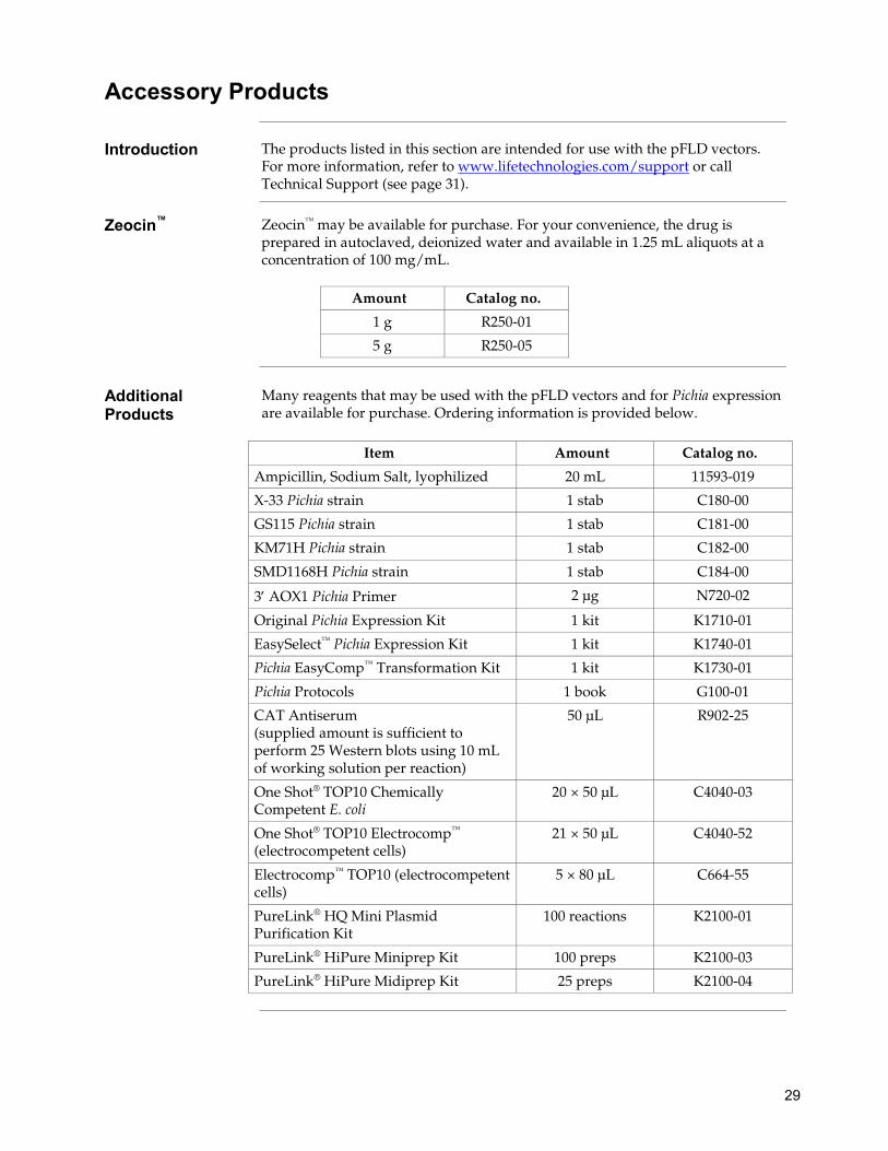
Accessory Products
Introduction The products listed in this section are intended for use with the pFLD vectors. For more information, refer to www.lifetechnologies.com/support or call Technical Support (see page 31).
Zeocin™ Zeocin™ may be available for purchase. For your convenience, the drug is prepared in autoclaved, deionized water and available in 1.25 mL aliquots at a concentration of 100 mg/mL.
Amount Catalog no.
1 g R250-01
5 g R250-05
Additional Products
Many reagents that may be used with the pFLD vectors and for Pichia expression are available for purchase. Ordering information is provided below.
Item Amount Catalog no.
Ampicillin, Sodium Salt, lyophilized 20 mL 11593-019
X-33 Pichia strain 1 stab C180-00
GS115 Pichia strain 1 stab C181-00
KM71H Pichia strain 1 stab C182-00
SMD1168H Pichia strain 1 stab C184-00
3′ AOX1 Pichia Primer 2 µg N720-02
Original Pichia Expression Kit 1 kit K1710-01
EasySelect™ Pichia Expression Kit 1 kit K1740-01
Pichia EasyComp™ Transformation Kit 1 kit K1730-01
Pichia Protocols 1 book G100-01
CAT Antiserum (supplied amount is sufficient to perform 25 Western blots using 10 mL of working solution per reaction)
50 μL R902-25
One Shot® TOP10 Chemically Competent E. coli
20 × 50 μL C4040-03
One Shot® TOP10 Electrocomp™ (electrocompetent cells)
21 × 50 μL C4040-52
Electrocomp™ TOP10 (electrocompetent cells)
5 × 80 μL C664-55
PureLink® HQ Mini Plasmid Purification Kit
100 reactions K2100-01
PureLink® HiPure Miniprep Kit 100 preps K2100-03
PureLink® HiPure Midiprep Kit 25 preps K2100-04

30
Accessory Products, Continued
Detecting Fusion Protein
A number of antibodies are available for purchase to detect expression of your fusion protein from the pFLD vector. Horseradish peroxidase (HRP) or alkaline phosphatase (AP)-conjugated antibodies allow one-step detection using colorimetric or chemiluminescent detection methods. The fluorescein isothiocyanate (FITC)-conjugated antibody allows one-step detection in immunofluorescence experiments. The amount of antibody supplied is sufficient for 25 Western blots or 25 immunostaining reactions (FITC-conjugated antibody only).
Antibody Epitope Catalog no.
Anti-V5 Detects 14 amino acid epitope derived from the P and V proteins of the paramyxovirus, SV5 (Southern et al., 1991). GKPIPNPLLGLDST
R960-25
Anti-V5-HRP R961-25
Anti-V5-AP Antibody R962-25
Anti-V5-FITC Antibody R963-25
Anti-His(C-term) Detects the C-terminal polyhistidine (6xHis) tag (requires the free carboxyl group for detection) (Lindner et al., 1997): HHHHHH-COOH
R930-25
Anti-His(C-term)-HRP R931-25
Positope™ Control Protein
R900-50
Purifying Fusion Protein
The polyhistidine (6xHis) tag allows purification of the recombinant fusion protein using metal-chelating resins such as ProBond™. Ordering information for ProBond™ resin is provided below.
Item Quantity Catalog no.
ProBond™ Purification System
Precharged ProBond™ resin and buffers plus 6 × 2 mL columns for native and denaturing purification
K850-01
ProBond ™ Purification System with Anti-V5-HRP Antibody
1 Kit (same as above plus 50 µL of antibody) The amount of antibody supplied is sufficient for 25 Westerns
K854-01
ProBond ™ Purification System with Anti-His(C-term)-HRP Antibody
1 Kit (same as above plus 50 µL of antibody) The amount of antibody supplied is sufficient for 25 Westerns
K853-01
ProBond™ Resin 50 mL R801-01
150 mL R801-15
Purification Columns 50 polypropylene columns R640-50

31
Technical Support
Obtaining support
For the latest services and support information for all locations, go to www.lifetechnologies.com/support. At the website, you can:
• Access worldwide telephone and fax numbers to contact Technical Support and Sales facilities
• Search through frequently asked questions (FAQs) • Submit a question directly to Technical Support ([email protected]) • Search for user documents, SDSs, vector maps and sequences, application
notes, formulations, handbooks, certificates of analysis, citations, and other product support documents
• Obtain information about customer training • Download software updates and patches
Safety Data Sheets (SDS)
Safety Data Sheets (SDSs) are available at www.lifetechnologies.com/support.
Certificate of Analysis
The Certificate of Analysis provides detailed quality control and product qualification information for each product. Certificates of Analysis are available on our website. Go to www.lifetechnologies.com/support and search for the Certificate of Analysis by product lot number, which is printed on the box.
Limited warranty Life Technologies Corporation is committed to providing our customers with high-quality goods and services. Our goal is to ensure that every customer is 100% satisfied with our products and our service. If you should have any questions or concerns about a Life Technologies product or service, contact our Technical Support Representatives. All Life Technologies products are warranted to perform according to specifications stated on the certificate of analysis. The Company will replace, free of charge, any product that does not meet those specifications. This warranty limits the Company’s liability to only the price of the product. No warranty is granted for products beyond their listed expiration date. No warranty is applicable unless all product components are stored in accordance with instructions. The Company reserves the right to select the method(s) used to analyze a product unless the Company agrees to a specified method in writing prior to acceptance of the order. Life Technologies makes every effort to ensure the accuracy of its publications, but realizes that the occasional typographical or other error is inevitable. Therefore the Company makes no warranty of any kind regarding the contents of any publications or documentation. If you discover an error in any of our publications, report it to our Technical Support Representatives. Life Technologies Corporation shall have no responsibility or liability for any special, incidental, indirect or consequential loss or damage whatsoever. The above limited warranty is sole and exclusive. No other warranty is made, whether expressed or implied, including any warranty of merchantability or fitness for a particular purpose.

32
Purchaser Notification
Limited Use Label License No: Research Use Only
The purchase of this product conveys to the purchaser the limited, non-transferable right to use the purchased amount of the product only to perform internal research for the sole benefit of the purchaser. No right to resell this product or any of its components is conveyed expressly, by implication, or by estoppel. This product is for internal research purposes only and is not for use in commercial applications of any kind, including, without limitation, quality control and commercial services such as reporting the results of purchaser's activities for a fee or other form of consideration. For information on obtaining additional rights, please contact [email protected] or Out Licensing, Life Technologies, 5791 Van Allen Way, Carlsbad, California 92008.
33
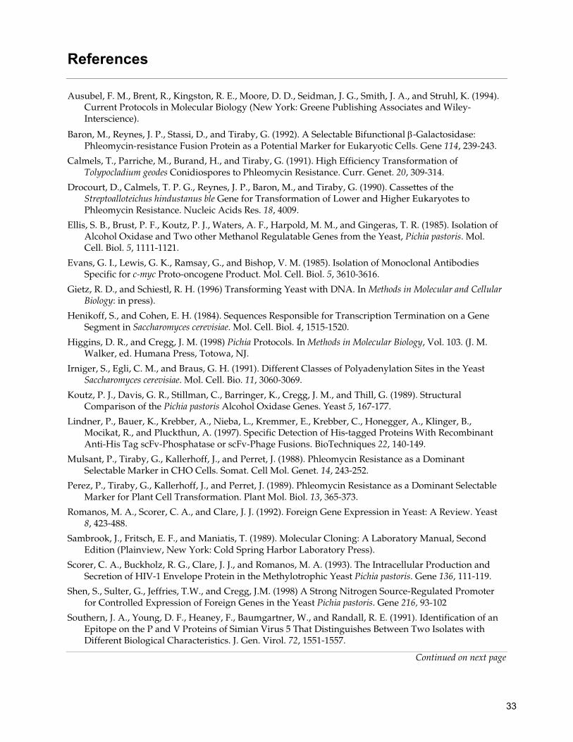
References Ausubel, F. M., Brent, R., Kingston, R. E., Moore, D. D., Seidman, J. G., Smith, J. A., and Struhl, K. (1994).
Current Protocols in Molecular Biology (New York: Greene Publishing Associates and Wiley-Interscience).
Baron, M., Reynes, J. P., Stassi, D., and Tiraby, G. (1992). A Selectable Bifunctional β-Galactosidase: Phleomycin-resistance Fusion Protein as a Potential Marker for Eukaryotic Cells. Gene 114, 239-243.
Calmels, T., Parriche, M., Burand, H., and Tiraby, G. (1991). High Efficiency Transformation of Tolypocladium geodes Conidiospores to Phleomycin Resistance. Curr. Genet. 20, 309-314.
Drocourt, D., Calmels, T. P. G., Reynes, J. P., Baron, M., and Tiraby, G. (1990). Cassettes of the Streptoalloteichus hindustanus ble Gene for Transformation of Lower and Higher Eukaryotes to Phleomycin Resistance. Nucleic Acids Res. 18, 4009.
Ellis, S. B., Brust, P. F., Koutz, P. J., Waters, A. F., Harpold, M. M., and Gingeras, T. R. (1985). Isolation of Alcohol Oxidase and Two other Methanol Regulatable Genes from the Yeast, Pichia pastoris. Mol. Cell. Biol. 5, 1111-1121.
Evans, G. I., Lewis, G. K., Ramsay, G., and Bishop, V. M. (1985). Isolation of Monoclonal Antibodies Specific for c-myc Proto-oncogene Product. Mol. Cell. Biol. 5, 3610-3616.
Gietz, R. D., and Schiestl, R. H. (1996) Transforming Yeast with DNA. In Methods in Molecular and Cellular Biology: in press).
Henikoff, S., and Cohen, E. H. (1984). Sequences Responsible for Transcription Termination on a Gene Segment in Saccharomyces cerevisiae. Mol. Cell. Biol. 4, 1515-1520.
Higgins, D. R., and Cregg, J. M. (1998) Pichia Protocols. In Methods in Molecular Biology, Vol. 103. (J. M. Walker, ed. Humana Press, Totowa, NJ.
Irniger, S., Egli, C. M., and Braus, G. H. (1991). Different Classes of Polyadenylation Sites in the Yeast Saccharomyces cerevisiae. Mol. Cell. Bio. 11, 3060-3069.
Koutz, P. J., Davis, G. R., Stillman, C., Barringer, K., Cregg, J. M., and Thill, G. (1989). Structural Comparison of the Pichia pastoris Alcohol Oxidase Genes. Yeast 5, 167-177.
Lindner, P., Bauer, K., Krebber, A., Nieba, L., Kremmer, E., Krebber, C., Honegger, A., Klinger, B., Mocikat, R., and Pluckthun, A. (1997). Specific Detection of His-tagged Proteins With Recombinant Anti-His Tag scFv-Phosphatase or scFv-Phage Fusions. BioTechniques 22, 140-149.
Mulsant, P., Tiraby, G., Kallerhoff, J., and Perret, J. (1988). Phleomycin Resistance as a Dominant Selectable Marker in CHO Cells. Somat. Cell Mol. Genet. 14, 243-252.
Perez, P., Tiraby, G., Kallerhoff, J., and Perret, J. (1989). Phleomycin Resistance as a Dominant Selectable Marker for Plant Cell Transformation. Plant Mol. Biol. 13, 365-373.
Romanos, M. A., Scorer, C. A., and Clare, J. J. (1992). Foreign Gene Expression in Yeast: A Review. Yeast 8, 423-488.
Sambrook, J., Fritsch, E. F., and Maniatis, T. (1989). Molecular Cloning: A Laboratory Manual, Second Edition (Plainview, New York: Cold Spring Harbor Laboratory Press).
Scorer, C. A., Buckholz, R. G., Clare, J. J., and Romanos, M. A. (1993). The Intracellular Production and Secretion of HIV-1 Envelope Protein in the Methylotrophic Yeast Pichia pastoris. Gene 136, 111-119.
Shen, S., Sulter, G., Jeffries, T.W., and Cregg, J.M. (1998) A Strong Nitrogen Source-Regulated Promoter for Controlled Expression of Foreign Genes in the Yeast Pichia pastoris. Gene 216, 93-102
Southern, J. A., Young, D. F., Heaney, F., Baumgartner, W., and Randall, R. E. (1991). Identification of an Epitope on the P and V Proteins of Simian Virus 5 That Distinguishes Between Two Isolates with Different Biological Characteristics. J. Gen. Virol. 72, 1551-1557.
Continued on next page

34
References, Continued
Tschopp, J. F., Brust, P. F., Cregg, J. M., Stillman, C., and Gingeras, T. R. (1987a). Expression of the lacZ Gene from Two Methanol Regulated Promoters in Pichia pastoris. Nucleic Acids Res. 15, 3859-3876.
Zaret, K. S., and Sherman, F. (1984). Mutationally Altered 3´ Ends of Yeast CYC1 mRNA Affect Transcript Stability and Translational Efficiency. J. Mol. Biol. 177, 107-136.
©2012 Life Technologies Corporation. All rights reserved. The trademarks mentioned herein are the properties of Life Technologies Corporation or their respective owners. Zeocin is a trademark of CAYLA, SA.

35
Notes

36
Notes


Headquarters5791 Van Allen Way | Carlsbad, CA 92008 USA | Phone +1 760 603 7200 | Toll Free in USA 800 955 6288For support visit www.invitrogen.com/support or email [email protected]
www.lifetechnologies.com