Peripheral Nerve Reconstruction With Autologous Grafts
16
3,350+ OPEN ACCESS BOOKS 108,000+ INTERNATIONAL AUTHORS AND EDITORS 115+ MILLION DOWNLOADS BOOKS DELIVERED TO 151 COUNTRIES AUTHORS AMONG TOP 1% MOST CITED SCIENTIST 12.2% AUTHORS AND EDITORS FROM TOP 500 UNIVERSITIES Selection of our books indexed in the Book Citation Index in Web of Science™ Core Collection (BKCI) Chapter from the book Basic Principles of Peripheral Nerve Disorders Downloaded from: http://www.intechopen.com/books/basic-principles-of-peripheral- nerve-disorders PUBLISHED BY World's largest Science, Technology & Medicine Open Access book publisher Interested in publishing with IntechOpen? Contact us at [email protected]
Transcript of Peripheral Nerve Reconstruction With Autologous Grafts

3,350+OPEN ACCESS BOOKS
108,000+INTERNATIONAL
AUTHORS AND EDITORS115+ MILLION
DOWNLOADS
BOOKSDELIVERED TO
151 COUNTRIES
AUTHORS AMONG
TOP 1%MOST CITED SCIENTIST
12.2%AUTHORS AND EDITORS
FROM TOP 500 UNIVERSITIES
Selection of our books indexed in theBook Citation Index in Web of Science™
Core Collection (BKCI)
Chapter from the book Basic Principles of Peripheral Nerve DisordersDownloaded from: http://www.intechopen.com/books/basic-principles-of-peripheral-nerve-disorders
PUBLISHED BY
World's largest Science,Technology & Medicine
Open Access book publisher
Interested in publishing with IntechOpen?Contact us at [email protected]
6
Peripheral Nerve Reconstruction with Autologous Grafts
Fabrizio Schonauer, Sergio Marlino, Stefano Avvedimento and Guido Molea
Chair of Plastic Surgery, University “Federico II”, Naples Italy
1. Introduction
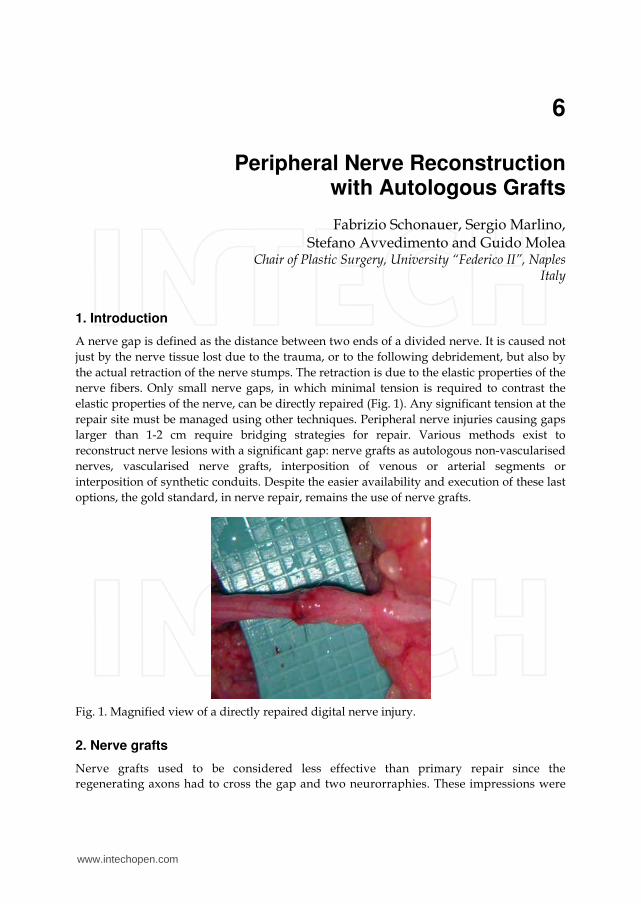
A nerve gap is defined as the distance between two ends of a divided nerve. It is caused not
just by the nerve tissue lost due to the trauma, or to the following debridement, but also by
the actual retraction of the nerve stumps. The retraction is due to the elastic properties of the
nerve fibers. Only small nerve gaps, in which minimal tension is required to contrast the
elastic properties of the nerve, can be directly repaired (Fig. 1). Any significant tension at the
repair site must be managed using other techniques. Peripheral nerve injuries causing gaps
larger than 1-2 cm require bridging strategies for repair. Various methods exist to
reconstruct nerve lesions with a significant gap: nerve grafts as autologous non-vascularised
nerves, vascularised nerve grafts, interposition of venous or arterial segments or
interposition of synthetic conduits. Despite the easier availability and execution of these last
options, the gold standard, in nerve repair, remains the use of nerve grafts.
Fig. 1. Magnified view of a directly repaired digital nerve injury.
2. Nerve grafts
Nerve grafts used to be considered less effective than primary repair since the
regenerating axons had to cross the gap and two neurorraphies. These impressions were
www.intechopen.com
Basic Principles of Peripheral Nerve Disorders
80
supported by the poor results which normally followed reconstructions using this
technique; various factors influenced the results in the early attempts.1 For a start, it was
thought that the longer the nerve graft, the worse the final result would be. This led
surgeons to perform all sorts of manoeuvres in order to reduce the distance between the
two nerve stumps, including flexing the joints, extensive nerve mobilisations and even
bone shortening. The results were disastrous because the disadvantage of the grafts (two
anastomosis to cross) was combined with the disadvantage of sutures under tension. We
now know that axonal regeneration takes place more easily when crossing two
anastomosis sites which are free of tension than across a neurorraphy carried out under
unfavourable conditions.
Another point of discussion was the source of the nerve grafts: the harvesting of rather thick
segments from nerve trunks of considerable size was thought necessary. However, a nerve
graft must first be re-vascularised before being repopulated by fibers. If the nerve graft is too
thick, its central part cannot be well re-vascularised and the results of the operation will be
poor. The introduction of thin nerve grafts has contributed greatly to the success of this
technique. 2
The principles and techniques for the use of nerve grafts are very similar to those used in
primary repair. The proximal and distal ends must be carefully prepared by transverse
section. The debate is whether it is better to postpone the graft reconstruction for a few
weeks or not. In fact, it may be risky to re-explore the region in complex traumas in which
multiple structures such as bone or blood vessels are involved. Under such circumstances,
intervention using a primary nerve graft may be justified. Should this be the choice,
thorough debridement must be carried out to ensure that the resection is well away from the
trauma zone. The dimensions of the defect are then measured with the joints extended to
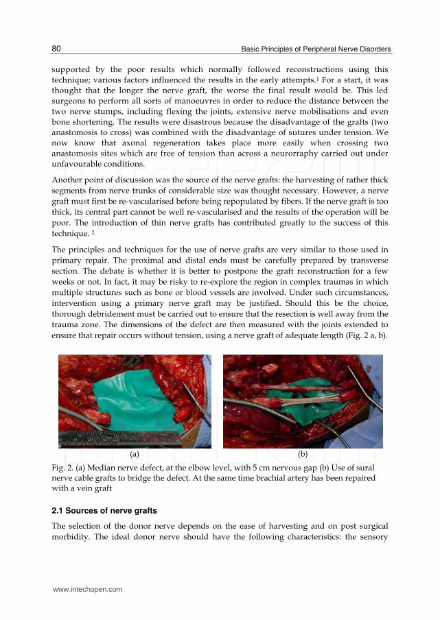
ensure that repair occurs without tension, using a nerve graft of adequate length (Fig. 2 a, b).
(a) (b)
Fig. 2. (a) Median nerve defect, at the elbow level, with 5 cm nervous gap (b) Use of sural nerve cable grafts to bridge the defect. At the same time brachial artery has been repaired with a vein graft
2.1 Sources of nerve grafts
The selection of the donor nerve depends on the ease of harvesting and on post surgical
morbidity. The ideal donor nerve should have the following characteristics: the sensory
www.intechopen.com
Peripheral Nerve Recontruction with Autologous Grafts
81
deficit caused by harvesting should occur in a non-critical cutaneous region; the donor
nerve should have sufficiently long segments with no lateral branches; it should be easy to
locate and surgically accessible; it should have a small overall diameter and well-developed
fascicles.3
Donor nerves for peripheral nerve reconstruction include: the medial antebrachial
cutaneous nerve (MABCN), the lateral antebrachial cutaneous nerve (LABCN), the
terminal sensitive branches of the posterior interosseous nerve and, traditionally, the sural
nerve.
The MABCN can provide up to a 10 cm graft. The resultant sensory deficit lies along the
medial aspect of the mid-forearm. The MABCN has also been reported as a donor graft for
repair of facial nerve defects. Higgins provided criteria for the selection of donor sites for
nerve harvest in digital nerve reconstruction.4 He investigated the cross-sectional area and
number of fascicles of both donor nerves and specific digital nerve segments.
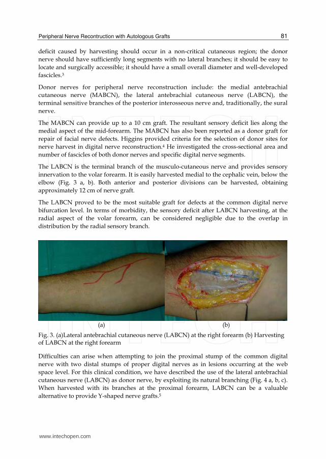
The LABCN is the terminal branch of the musculo-cutaneous nerve and provides sensory
innervation to the volar forearm. It is easily harvested medial to the cephalic vein, below the
elbow (Fig. 3 a, b). Both anterior and posterior divisions can be harvested, obtaining
approximately 12 cm of nerve graft.
The LABCN proved to be the most suitable graft for defects at the common digital nerve
bifurcation level. In terms of morbidity, the sensory deficit after LABCN harvesting, at the
radial aspect of the volar forearm, can be considered negligible due to the overlap in
distribution by the radial sensory branch.
(a) (b)
Fig. 3. (a)Lateral antebrachial cutaneous nerve (LABCN) at the right forearm (b) Harvesting of LABCN at the right forearm
Difficulties can arise when attempting to join the proximal stump of the common digital
nerve with two distal stumps of proper digital nerves as in lesions occurring at the web
space level. For this clinical condition, we have described the use of the lateral antebrachial
cutaneous nerve (LABCN) as donor nerve, by exploiting its natural branching (Fig. 4 a, b, c).
When harvested with its branches at the proximal forearm, LABCN can be a valuable
alternative to provide Y-shaped nerve grafts.5
www.intechopen.com

Basic Principles of Peripheral Nerve Disorders
82
(a) (b)
(c)
Fig. 4. (a) Right hand circular saw injury with disruption of all digital nerves in the palm with exception of the digital nerves to the thumb. (b) Use of standard and Y-shaped nerve grafts harvested from the right forearm (c) Use of LABCN and its branchings to mimic common digital nerve bifurcations.
The terminal branch of the posterior interosseus nerve has been used for distal digital nerve
graft or as a single fascicular strand that can be used to replace one fascicle of the digital
nerve. Today, the posterior interosseus nerve is considered a good choice for digital nerve
grafting. The use of this nerve is limited, but there is no functional deficit from harvesting
the nerve, as it is an articular branch. The posterior interosseous nerve is found at the wrist
level, lying on the interosseous membrane deep to the extensor tendons. As it branches
distally, it usually lies just ulnar and deep to the pollicis longus tendon and muscle. This
nerve is obtained by a longitudinal dorsal wrist incision. After opening the deep fascia,
retraction of the extensor tendons reveals the nerve lying on the interosseous membrane.
Care should be taken to preserve the extensor retinaculum. 6
www.intechopen.com
Peripheral Nerve Recontruction with Autologous Grafts
83
(a) (b)
(c) (d)
(e) (f)
(g) (h)
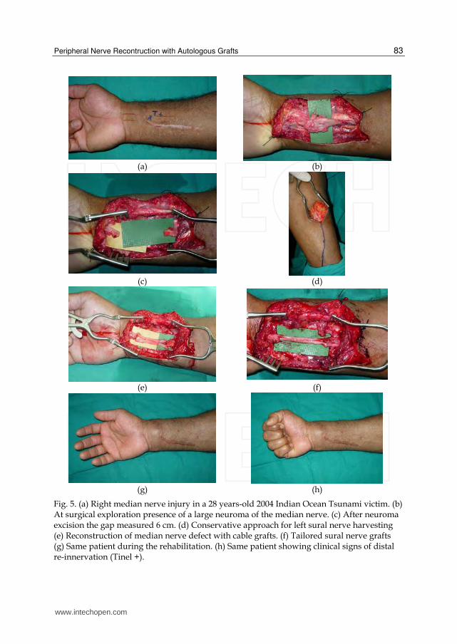
Fig. 5. (a) Right median nerve injury in a 28 years-old 2004 Indian Ocean Tsunami victim. (b) At surgical exploration presence of a large neuroma of the median nerve. (c) After neuroma excision the gap measured 6 cm. (d) Conservative approach for left sural nerve harvesting (e) Reconstruction of median nerve defect with cable grafts. (f) Tailored sural nerve grafts (g) Same patient during the rehabilitation. (h) Same patient showing clinical signs of distal re-innervation (Tinel +).
www.intechopen.com
Basic Principles of Peripheral Nerve Disorders
84
The sural nerve complex usually consists of three components: the medial sural cutaneous nerve, the peroneal communicating branch and the “proper” sural nerve. In 80% of dissections the sural nerve is formed by the union of the medial cutaneous nerve and the peroneal communicating branch. In 20% of cases the peroneal communicating branch is absent.7 The nerve is found adjacent to the lesser saphenous vein at the lateral malleolus. When the length of nerve graft need is limited, the peroneal communicating branch can be harvested alone and the medial sural cutaneous nerve can be saved.
Harvest of the sural nerve is usually performed with the patient in a supine position, with the lower extremity flexed and internally rotated at the hip, flexed about 40 degrees at the knee and the ankle dorsiflexed. The sural nerve in the adult can provide 30 to 40 cm of nerve graft. (Fig. 5 a, b, c, d, e, f, g, h)
Sural nerve harvest has traditionally been performed through a “stocking-seam” posterior lower leg incision. This longitudinal approach, that can theoretically extend from the lateral aspect of the ankle to the popliteal fossa, has the advantage of providing excellent visualization of the entire sural nerve anatomy. 8
The resulting donor-site scar has a tendency to thicken and widen and can be aesthetically unsatisfactory.9 Alternatively multiple step incisions can be used to harvest the sural nerve.10 This method yields better donor-site scars, but the limited visualization makes the nerve vulnerable to injury.11 The use of a tendon stripper with a limited incision at the ankle has also been described.12-13
To minimize donor-site scars and maximize visualization, endoscopic harvest of the sural nerve has been proposed by several authors (Kobayashi et al.14, Capek et al.9 and Eich and Fix15). Endoscopic harvest of the sural nerve offers better results in terms of scars but requires more operating time, familiarity with endoscopic techniques and proper instruments, including long scissors, a nerve retractor and a tendon or nerve stripper device. However, the increase in time as compared with conventional open methods could be counterbalanced by shorted time in suturing the wounds.16
3. Vascularized nerve grafts
The theoretical advantage of a vascularized nerve graft (VNG) lies in being able to immediately supply intra-neural perfusion in a poorly vascularized bed in the presence of very wide gaps. Their clinical role has not been well defined despite the fact that they were introduced over two decades ago. Taylor and Ham introduced free vascularized nerve grafts in an attempt to prevent ischemic graft failure.17 Initial experience with this technique in scarred beds was promising, however results and indications still remain controversial. In general, it may be said that vascularized nerve grafts are indicated when a long graft is required in a poorly vascularized bed, or when a graft with a large diameter is desired, in order to provide it with independent vascularization. Bonney et al 18 reviewed 12 cases of brachial plexus reconstruction with microsurgically revascularization nerve grafts and could only report an “impression” of improved recovery; Gilbert found no superiority of free vascularized grafting over routine sural grafts 19
More recently VNG have gained a new popularity; clinical studies with VNG of sural nerve have been conducted revealing their usefulness especially in lower limb nerve reconstruction.20
www.intechopen.com
Peripheral Nerve Recontruction with Autologous Grafts
85
4. Nerve conduits
Nerve gaps which are too large to be repaired using tension-free sutures are usually reconstructed using nerve grafts (interpositional nerve grafts). Research on the alternative use of conduits is in continual expansion 21. The advantage lies in avoiding the donor nerve sacrifice. The first works date back to 1880; since then, nerve conduits have been constructed using biological materials: bone, vein, muscle, artery and synthetic materials (silicon, polyglycolic acid and polyglactin).
For structural reasons, blood vessels were used as an obvious choice, due to the natural presence of a lumen. They were used both to protect the site of nerve anastomoses and as nerve conduits, in a number of modifications. In the past, the use of nerve conduits in nerve reconstruction has provided results inferior to those obtained using nerve grafts; today we see a renewed interest both in venous grafts22-25 and arterial grafts26, especially for short gap reconstruction (<3 cm).
4.1 Vein grafts
As we already stated the superior regenerative performance of autologous peripheral nerve grafts has resulted in a wide acceptance as the ‘‘gold standard’’ for peripheral nerve repair. Nevertheless, a number of issues pushed the research towards alternative bridging materials for the repair of peripheral nerve injuries. Such issues included the comorbidity at the site of donor nerve harvesting, the obvious limitations to the amount of donor nerve that can be used and the unsatisfactory functional outcomes in some of the patients receiving nerve autografts.
Vein conduit grafting has not been accepted as an alternative to nerve grafting until 1982, when Chiu et al. proved that it produced regeneration of nerve fascicles and recovery of distal sensation.27 The vein conduit graft acts as a guide for axonal sprouting and as a barrier against scar tissue ingrowth, maintaining an internal milieu for nerve regeneration.
Vein conduit grafts have two advantages compared to nerve grafts: they are readily accessible and they cause minimal donor-site morbidity.
Walton et al. reported the recovery of two-point discrimination in 12 of 18 digital nerves reconstructed with vein conduit grafts.28
Chiu and Strauch found that nerve gaps of 3 cm or less could be repaired successfully with vein conduit grafting and demonstrated their efficacy in both acute and delayed digital nerve repair.22
Tang et al. repaired nerve defects less then 3 cm long with vein conduit graft and the results were good to excellent in 61.1% of the injured digital nerves.29
A potential drawback with vein grafting is that the vein wall may collapse. However, Tseng
et al. have demonstrated, in the rat, that hematoma and thrombin within the vein keep the
conduit patent. 30
A limitation of autogenous venous nerve conduit is that its success has only been demonstrated in peripheral sensory nerves with small defects (<3 cm). When nerve defects exceed 3 cm, a vein conduit graft is less effective in maintaining the growth of nerve axons.31
www.intechopen.com
Basic Principles of Peripheral Nerve Disorders
86
In a rabbit model it has been shown that nerve regeneration is enhanced by the distal stump
of the transected nerve. This happens mainly because the distal stump of the nerve produces
some key factors that promote nerve regeneration as nerve growth factor (NGF). In a rat
model, the outcome of an NGF-treated vein graft was better than that of a saline-treated
vein graft.32 Human study also showed good functional recovery in digital, median, ulnar,
and superficial radial nerve defects longer than 3 cm with nerve tissue interposed into the
vein conduit grafts.33 Although NGF is present in the intima and adventitia of the femoral
veins of rats, the amount of it in a vein graft may be insufficient to promote nerve healing.34
The 3 cm limit of the vein conduit graft might be overcome by adding NGF to the wound to
create a more suitable milieu for nerve regeneration
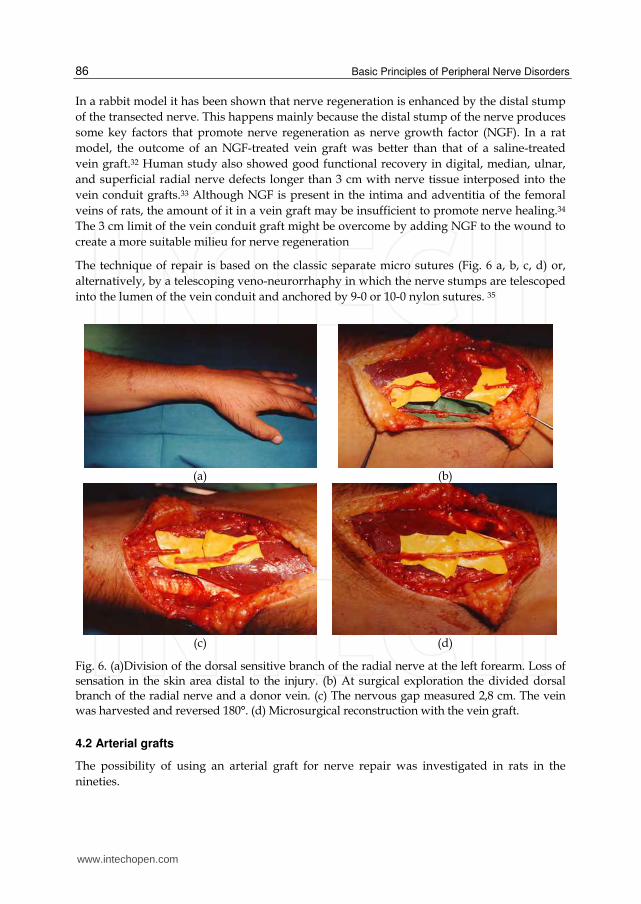
The technique of repair is based on the classic separate micro sutures (Fig. 6 a, b, c, d) or,
alternatively, by a telescoping veno-neurorrhaphy in which the nerve stumps are telescoped
into the lumen of the vein conduit and anchored by 9-0 or 10-0 nylon sutures. 35
(a) (b)
(c) (d)
Fig. 6. (a)Division of the dorsal sensitive branch of the radial nerve at the left forearm. Loss of sensation in the skin area distal to the injury. (b) At surgical exploration the divided dorsal branch of the radial nerve and a donor vein. (c) The nervous gap measured 2,8 cm. The vein was harvested and reversed 180°. (d) Microsurgical reconstruction with the vein graft.
4.2 Arterial grafts
The possibility of using an arterial graft for nerve repair was investigated in rats in the
nineties.
www.intechopen.com
Peripheral Nerve Recontruction with Autologous Grafts
87
Itoh et al. failed to find consistent nerve fiber regeneration with artery graft. They supposed
that their failure was caused by a laminin deficiency in the endothelial layer of artery wall.36
They concluded that arteries should not be used for tubulization.
Many authors reported the beneficial effects of laminin and collagen in the enhancement of
peripheral nerve regeneration. The artery wall has three layers: the endothelial layer
contains a laminin-rich basal lamina, the media is a muscle layer that is also rich in laminin
and the adventitia is rich in collagen. De Castro et al. turned the artery graft inside-out,
having these anatomical layers reversed. The resulting conduit exposed regenerating axons
directly to the adventitia. De Castro concluded that both the inside-out artery graft and
standard artery graft were valuable techniques for the repair of sensory peripheral nerves in
rats. 37
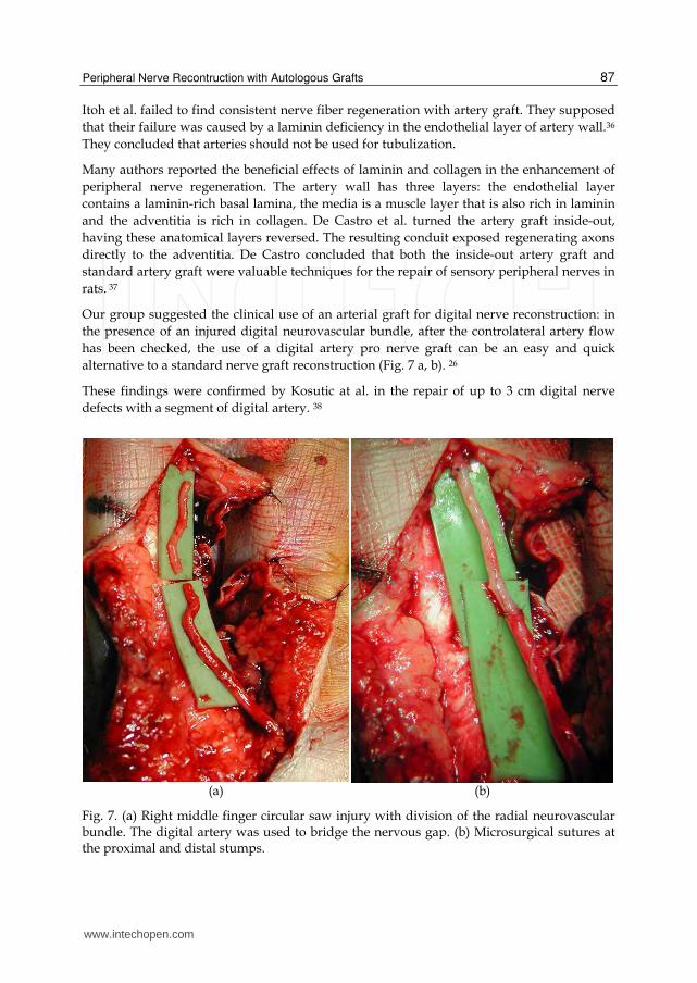
Our group suggested the clinical use of an arterial graft for digital nerve reconstruction: in
the presence of an injured digital neurovascular bundle, after the controlateral artery flow
has been checked, the use of a digital artery pro nerve graft can be an easy and quick
alternative to a standard nerve graft reconstruction (Fig. 7 a, b). 26
These findings were confirmed by Kosutic at al. in the repair of up to 3 cm digital nerve
defects with a segment of digital artery. 38
(a) (b)
Fig. 7. (a) Right middle finger circular saw injury with division of the radial neurovascular bundle. The digital artery was used to bridge the nervous gap. (b) Microsurgical sutures at the proximal and distal stumps.
www.intechopen.com
Basic Principles of Peripheral Nerve Disorders
88
4.3 Muscle grafts
In theory, any biological tissue containing basal lamina may be a candidate to be used as a
bridge for nerve regeneration.35
The use of muscle conduits has been recorded by multiple authors. The rationale is that the
longitudinally oriented basal lamina in skeletal muscle and the extracellular matrix
components are sufficient to direct and enhance nerve regeneration. This is based on
similarities between the basal lamina of muscle and the endoneural tubes of degenerating
nerves.39-40
Studies in animals and humans have demonstrated that both fresh and denatured muscle
conduits can provide successful regeneration and even lead to superior results when
compared with end-to-end sutures.
Skeletal muscle may be pretreated to produce an acute necrosis of myocytes, intramuscular
nerves and other cellular components of the skeletal muscle fibers. The cellular debris is
removed by macrophages, producing a network of coaxially directed tubes of muscle basal
lamina, lowering physical resistance and facilitating nerve regeneration.41
Numerous studies have confirmed the efficiency of frozen and thawed muscle grafting in
experimental models and animals.42
The first clinical trials to repair digital nerves and mixed sensory-motor nerves were
encouraging as well. However, motor nerve recovery remained poor in those studies,
probably because of considerable delay in repair.
The main concern regarding muscle grafts is the risk that nerve fibers can grow out of the
muscle tissue during regeneration, resulting in a decreased number of axons reaching the
end organ.41
Muscle can be harvested at the time of nerve repair by excising a rectangular muscle block
from the inferior border of the great pectoral muscle in the anterior axillary fold. The length
of harvested muscle can be approximately twice the nerve gap, to allow for shrinkage
during preparation. The block of muscle is frozen in liquid nitrogen until thermal
equilibrium has been obtained and then thawed in sterile distilled water. The graft is then
trimmed to the appropriate size to bridge the gap, keeping its long axis aligned and leaving
it about 2 mm wider in each plane than the diameter of the nerve. The prepared graft is
positioned using 4 to 5 monofilament interrupted nylon stitches (Ethilon 8-0) to join the
epinevrium of the nerve and the extreme periphery of the muscle graft. 33
4.3.1 Vein grafts filled with muscle
It is accepted that the use of vein or muscle graft alone is a good solution for nerve
reconstruction but suitable only for bridging limited nerve defects (<3 cm). Conversely,
these two techniques present important disadvantages for longer distances, such as the
collapse of the vein or dispersion of the regenerating axons out of the muscle. To allow for
the reconstruction of longer nerve defects and avoid vein collapse, filling of the vein with
muscle, or even slices of nerve has been suggested.
www.intechopen.com
Peripheral Nerve Recontruction with Autologous Grafts
89
In 1993, Brunelli et al. described the use of vein conduits, filled with fresh skeletal muscle, to
bridge nerve defects in rats, with functional results better than those obtained with muscle
or vein grafts alone and similar to those found with nerve graft.43 This technique was
abandoned for 7 years until Battiston et al. published a clinical report in 200044 and a review
in 2005.45 To avoid the dispersion of regenerating axons from the muscle guide, it may be
advisable to insert the muscle into the vein or even into artificial conduits.
Even though early experimental and clinical results were good, it is not clear why muscle-
in-vein conduit did not convince hand surgeons to proceed with further clinical application
of this technique. Perhaps lack of clear indications and limits of use (motor or sensory
nerves, or both) or difficulty in assembling the conduit itself could explain the rare
application of this technique.
5. Conclusion
It has been clearly demonstrated that the influence of the distal stump can be equally well
exerted using a conduit or a nerve graft over short distances less than 3 cm. A vein conduit
graft is a good alternative to a nerve graft. Limitations to success are probably due to the
medium inside the conduit. Substrates containing components of the extracellular matrix,
fibronectin and laminin have been shown to be important as scaffolding for supporting
axonal regeneration. Furthermore, several neurotrophic factors and some of their receptors
have been shown to be capable of facilitating nerve regeneration. With the advancement of
knowledge in the field of neurobiology, researchers are starting to report successes in the
progress of regeneration beyond the critical 3 cm gap barrier.
In clinical practice, conduits can be safely used only for gaps of less than 3 cm in
reconstructions of exclusively sensory nerves in patients who refuse harvesting of
autologous nerve grafts. However, nerve conduits will certainly play a much more
important clinical role in the future.
6. References
[1] Bunnell S. Surgery of nerves of the hand. Gynaecology and Obstetrics, 1927, 44, 145-152
[2] Beazley WC, Milek MA and Reiss BH. Results of nerve grafting in severe soft tissue
injuries. Clinical Orthopaedics and Related Research 1984 Sep;(188):208-12.
[3] Sunderland S, Ray L. The selection and use of autografts for bridging gaps in injured
nerves. Brain 1947 Mar;70(1):75-92.
[4] Higgins JP, Fisher S, Serletti JM, Orlando GS. Assessment of nerve graft donor sites used
for reconstruction of traumatic digital nerve defects. J Hand Surg Am. 2002
Mar,27(2):286-92.
[5] Schonauer F., Taglialatela Scafati S., LaRusca I, Molea G. Digital nerve reconstruction by
multiple Y-shaped nerve grafts at the metacarpophalangeal joint level. Plast.
Reconstr Surg. 2006 Apr. 15; 117 (5); 1661-2.
[6] Dellon AL, Seif SS. Anatomic dissections relating the posterior interosseous nerve to the
carpus, and the etiology of dorsal wrist ganglion pain. J Hand Surg Am. 1978 Jul; 3
(4):326-32.
www.intechopen.com
Basic Principles of Peripheral Nerve Disorders
90
[7] Ortigüela ME, Wood MB, Cahill DR. Anatomy of the sural nerve complex. J Hand Surg
Am. 1987 Nov;12 (6):1119-23.
[8] Matsuyama T, Mackay M, Midha R. Peripheral nerve repair and grafting techniques: a
review. Neurol Med Chir (Tokyo). 2000 Apr; 40(4): 187-99.
[9] Capek L, Clarke HM, Zuker RM. Endoscopic sural nerve harvest in the pediatric patient.
Plast Reconstr Surg. 1996 Oct; 98(5): 884-8.
[10] Chang DW. Minimal incision technique for sural nerve graft harvest experience with 61
patients. J Reconstr Microsur. 2002 Nov, 18(8): 671-6.
[11] Rindell K. and Telaranta T. A new atraumatic and simple method of taking sural nerve
grafts. Ann. Chir. Gynaecol. 1984, 73:40.
[12] Hill HL, Vasconez LO, and Jurkiewicz MJ. Method for obtaining a sural nerve graft.
Plast. Reconstr. Surg. 1978, 61:177.
[13] Hankin, F. M., Jaeger, S. H., and Beddings, A. Autogenous sural nerve grafts: A
harvesting technique. Orthopedics 1985, 8:1160.
[14] Kobayashi, S., Akizuki, T., Sakai, Y., and Ohmori, K. Harvest of sural nerve grafts using
the endoscope. Ann. Plast. Surg. 1995, 35:249.
[15] Eich, B. S., II, and Fix, R. J. New technique for endoscopic sural nerve graft. J. Reconstr.
Microsurg. 2000, 16:329.
[16] Lin CH, Mardini S, Levin SL, Lin YT, Yeh JT. Endoscopically assisted sural nerve
harvest for upper extremity posttraumatic nerve defects: an evaluation of
functional outcomes. Plast Reconstr Surg. 2007 Feb; 119(2):616-26.
[17] Taylor G, Ham F. The free vascularized nerve graft. Plast Reconstr Surg 1976; 57:
413-26
[18] Bonney G, Birch R, Jamieson AM, Eames RA. Experience with vascularized nerve graft.
Clin Plast Surg. 1984 Jan;11(1):137-42
[19] Gilbert A. Vascularized sural nerve graft. Clin Plast Surg. 1984 Jan;11(1):73-7
[20] Terzis JK, Kostopolous VK. Vascularized nerve grafts for lower extremity nerve reconstruction. Ann Plast Surg. 2010 Feb;64(2):169-76
[21] Taras JS, Nanavati V, Steelman P. Nerve conduits. J Hand Ther 2005 Apr-Jun; 18(2):
191-7.
[22] Suematsu N, Atsuta Y, Hirayama T. Vein graft for repair of peripheral nerve gap. J
Reconstr Microsurg. 1988;4:313-8.
[23] Chiu DT, Strauch B. A prospective clinical evaluation of autogenous vein grafts used as
a nerve conduit for distal sensory nerve defects of 3 cm or less. Plast Reconstr Surg.
1990 Nov;86(5):928-34.
[24] Chiu D.T. Autogenous venous nerve conduits. A review Hand Clinics 1999 15, 667-71
[25] Risitano G, Cavallaro G, Merrino T, Coppolino S, Ruggeri F. Clinical results and
thoughts on sensory nerve repair by autologous vein graft in emergency hand
reconstruction. Chir Main. 2002; 21: 194-7
[26] Schonauer F, La Rusca I, Molea G. Homolateral digital artery nerve graft. Plast Reconstr
Surg. 2006 Apr 15;117(5):1661-2.
[27] Chiu DT, Janecka I, Krizek TJ, Wolff M, Lovelace RE. Autogenous vein graft as a
conduit for nerve regeneration. Surgery 1982;91:226–
www.intechopen.com
Peripheral Nerve Recontruction with Autologous Grafts
91
[28] Walton RL, Brown RE, Matory WE Jr, Borah GL, Dolph JL. Autogenous vein graft
repair of digital nerve defects in the finger: A retrospective clinical study. Plast
Reconstr Surg 1989;84:944–949
[29] Tang JB, Gu YQ, Song YS. Repair of digital nerve defect with autogenous vein graft
during flexor tendon surgery in zone 2. J Hand Surg B 1993;18:449–453.
[30] Tseng CY, Hu G, Ambron RT, Chiu DT. Histologic analysis of Schwann cell migration
and peripheral nerve regeneration in the autogenous venous nerve conduit
(AVNC). J Reconstr Microsurg. 2003;19:331–340.
[31] Strauch B, Ferder M, Lovelle-Allen S, Moore K, Kim DJ, Llena J. Determining the
maximal length of a vein conduit used as an interposition graft for nerve
regeneration. J Reconstr Microsurg 1996;12:521–527.
[32] Pu LL, Syed SA, Reid M, Patwa H, Goldstein JM, Forman DL, Thomson JG. Effects of
nerve growth factor on nerve regeneration through a vein graft across a gap. Plast
Reconstr Surg 1999;104: 1379–1385
[33] Tang JB. Vein conduits with interposition of nerve tissue for peripheral nerve defects. J
Reconstr Microsurg 1995;11:21–26.
[34] Levine MH, Yates KE, Kaban LB. Nerve growth factor is expressed in rat femoral vein. J
Oral Maxillofac Surg 2002;60:729–733
[35] Dahlin LB, Lundborg G Use of tubes in peripheral nerve repair. Neurosurg Clin N Am
2001; 12: 341–352
[36] Itoh S, Shinomiya K, Samejima H, Ohta T, Ishizuki M, Ichinose S. Experimental study
on nerve regeneration through the basement membrane tubes of the nerve, muscle,
and artery. Microsurgery 1996;17: 525–534
[37] Rodrigues Ade C, Silva MD. Inside-out versus standard artery graft to repair a sensory
nerve in rats. Microsurgery 2001;21(3):102-7.
[38] Kosutic D, Krajnc I, Pejkovic B, Solman L. Autogenous digital artery graft for repair of
digital nerve defects in emergency hand reconstruction: two-year follow-up. J Plast
Reconstr Aesthet Surg. 2009 Apr;62(4):553. Epub 2008 Dec 30
[39] Chen LE, Seaber AV, Urbaniak JR, Murrel GA. Denatured muscle as a nerve conduit: a
functional, morphologic, and electrophysiologic evaluation. J Reconstr Microsurg
1994; 10: 137–144
[40] Roganovic Z., Ilic S, Savic M. Acta Radial nerve repair using an autologous denatured
muscle graft: comparison with outcomes of nerve graft repair. Neurochir (Wien)
(2007) 149: 1033–1039
[41] Meek MF, Varejao AS, Geuna S. Use of skeletal muscle tissue in peripheral nerve repair:
review of the literature. Tissue Eng 2004; 10: 1027– 1036
[42] Glasby MA, Gschmeissner SE, Huang CL, De Souza BA. Degenerated muscle grafts
used for peripheral nerve repair in primates. J Hand Surg 1986; 11: 347–351
[43] Brunelli G, Battiston B, Vigasio A, Brunelli G, Marocolo D. Bridging nerve defects with
combined skeletal muscle and vein conduits. Microsurgery 1993;14:247–251.
[44] Battiston B, Tos P, Cushway TR, Geuna S. Nerve repair by means of vein filled with
muscle grafts. I. Clinical results. Microsurgery 2000; 20:32–36.
www.intechopen.com

Basic Principles of Peripheral Nerve Disorders
92
[45] Battiston B, Geuna S, Ferrero M, Tos P. Nerve repair by means of tubulization: literature
review and personal clinical experience comparing biological and synthetic
conduits for sensory nerve repair. Microsurgery 2005;25:258 –267.
www.intechopen.com
Basic Principles of Peripheral Nerve DisordersEdited by Dr. Seyed Mansoor Rayegani
ISBN 978-953-51-0407-0Hard cover, 278 pagesPublisher InTechPublished online 28, March, 2012Published in print edition March, 2012
InTech EuropeUniversity Campus STeP Ri Slavka Krautzeka 83/A 51000 Rijeka, Croatia Phone: +385 (51) 770 447 Fax: +385 (51) 686 166www.intechopen.com
InTech ChinaUnit 405, Office Block, Hotel Equatorial Shanghai No.65, Yan An Road (West), Shanghai, 200040, China
Phone: +86-21-62489820 Fax: +86-21-62489821
Peripheral nerve disorders are comprising one of the major clinical topics in neuromusculoskeletal disorders.Sharp nerve injuries, chronic entrapment syndromes, and peripheral neuropathic processes can be classifiedin this common medical topic. Different aspects of these disorders including anatomy, physiology,pathophysiology, injury mechanisms, and different diagnostic and management methods need to beaddressed when discussing this topic. The goal of preparing this book was to gather such pertinent chapters tocover these aspects.
How to referenceIn order to correctly reference this scholarly work, feel free to copy and paste the following:
Fabrizio Schonauer, Sergio Marlino, Stefano Avvedimento and Guido Molea (2012). Peripheral NerveReconstruction with Autologous Grafts, Basic Principles of Peripheral Nerve Disorders, Dr. Seyed MansoorRayegani (Ed.), ISBN: 978-953-51-0407-0, InTech, Available from: http://www.intechopen.com/books/basic-principles-of-peripheral-nerve-disorders/peripheral-nerve-reconstruction-with-autologous-grafts


![Functional Outcomes of Multiple Sural Nerve Grafts for Facial Nerve … · 2019-08-13 · Facial Nerve Disorders Committee [12]. In addition, two of the 12 patients were assessed](https://static.fdocuments.us/doc/165x107/5edf2bbaad6a402d666a853e/functional-outcomes-of-multiple-sural-nerve-grafts-for-facial-nerve-2019-08-13.jpg)