PDF (2.16 MB) - IOPscience
10
Science and Technology of Advanced Materials PAPER • OPEN ACCESS Probing cellular behaviors through nanopatterned chitosan membranes To cite this article: Chung-Yao Yang et al 2013 Sci. Technol. Adv. Mater. 14 044406 View the article online for updates and enhancements. You may also like Microfluidic fabrication of stable collagen microgels with aligned microstructure using flow-driven co-deposition and ionic gelation Santiago O Correa, Xiaolong Luo and Christopher B Raub - Transport phenomena in chitosan synthetic membranes with emphasis on the effect of variations in the ratio of matrix/solvent N Nyoman Rupiasih, Yayuk Eka Puspita and Made Sumadiyasa - Chitosan membrane technology as adsorbent media: management of Tallo River Basin Makassar City, South Sulawesi, Indonesia Hamsina Hamsina, B Surya, M Muhammadiah et al. - This content was downloaded from IP address 222.113.13.203 on 13/02/2022 at 15:56
Transcript of PDF (2.16 MB) - IOPscience

Science and Technology ofAdvanced Materials
PAPER • OPEN ACCESS
Probing cellular behaviors through nanopatternedchitosan membranesTo cite this article: Chung-Yao Yang et al 2013 Sci. Technol. Adv. Mater. 14 044406
View the article online for updates and enhancements.
You may also likeMicrofluidic fabrication of stable collagenmicrogels with aligned microstructureusing flow-driven co-deposition and ionicgelationSantiago O Correa, Xiaolong Luo andChristopher B Raub
-
Transport phenomena in chitosansynthetic membranes with emphasis onthe effect of variations in the ratio ofmatrix/solventN Nyoman Rupiasih, Yayuk Eka Puspitaand Made Sumadiyasa
-
Chitosan membrane technology asadsorbent media: management of TalloRiver Basin Makassar City, SouthSulawesi, IndonesiaHamsina Hamsina, B Surya, MMuhammadiah et al.
-
This content was downloaded from IP address 222.113.13.203 on 13/02/2022 at 15:56

IOP PUBLISHING SCIENCE AND TECHNOLOGY OF ADVANCED MATERIALS
Sci. Technol. Adv. Mater. 14 (2013) 044406 (9pp) doi:10.1088/1468-6996/14/4/044406
Probing cellular behaviors throughnanopatterned chitosan membranesChung-Yao Yang, Chun-Yen Sung, Hung-Hsun Shuai, Chao-Min Chengand J Andrew Yeh
Institute of Nanoengineering and Microsystems, National Tsing Hua University, Hsinchu 30013, Taiwan
E-mail: [email protected] and [email protected]
Received 26 May 2013Accepted for publication 18 July 2013Published 13 August 2013Online at stacks.iop.org/STAM/14/044406
AbstractThis paper describes a high-throughput method for developing physically modified chitosanmembranes to probe the cellular behavior of MDCK epithelial cells and HIG-82 fibroblastsadhered onto these modified membranes. To prepare chitosan membranes withmicro/nanoscaled features, we have demonstrated an easy-to-handle, facile approach thatcould be easily integrated with IC-based manufacturing processes with mass productionpotential. These physically modified chitosan membranes were observed by scanning electronmicroscopy to gain a better understanding of chitosan membrane surface morphology. AfterMDCK cells and HIG-82 fibroblasts were cultured on these modified chitosan membranes forvarious culture durations (i.e. 1, 2, 4, 12 and 24 h), they were investigated to decipher cellularbehavior. We found that both cells preferred to adhere onto a flat surface rather than on ananopatterned surface. However, most (>80%) of the MDCK cells showed roundedmorphology and would suspend in the cultured medium instead of adhering onto the planarsurface of negatively nanopatterned chitosan membranes. This means different cell types (e.g.fibroblasts versus epithelia) showed distinct capabilities/preferences of adherence formaterials of varying surface roughness. We also showed that chitosan membranes could bere-used at least nine times without significant contamination and would provide us consistencyfor probing cell–material interactions by permitting reuse of the same substrate. We believethese results would provide us better insight into cellular behavior, specifically, microscopicproperties and characteristics of cells grown under unique, nanopatterned cell-interfaceconditions.
Keywords: nanopatterned chitosan membrane, cell–material interfaces, actin cytoskeleton,HIG-82, fibroblast, cell structure
1. Introduction
This paper describes an easy-to-handle, facile approach—acombination of multiple IC-based manufacturingprocesses—to prepare nanopatterned chitosan membranesubstrates (with two alternatives) to probe the cellularbehavior of two types of mammalian cells—Madin-Darbycanine kidney (MDCK) epithelial cells and HIG-82 rabbit
Content from this work may be used under the terms of theCreative Commons Attribution-NonCommercial-ShareAlike
3.0 licence. Any further distribution of this work must maintain attribution tothe author(s) and the title of the work, journal citation and DOI.
synobyl fibroblasts. Individual cells were exposed to variousphysical stimuli, such as fluidic shear stress or substraterigidity [1–3]. Subsequent intracellular signals, in turn,modulate the dynamic responses of intracellular proteins,causing observable, measurable cellular behavior notedherein. Cells are also influenced by numerous non-specificexternal stimuli (i.e. non-physically based stimuli) such aschanges in either pH value or temperature, electrical fieldsand the surface chemistry of the adhesive substrate [1]. Tofully understand how single mammalian cells sense andthen respond to these stimuli, whether multiple or singularin nature, has been a longstanding but interesting subject
1468-6996/13/044406+09$33.00 1 © 2013 National Institute for Materials Science Printed in the UK

Sci. Technol. Adv. Mater. 14 (2013) 044406 C-Y Yang et al
in multiple research communities such as biomaterials, cellbiology, tissue engineering and mechanobiology [4, 5].
Substrate properties influence cellular behavior includinga cell’s protein reconstruction; this offers opportunities forspecific ligand–receptor interactions (e.g. actin filamentremodeling or integrin redistribution) [1, 6] and optionsfor leveraging general surface chemistry properties suchas relative surface charge and hydrophilicity [7, 8].Cell–material interfaces and corresponding responsescan alter the physiological functionality of singlemammalian cells, significantly affecting the success ofimplantable medical devices. For example, modified siliconemicrotopography (10 µm vertical projections) has beenshown to inhibit the over-proliferation of rat neonatalcardiac fibroblasts when co-culturing cardiac myocyteswith rat neonatal cardiac fibroblasts [9]. In this study byBoateng et al [9], excessive fibroblast proliferation in cardiacmyocyte cultures was decreased by 50% after 5 days ofculture, indicating that microtopographic properties (i.e.variations from planar to raised/uneven) can effectivelyprevent non-myocyte proliferation. Numerous micro- andnano-engineering approaches have been used to controlcell–substrate interactions in vitro by presenting specificmolecules to cells [10–16]. For example, one of the firstapplications using a microengineering approach to controlcell microenvironment was demonstrated by Whitesideset al [10–13]. In their studies, they used soft lithography tofabricate microstructures to generate micropatterned surfacescomprised of adhesive regions to which cells could attach.In addition, self-assembled monolayers or self-assembledmulti-layer structures have been shown to affect the behaviorof neurons [14], myoblasts [15] and fibroblasts [16]. Thesurface chemistry and topography of the substrate may beimportant in directing cellular behavior such as proliferation,migration and matrix deposition. Flemming et al [17],provided an extensive review of the effects of surfacetopography on cellular behavior. In the last decade, the abilityto fabricate and evaluate the effect of nanosized topographicalfeatures similar to the extracellular matrix (ECM) has allowedresearchers to more accurately represent and examine cells ina physiological environment.
Most previous in vitro studies have examined cellscultured on a two-dimensional flat and rigid substrates; invivo, however, most cells are exposed to a three-dimensionalmicroenvironment with complex topographical features.Using some advanced nanofabrication methods, nanoscaletopographical features can be incorporated into the in vitroexperimental platform to mechanically and structurally mimicvarious in vivo three-dimensional ECM environments. Qiet al studied the interactions between biological cells andvertically aligned silicon nanowire (SiNW) arrays (fabricatedby self-assembling nanoelectrochemistry). They focused onunderstanding how SiNW arrays affect cellular behaviorsuch as cell adhesion and spreading [18]. The results ofthis study show that SiNW arrays can not only enhance thecell–substrate adhesion force but also restrict cell spreading.Pesen et al [19] used electron beam lithography to patternnanostructures and two different combinations of applied
proteins to examine cellular mechanisms associated withthe formation and organization of focal adhesions. It wasfound that the very shape of nanoscale surface patterns canbe used to modulate focal adhesion complex organization anddetermine the organization of the adjoining cytoskeleton. Haqet al [20] used nanopillars and nanopores with dimensionscomparable too, but slightly larger than, those of filopodia(∼100–150 nm) to investigate the electrophysiologicalactivities of neurons. They found that PC 12 cells exhibited ahigher density and shorter neurite extension on nanopillars,but a lower density and intermediate neurite extension whengrown on nanopores. Nanopillars had a greater inhibiting eff-ect on neurite outgrowth than nanopores. Cecchini et al [21]used nanogratings to analyze the differentiation of PC 12cells. He concluded that topographic cues were moreeffective in determining axon outgrowth direction thancell–cell interactions. His results showed unstable synapticconnections and cell-body polarization, and further suggestedthat the substrate topography can act cooperatively withnerve growth factor signaling to regulate cell differentiationand neurite outgrowth. These approaches have allowedresearchers to probe biologically relevant issues usingadvanced nanotechnologies. However, the main drawbacksof these approaches include the following: (i) the challengesand time required to fabricate nanotopography (e.g. it istime consuming to fabricate nanostructures by using eitherelectron beam or electrospinning); and (ii) difficultieswith mass production for further applications (e.g. tissueengineering).
The challenges in tissue engineering include thefollowing: (i) the generation of vascularized tissues andcomplex geometries is difficult due to our limited ability tocontrol the cellular environment at the micro/nanoscale; (ii)the lack of suitable materials with the desired degradationrates and the mechanical properties for the desired tissue;(iii) the optimization of scaffold architecture, includingpore size, bioactivity, surface topography and morphology.Therefore, new and optimized processing methods mustbe developed to address issues related to cell seeding,vascularization and scale up into three-dimensional structures.Chitosan-based materials (or chitin-based materials) originatefrom nature, they have been approved by the US Foodand Drug Administration (FDA) and EnvironmentalProtection Agency (EPA) and are used in a variety ofbiotechnologically relevant applications, such as drug carriersand nanoparticles [22, 23]. However, it is still challengingto produce surface modifications, such as nanostructures,on chitosan membranes. Herein, extending our previouswork [1–3, 8, 24], we have developed a robust and easy tobuild and operate method to fabricate nanoscale topographicalfeatures and mesa structures (positive and negative forms)on chitosan surfaces to study behavior of MDCK cells andHIG-82 fibroblasts. In this study, the physical nanofabricationapproach was developed through the following steps: (i)mesa structures were defined on monocrystalline siliconwafers using a UV-photolithography process followed bytreatment with Ag-nanoparticles (AgNPs assisted etching)to form nanopatterns [8]; (ii) the positively and negatively
2

Sci. Technol. Adv. Mater. 14 (2013) 044406 C-Y Yang et al
nanopatterned chitosan membranes were prepared througha combination of solution casting, drying and peelingoff from silicon nanopatterns; and, (iii) the mammaliancells—MDCK cells and HIG-82 fibroblasts—were culturedon nanopatterned chitosan membranes to probe cellularbehavior.
2. Materials and methods
2.1. Preparation of positively and negatively nanopatternedsilicon molds
First, the positive silicon mold was made by photolithographyto create desired patterns on a 4-inch monocrystalline siliconwafer. The patterned silicon wafer with photoresist was thendipped into a 0.01 M silver nitrate solution for 5 min. Thenumber of silver ions (Ag+) decreased on the Si surfacebecause of the higher electronegativity of Ag than Si. Afterformation of a metallic catalyst layer, the wafer was immersedin an etchant comprising hydrogen fluoride (HF, 49 wt%) andhydrogen peroxide (H2O2, 30 wt%), with a mixture ratio of3:1 (v/v), for 1.5 min at room temperature. After etching,the positively nanopatterned silicon mold was prepared byphotoresist removal with acetone and rinsed with de-ionizedwater (figure 1(a)).
The negative silicon mold was made by dipping a4-inch monocrystalline silicon wafer into a 0.01 M silvernitrate solution for 5 min and then immersing in an etchantcomprising HF (49 wt%) and H2O2 (30 wt%), with a mixtureratio of 3:1 (v/v), for 1.5 min at room temperature. The desiredpatterns were completed using a photolithography processafter coating photoresist on nanopatterned silicon wafer.The negatively nanopatterned silicon mold was prepared byreactive ion etching (P300, Integrated Plasma IncorporatedLimited Co., Taiwan) for 6 min and photoresist removalby an oxygen plasma stripper system (P300, IntegratedPlasma Incorporated Limited Co., Taiwan) for 8 min(figure 1(b)).
2.2. Preparation of positively and negatively nanopatternedchitosan membranes
The 1% (w/v) chitosan solution was prepared by dissolvingmedium molecular weight chitosan powder (190–310 kDa;Sigma-Aldrich; St Louis, MO, USA) in 1% (v/v) aceticacid solution. The solution was continuously stirred with asterile magnetic bar for 6 h, and undissolved impurities andfoam were removed by vacuum filtration and a Whatman no.541 filter paper (0.22 µm pore size). After casting chitosansolution on positively and negatively nanopatterned siliconmolds, the nanopatterned chitosan membranes were carriedout by drying in an oven at 60 ◦C for 6 h and peeling offfrom silicon molds by immersing into 0.1 M sodium hydroxylsolution for 4 h (figure 1(c)). The nanopatterned chitosanmembranes were finally rinsed with de-ionized water untilneutral pH value was achieved.
2.3. Cell culture
Rabbit synobyl fibroblasts (HIG-82) and MDCK cellswere maintained in normal tissue culture T-25 flasks withHam’s F-12 medium (Invitrogen, Carlsbad, CA, USA) forHIG-82 fibroblasts and Dulbecco’s Modified Eagle’s Medium(Invitrogen, Carlsbad, CA, USA) for MDCK epithelial cells.Both media were supplemented with 10% fetal bovineserum (FBS; HyClone Laboratories Inc., Rockford, IL, USA),4 mM l-glutamine, 100 U mL−1 penicillin and 1.5 g L−1
sodium bicarbonate, and culture were maintained at 37 ◦Cin a humidified 5% CO2 incubator. Cells were trypsinizedwith 0.25% trypsin–ethylenediaminetetraacetic acid (EDTA)and re-seeded onto the sterilized nanopatterned chitosanmembranes (positive and negative membranes) at a densityof 5 × 10⁴ cells mL−1 for 12 h to perform experiments andexaminations.
2.4. Scanning electron microscopy (SEM)
The morphology of silicon molds and chitosan membraneswas examined with a high-resolution field-emissionSEM—Ultra 55 (Carl Zeiss SMT AG, Germany). Thechitosan membranes were dried in air and then sputteredwith gold at a current of 15 mA for 3 min by using an ionsputterer (E-1010; Hitachi, Japan). The MDCK cells andHIG-82 fibroblasts adhered onto positively nanopatternedchitosan membranes visualized with SEM were first fixedwith a fixative containing 2.5% glutaraldehyde per 0.1 MPBS at 4 ◦C for 30 min. After washing twice with 0.1 MPBS, the cells were post-fixed with 1% osmium tetroxide(Sigma-Aldrich, St Louis, MO, USA) at room temperaturefor 30 min. The cells were then washed twice again with0.1 M PBS, dehydrated through serial gradients of ethanol(10 min for each gradient, 50, 70, 80, 90 and 100), and finallydried out in a critical point dryer (HCP-2; Hitachi, Japan).The cells on positively nanopatterned chitosan membraneswere sputtered with gold at a current of 15 mA for 3 min byan ion sputterer.
2.5. Epi-fluorescence microscopy
MDCK cells and HIG-82 fibroblasts were cultured onpositively nanopatterned chitosan membranes and thenwashed twice with PBS. The cells were fixed with fresh 4%paraformaldehyde for 15 min at room temperature. Afterwashing with PBS two more times, the substrates wereimmersed in 0.1% Triton X-100 for 10 min to permeabilizecell membranes. Rhodamine-conjugated phalloidin(Invitrogen, Carlsbad, CA, USA) was used to label actinfilaments in living cells, while 4′,6-diamidino-2-phenylindole(DAPI; Invitrogen, Carlsbad, CA, USA) was used to stainDNA in nuclei. The cells were mounted with Fluoromount-G(Southernbiotech, Birmingham, AL, USA), and sealed undera coverslip. The cells were examined under an epi-fluorescentmicroscope (Zeiss Axio Observer, Germany) equipped witha 10× eyepiece and 63 × (NA1.4) objective to image theirmorphology and actin filaments.
3
Sci. Technol. Adv. Mater. 14 (2013) 044406 C-Y Yang et al
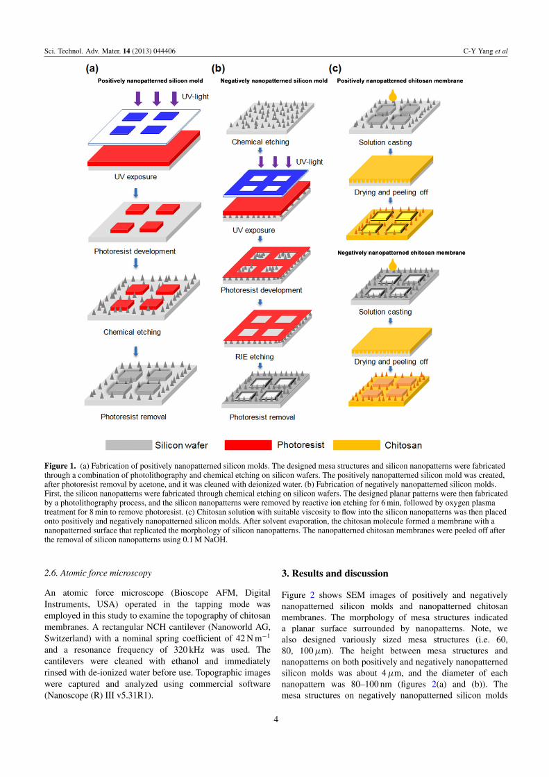
Figure 1. (a) Fabrication of positively nanopatterned silicon molds. The designed mesa structures and silicon nanopatterns were fabricatedthrough a combination of photolithography and chemical etching on silicon wafers. The positively nanopatterned silicon mold was created,after photoresist removal by acetone, and it was cleaned with deionized water. (b) Fabrication of negatively nanopatterned silicon molds.First, the silicon nanopatterns were fabricated through chemical etching on silicon wafers. The designed planar patterns were then fabricatedby a photolithography process, and the silicon nanopatterns were removed by reactive ion etching for 6 min, followed by oxygen plasmatreatment for 8 min to remove photoresist. (c) Chitosan solution with suitable viscosity to flow into the silicon nanopatterns was then placedonto positively and negatively nanopatterned silicon molds. After solvent evaporation, the chitosan molecule formed a membrane with ananopatterned surface that replicated the morphology of silicon nanopatterns. The nanopatterned chitosan membranes were peeled off afterthe removal of silicon nanopatterns using 0.1 M NaOH.
2.6. Atomic force microscopy
An atomic force microscope (Bioscope AFM, DigitalInstruments, USA) operated in the tapping mode wasemployed in this study to examine the topography of chitosanmembranes. A rectangular NCH cantilever (Nanoworld AG,Switzerland) with a nominal spring coefficient of 42 N m−1
and a resonance frequency of 320 kHz was used. Thecantilevers were cleaned with ethanol and immediatelyrinsed with de-ionized water before use. Topographic imageswere captured and analyzed using commercial software(Nanoscope (R) III v5.31R1).
3. Results and discussion
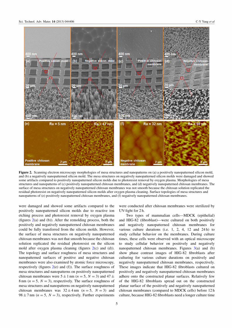
Figure 2 shows SEM images of positively and negativelynanopatterned silicon molds and nanopatterned chitosanmembranes. The morphology of mesa structures indicateda planar surface surrounded by nanopatterns. Note, wealso designed variously sized mesa structures (i.e. 60,80, 100 µm). The height between mesa structures andnanopatterns on both positively and negatively nanopatternedsilicon molds was about 4 µm, and the diameter of eachnanopattern was 80–100 nm (figures 2(a) and (b)). Themesa structures on negatively nanopatterned silicon molds
4
Sci. Technol. Adv. Mater. 14 (2013) 044406 C-Y Yang et al
Figure 2. Scanning electron microscopy morphologies of mesa structures and nanopatterns on (a) a positively nanopatterned silicon mold,and (b) a negatively nanopatterned silicon mold. The mesa structures on negatively nanopatterned silicon molds were damaged and showedsome artifacts compared to positively nanopatterned silicon molds due to photoresist removal by oxygen plasma. Morphologies of mesastructures and nanopatterns of (c) positively nanopatterned chitosan membranes, and (d) negatively nanopatterned chitosan membranes. Thesurface of mesa structures on negatively nanopatterned chitosan membranes was not smooth because the chitosan solution replicated theresidual photoresist on negatively nanopatterned silicon molds after oxygen plasma cleaning. Surface topologies of mesa structures andnanopatterns of (e) positively nanopatterned chitosan membranes, and (f) negatively nanopatterned chitosan membranes.
were damaged and showed some artifacts compared to thepositively nanopatterned silicon molds due to reactive ionetching process and photoresist removal by oxygen plasma(figures 2(a) and (b)). After the remolding process, both thepositively and negatively nanopatterned chitosan membranescould be fully transferred from the silicon molds. However,the surface of mesa structures on negatively nanopatternedchitosan membranes was not that smooth because the chitosansolution replicated the residual photoresist on the siliconmold after oxygen plasma cleaning (figures 2(c) and (d)).The topology and surface roughness of mesa structures andnanopatterned surfaces of positive and negative chitosanmembranes were also examined by atomic force microscopy,respectively (figures 2(e) and (f)). The surface roughness ofmesa structures and nanopatterns on positively nanopatternedchitosan membranes were 5 ± 1 nm (n = 5, N = 3) and 47 ±
8 nm (n = 5, N = 3), respectively. The surface roughness ofmesa structures and nanopatterns on negatively nanopatternedchitosan membranes was 32 ± 4 nm (n = 5, N = 3) and98 ± 7 nm (n = 5, N = 3), respectively. Further experiments
were conducted after chitosan membranes were sterilized byUV-light for 2 h.
Two types of mammalian cells—MDCK (epithelial)and HIG-82 (fibroblast)—were cultured on both positivelyand negatively nanopatterned chitosan membranes forvarious culture durations (i.e. 1, 2, 4, 12 and 24 h) tostudy cellular behavior on the membranes. During culturetimes, these cells were observed with an optical microscopeto study cellular behavior on positively and negativelynanopatterned chitosan membranes. Figures 3(a) and (b)show phase contrast images of HIG-82 fibroblasts afterculturing for various culture durations on positively andnegatively nanopatterned chitosan membranes, respectively.These images indicate that HIG-82 fibroblasts cultured onpositively and negatively nanopatterned chitosan membranesadhere onto the constructed planar surfaces. Relatively fewof the HIG-82 fibroblasts spread out on the constructedplanar surface of the positively and negatively nanopatternedchitosan membranes (compared to MDCK cells) before 12 hculture, because HIG-82 fibroblasts need a longer culture time
5

Sci. Technol. Adv. Mater. 14 (2013) 044406 C-Y Yang et al
Figure 3. Phase contrast images of HIG-82 fibroblasts after 1, 2, 4, 12 and 24 h cultures on (a) positively nanopatterned chitosanmembranes, and (b) negatively nanopatterned chitosan membranes. Phase contrast images of MDCK cells after 1, 2, 4, 12 and 24 h cultureson (c) positively nanopatterned chitosan membranes, and (d) negatively nanopatterned chitosan membranes. Both cells preferred to adhereonto a flat/planar surface rather than on nanopatterns of positively nanopatterned chitosan membranes. HIG-82 fibroblasts could adhere ontorough surfaces better than MDCK cells could, based on the morphology of flat surfaces on negative nanopatterned chitosan membranes. (e)The ratio of MDCK cells and HIG-82 fibroblasts adhered onto a flat surface versus total cells with various widths of positively andnegatively nanopatterned chitosan membranes. Data are mean ± standard deviation (N = 5, n = 10).
to proliferate and spread (i.e. 24 h culture). Figures 3(c) and(d) show phase contrast images of MDCK cells after culturingfor various culture durations on positively and negativelynanopatterned chitosan membranes, respectively. The imagesshow that MDCK cells cultured on positively nanopatterned
chitosan membranes adhere and spread out at the constructedplanar surface within 4 h. However, most (>80%) of theMDCK cells showed rounded morphology and would suspendin the culture medium instead of adhering onto the planarsurface of negatively nanopatterned chitosan membranes.
6

Sci. Technol. Adv. Mater. 14 (2013) 044406 C-Y Yang et al
Figure 4. Scanning electron microscopy images of (a) MDCK cells, and (b) HIG-82 fibroblasts after 12 h of culture on positivelynanopatterned chitosan membranes. The MDCK cells showed spread morphology, however, HIG-82 fibroblasts showed rounded shape withless spreading. Phase contrast images combined with fluorescence images of actin filaments (red) and nuclei (blue) throughrhodamine-conjugated phalloidin and DAPI of (c) MDCK cells and (d) HIG-82 fibroblasts. (e) Schematic and phase contrast images ofMDCK cells cultured on reusable nanopatterned chitosan membranes nine times. At the ninth cycle, apoptotic cells were labeled withAnnexin V-FITC (green) and all cells were stained for nuclei (blue). In the ninth cycle, only around 3% ± 1% (N = 3, n = 10) MDCK cellsunderwent apoptosis which implied that no significant defects or contamination were found on these membranes.
Figure 3(e) shows the percentage of HIG-82 fibroblastsand MDCK cells adhered onto flat surfaces with varioussizes of the positively and negatively nanopatterned chitosanmembranes at various culture durations. Results indicatethat, for HIG-82 fibroblasts cultured on both positively andnegatively nanopatterned chitosan membranes, cells preferredto adhere onto the flat surfaces rather than onto nanopatternedsurfaces (>90%); however, MDCK cells preferred to adhereonto the flat surface of positively nanopatterned chitosanmembranes. Surface roughness (or topography) is one of theimportant factors influencing cell adhesion and proliferation.
Indeed, roughness has been shown to modulate the biologicalresponse of tissues in contact with implants [25–27]. Thesurface roughness has a direct influence in vitro as well asin vivo on cellular morphology, proliferation and phenotypeexpression. Depending on the scale of irregularities of thematerial surface, surface roughness can be divided intomacro-roughness (100 µm–millimeters), micro-roughness(100 nm–100 µm) and nano-roughness (less than 100 nm),each with its specific influence [27]. Macro-roughnessis favorable for cells, because it is larger than the cellsize and is not usually felt by cells. Micro-roughness
7

Sci. Technol. Adv. Mater. 14 (2013) 044406 C-Y Yang et al
is more controversial, because different cell types (e.g.epithelia versus fibroblasts) respond differently dependingon the scale of surface roughness (or topography) [28–31].Nano-roughness has been found to have significant effectson cell response, such as cell adhesion and proliferation[8, 18, 19]. The topography of the mesa structures onnegatively nanopatterned chitosan membranes (figure 2(d),surface roughness ∼32 nm (<40 nm)) could be suitable forHIG-82 fibroblasts to adhere [32], but not MDCK cells.The topography of nanopatterns on both nanopatternedchitosan membranes (figures 2(c) and (d), surface roughness∼50–100 nm (>50 nm)) could be unsuitable for MDCK cellsand HIG-82 fibroblasts to attach and restrict cell spreading.
Figures 4(a) and (b) show SEM images of MDCK cellsand HIG-82 fibroblasts cultured on positively nanopatternedchitosan membranes for 12 h. The MDCK cells spreadafter 12 h culture; however, HIG-82 fibroblasts showed arounded shape with less spreading. We also examinedcell morphology using actin filament staining as shownin figures 4(c) and (d). Figures 4(c) and (d) show phasecontrast images combined with fluorescence images ofMDCK cells and HIG-82 fibroblasts with labeled cytoskeletonand nuclei cultured on positively nanopatterned chitosanmembranes for 12 h, respectively. After culturing cells onpositively nanopatterned chitosan membranes of suitable sizeor geometry, we can investigate the interaction between singlecells and surrounding microenvironments (figure 4(d)). Wealso examined the reusable capacity of our manufacturedchitosan membranes through the following steps: (i) MDCKcells were cultured on positively nanopatterned chitosanmembrane for 24 h to form the desired patterns; (ii) thechitosan membrane was rinsed twice with PBS after MDCKcells were detached by trypsin-EDTA; and, (iii) MDCK cellswere re-seeded on the chitosan membrane for another 24 hof culture. We found that our chitosan membranes could bere-used at least nine times (figure 4(e)). In addition, we usedAnnexin-V conjugated fluorescein isothiocyanate (FITC) tolabel the apoptotic cells to examine whether these re-usedchitosan membranes were contaminated. In the ninth cycle,approximately 3% ± 1% (n = 10, N = 3) of MDCK cellsunderwent apoptosis, implying that no significant defectsor contamination were found on these membranes. Thisapplication can provide us the consistency for probingcell–material interactions by reusing the same substrate. Forexample, the ideal stem cell culture platform would supportlong-term expansion (>20 passages) of undifferentiated stemcells, maintain efficacy in defined media, has compatibilitywith common sterilization techniques, results from a processthat is scalable, reusable and relatively inexpensive, anddemonstrates efficacy for multiple stem cell lines and types.Furthermore, we can also examine various cellular behaviorat specific locations on the same substrate by detachingand reseeding different cells on these reusable chitosanmembranes.
4. Conclusions
In this experiment, we developed an approach to createnanopatterns on chitosan membranes, one of the US
FDA/US EPA-approved biomaterials. These newly developednanopatterned chitosan membranes would provide us a stablein vitro cell culture platform in order to obtain morecomprehensive insight into cellular behavior and structuralresponses. In this study, we have summarized the following:(i) both HIG-82 fibroblasts and MDCK cells cultured onpositively nanopatterned chitosan membranes would attachonto mesa structures, indicating that both cells preferred toadhere onto a flat surface rather than a nanopatterned surface;(ii) different cell types (e.g. fibroblasts versus epithelia)showed distinct capabilities/preferences for adherence ontomaterials of varying surface roughness; and, (iii) theconstructed chitosan membranes could be re-used at leastnine times without contamination. These promising results,we believe, would have a wide range of potential applicationssuch as the development of naturally derived biomaterials,the design and manufacture of biomedical devices, in vitrodiagnostic devices or platforms, and, ultimately, tissueengineering.
Acknowledgments
We thank the National Science Council of Taiwanfor financially supporting this research under contractno. NSC 101-2623-E-007-005-ET (to JAY), NSC101-2628-E-007-011-MY3 (to C-MC), and the Grantfor Interactive Nano/MicroElectroMechanical Componentsand Systems from National Tsing Hua University, Taiwan (toJAY and C-MC).
References
[1] Cheng C-M, Steward R L Jr and LeDuc P R 2009 J. Biomech.42 187
[2] Chou S-Y, Cheng C-M and LeDuc P R 2009 Biomaterials30 3136
[3] Chou S-Y, Cheng C-M, Chen C-C and LeDuc P R 2011 SoftMatter 7 9871
[4] Vermesh U, Vermesh O, Wang J, Kwong G A, Ma C, Hwang Kand Heath J R 2011 Angew. Chem. Int. Edn Engl. 50 7378
[5] Cheng C-M, Lin Y-W, Bellin R M, Steward R L Jr, Cheng Y-R,LeDuc P R and Chen C-C 2010 Nature Protocol 5 714
[6] Bellin R M et al 2009 Proc. Natl. Acad. Sci. USA. 106 22102[7] Webb K, Hlady V and Tresco P A 2009 J. Biomed. Mater. Res.
41 422[8] Yang C-Y, Huang L-Y, Shen T-L and Yeh J A 2010 Eur. Cell
Mater. 20 415[9] Boateng S Y, Hartman T J, Ahluwalia N, Vidula H, Desai T A
and Russell B 2003 Am. J. Physiol. Cell Physiol. 285 C171[10] Chen C S, Mrksich M, Huang S, Whitesides G M and
Ingber D E 1997 Science 276 1425[11] Zhao X-M, Xia Y and Whitesides G M 1997 J. Mater. Chem.
7 1069[12] Xia Y and Whitesides G M 1998 Annu. Rev. Mater. Sci. 28 153[13] Whitesides G M, Ostuni E, Takayama S, Jiang X and
Ingber D E 2001 Annu. Rev. Biomed. Eng. 3 335[14] Romanova E V, Oxley S P, Rubakhin S S, Bohn P W and
Sweedler J V 2006 Biomaterials 27 1665[15] Lan M A, Gersbach C A, Michael K E, Keselowsky B G and
Garcia A J 2005 Biomaterials 26 4523[16] Hwang J J, Iyer S N, Li L S, Claussen R, Harrington D A and
Stupp S I 2002 Proc. Natl Acad. Sci. USA 99 9662
8

Sci. Technol. Adv. Mater. 14 (2013) 044406 C-Y Yang et al
[17] Flemming R G, Murphy C J, Abrams G A, Goodman S L andNealey P F 1999 Biomaterials 20 573
[18] Qi S, Yi C, Fong C C and Yang M 2009 Appl. Mater.Interfaces 1 30
[19] Pesen D and Haviland D B 2009 Appl. Mater. Interfaces 1 543[20] Haq F, Anandan V, Keith C and Zhang G 2007 Int. J.
Nanomedicine 2 107[21] Cecchini M, Ferrari A and Beltram F 2008 J. Phys.: Conf. Ser.
100 012003[22] Koping-Hoggård M, Mel’nikova Y-S, Vårum K-M,
Lindman B and Artursson P 2003 J. Gene Med. 5 130[23] Kim I-Y, Seo S-J, Moon H-S, Yoo M-K, Park I-Y, Kim B-C
and Cho C-S 2008 Biotechnol. Adv. 26 1[24] Shuai H-H, Yang C-Y, Harn I-C, York R L, Liao T-C,
Chen W-S, Yeh J A and Cheng C-M 2013 Chem. Sci. 4 3058[25] Dalby M J, Childs S, Riehle M O, Johnstone H J,
Affrossman S and Curtis A S G 2003 Biomaterials 24 927
[26] Dalby M J, Riehle M O, Johnstone H, Affrossman S andCurtis A S G 2002 Biomaterials 23 2945
[27] Vagaska B, Bacakova L, Filova E and Balık K 2010 Physiol.Res. 59 309
[28] Chung T W, Liu D Z, Wang S Y and Wang S S 2003Biomaterials 24 4655
[29] Lee S J, Choi J S, Park K S, Khang G, Lee Y M and Lee H B2004 Biomaterials 25 4699
[30] Donoso M G, Mendez-Vilas A, Bruque J M andGonzalez-Martin M L 2007 Int. Biodeter. Biodegr.59 245
[31] De Bartolo L, Rende M, Morelli S, Giusi G, Salerno S,Piscioneri A, Gordano A, di Vito A, Canonaco M andDrioli E 2008 J. Membr. Sci. 325 139
[32] Großner-Schreiber B, Herzog M, Hedderich J, Duck A,Hannig M and Griepentrog M 2006 Clin. Oral Impl. Res.17 736
9