
PDF (1.99 MB) - KoreaMed Synapse
11
www.kjlm.org 61 e Path to Clinical Proteomics Research: Integration of Proteomics, Genomics, Clinical Laboratory and Regulatory Science Emily S. Boja, Ph.D., and Henry Rodriguez, Ph.D. Office of Cancer Clinical Proteomics Research, National Cancer Institute, National Institutes of Health, Bethesda, Maryland, USA Better biomarkers are urgently needed to cancer detection, diagnosis, and prognosis. While the genomics community is making significant advances in understanding the molecular basis of disease, proteomics will delineate the functional units of a cell, pro- teins and their intricate interaction network and signaling pathways for the underlying disease. Great progress has been made to characterize thousands of proteins qualitatively and quantitatively in complex biological systems by utilizing multi-dimensional sample fractionation strategies, mass spectrometry and protein microarrays. Comparative/quantitative analysis of high-quality clinical biospecimen (e.g., tissue and biofluids) of human cancer proteome landscape has the potential to reveal protein/peptide biomarkers responsible for this disease by means of their altered levels of expression, post-translational modifications as well as dif- ferent forms of protein variants. Despite technological advances in proteomics, major hurdles still exist in every step of the bio- marker development pipeline. The National Cancer Institute’s Clinical Proteomic Technologies for Cancer initiative (NCI-CPTC) has taken a critical step to close the gap between biomarker discovery and qualification by introducing a pre-clinical “verification” stage in the pipeline, partnering with clinical laboratory organizations to develop and implement common standards, and devel- oping regulatory science documents with the US Food and Drug Administration to educate the proteomics community on analyti- cal evaluation requirements for multiplex assays in order to ensure the safety and effectiveness of these tests for their intended use. Key Words: Quantitative proteomics, Biomarker, Multiplex protein assays, MRM-MS, Immunoassays Received: December 23, 2010 Manuscript No: KJLM-10-176 Revision received: January 21, 2011 Accepted: February 23, 2011 Corresponding author : Henry Rodriguez, Ph.D. Office of Cancer Clinical Proteomics Research, Office of the Director, National Cancer Institute, National Institutes of Health, 31 Center Drive, MSC 2580, Bethesda, Maryland 20892, USA Tel: +1-301-451-8883, Fax:+1-301-496-7807 E-mail: [email protected] ISSN 1598-6535 © The Korean Society for Laboratory Medicine. This is an Open Access article distributed under the terms of the Creative Commons Attribution Non-Commercial License (http://creativecommons.org/licenses/by-nc/3.0) which permits unrestricted non-commercial use, distribution, and reproduction in any medium, provided the original work is properly cited. Korean J Lab Med 2011;31:61-71 DOI 10.3343/kjlm.2011.31.2.61 Review Clinical Chemistry KJLM INTRODUCTION 1. Advances in clinical proteomics research In the post-genome era, the field of proteomics sparked great interest in the pursuit of protein/peptide biomarkers especially aſter mass spectrometry (MS) demonstrated the capability of characterizing a large number of proteins and their post-translational modifications (PTMs) in complex biological systems [1-12]. Technological advances in protein science such as protein/antibody chips, depletion of multi- ple high abundance proteins by affinity columns, and affin- ity enrichment of targeted protein analytes as well as multi- dimensional chromatographic fractionation, all of which expanded the dynamic range of detection for low abun- dance proteins by several orders of magnitude in serum or plasma, have made it possible for the detection of disease- relevant proteins in these complex biological matrices [13- 21]. In fact, proteomics has been widely applied in various areas of science, ranging from the deciphering of molecular pathogenesis of diseases, the characterization of novel drug targets, to the discovery of potential diagnostic and prog- nostic biomarkers, where the technology is able to identify and quantify proteins associated with a particular disease by means of their altered levels of expression [22-24] and/or PTMs [25-27] between the control and disease states (i.e., biomarker candidates). This type of comparative (semi- quantitative) analysis enables correlations to be drawn be- tween the range of proteins, their variations and modifica- tions produced by a cell, tissue and biofluids and the initia- tion, progression, therapeutic monitoring or remission of a
Transcript of PDF (1.99 MB) - KoreaMed Synapse
www.kjlm.org 61
The Path to Clinical Proteomics Research: Integration of Proteomics, Genomics, Clinical Laboratory and Regulatory Science
Emily S. Boja, Ph.D., and Henry Rodriguez, Ph.D.
Office of Cancer Clinical Proteomics Research, National Cancer Institute, National Institutes of Health, Bethesda, Maryland, USA
Better biomarkers are urgently needed to cancer detection, diagnosis, and prognosis. While the genomics community is making significant advances in understanding the molecular basis of disease, proteomics will delineate the functional units of a cell, pro-teins and their intricate interaction network and signaling pathways for the underlying disease. Great progress has been made to characterize thousands of proteins qualitatively and quantitatively in complex biological systems by utilizing multi-dimensional sample fractionation strategies, mass spectrometry and protein microarrays. Comparative/quantitative analysis of high-quality clinical biospecimen (e.g., tissue and biofluids) of human cancer proteome landscape has the potential to reveal protein/peptide biomarkers responsible for this disease by means of their altered levels of expression, post-translational modifications as well as dif-ferent forms of protein variants. Despite technological advances in proteomics, major hurdles still exist in every step of the bio-marker development pipeline. The National Cancer Institute’s Clinical Proteomic Technologies for Cancer initiative (NCI-CPTC) has taken a critical step to close the gap between biomarker discovery and qualification by introducing a pre-clinical “verification” stage in the pipeline, partnering with clinical laboratory organizations to develop and implement common standards, and devel-oping regulatory science documents with the US Food and Drug Administration to educate the proteomics community on analyti-cal evaluation requirements for multiplex assays in order to ensure the safety and effectiveness of these tests for their intended use.
Key Words: Quantitative proteomics, Biomarker, Multiplex protein assays, MRM-MS, Immunoassays
Received: December 23, 2010 Manuscript No: KJLM-10-176Revision received: January 21, 2011Accepted: February 23, 2011Corresponding author: Henry Rodriguez, Ph.D.Office of Cancer Clinical Proteomics Research, Office of the Director, National Cancer Institute, National Institutes of Health, 31 Center Drive, MSC 2580, Bethesda, Maryland 20892, USATel: +1-301-451-8883, Fax:+1-301-496-7807E-mail: [email protected]
ISSN 1598-6535 © The Korean Society for Laboratory Medicine.This is an Open Access article distributed under the terms of the Creative Commons Attribution Non-Commercial License (http://creativecommons.org/licenses/by-nc/3.0) which permits unrestricted non-commercial use, distribution, and reproduction in any medium, provided the original work is properly cited.
Korean J Lab Med 2011;31:61-71DOI 10.3343/kjlm.2011.31.2.61
Review Clinical Chemistry KJLM
INTRODUCTION
1. Advances in clinical proteomics researchIn the post-genome era, the field of proteomics sparked
great interest in the pursuit of protein/peptide biomarkers especially after mass spectrometry (MS) demonstrated the capability of characterizing a large number of proteins and their post-translational modifications (PTMs) in complex biological systems [1-12]. Technological advances in protein
science such as protein/antibody chips, depletion of multi-ple high abundance proteins by affinity columns, and affin-ity enrichment of targeted protein analytes as well as multi-dimensional chromatographic fractionation, all of which expanded the dynamic range of detection for low abun-dance proteins by several orders of magnitude in serum or plasma, have made it possible for the detection of disease-relevant proteins in these complex biological matrices [13-21]. In fact, proteomics has been widely applied in various areas of science, ranging from the deciphering of molecular pathogenesis of diseases, the characterization of novel drug targets, to the discovery of potential diagnostic and prog-nostic biomarkers, where the technology is able to identify and quantify proteins associated with a particular disease by means of their altered levels of expression [22-24] and/or PTMs [25-27] between the control and disease states (i.e., biomarker candidates). This type of comparative (semi-quantitative) analysis enables correlations to be drawn be-tween the range of proteins, their variations and modifica-tions produced by a cell, tissue and biofluids and the initia-tion, progression, therapeutic monitoring or remission of a
62 www.kjlm.org
Boja ES, et al. • The Path to Clinical Proteomics Research
DOI 10.3343/kjlm.2011.31.2.61
KJLM
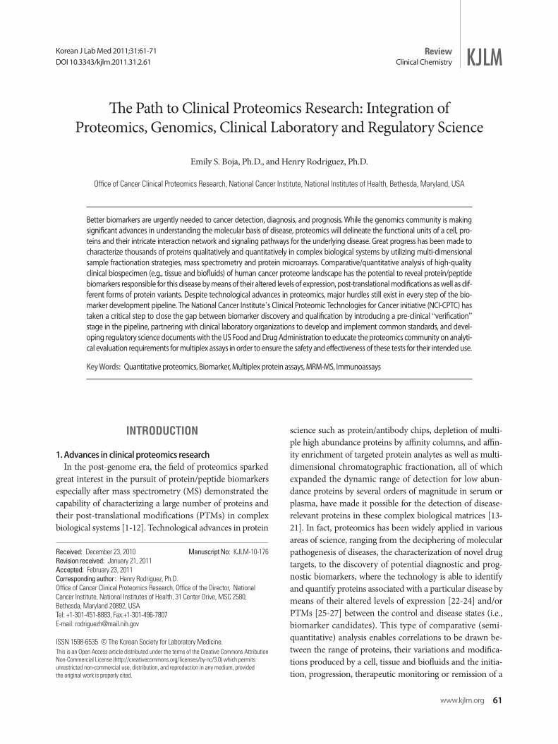
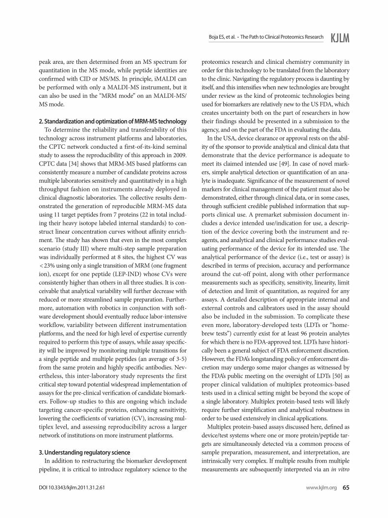
disease state. Post-translational modifications including phosphorylation, glycosylation, acetylation and oxidation, in particular, have been of great interest in this field as it has been demonstrated to be linked to disease pathology and useful targets for therapeutics. In addition to MS-based large-scale protein and peptide sequencing, other innova-tive approaches including self-assembling protein microar-rays [28] and bead-based flow cytometry [29] to identify and quantify proteins and protein-protein interaction in a high throughput manner have furthered our understanding in the molecular mechanisms involved in diseases. In sum-mary, clinical proteomics has come a long way in the past decade in terms of technology/platform development, pro-tein chemistry, and bioinformatics to identify molecular signatures of diseases based on protein pathways and sig-naling cascades. Hence, it undoubtedly holds great promise for disease diagnosis, prognosis, and prediction of thera-peutic outcome on an individualized basis. However, with-out proper study design and implementation of robust ana-lytical techniques, the efforts and expectations to make bio-markers a useful reality in the near future can easily be hampered. This is clearly manifested by the stagnant rate of clearance or approval of protein biomarkers for all diseases by the Food and Drug Administration (FDA) in the US (i.e., averaging ~1.5 protein/yr in the past 15 yr [30]), in contrast to over 1,200 biomarker candidates reported in the scien-tific literature for cancer alone. The question arises as to what has caused such a huge disconnect between biomarker discovery using modern proteomic technologies and bio-marker qualification requiring much more stringent analyt-ical and clinical criteria. Several major barriers have been postulated to be responsible for this discrepancy, including: (1) technological variability within/across proteomic plat-forms; (2) improper biospecimen collection, handling, stor-age and processing; (3) incapability of credentialing bio-marker candidates prior to costly and time-consuming clinical qualification studies using well-established method-ologies; (4) a lack of knowledge in the evaluation criteria re-quired for these distinct processes in the pipeline and in regulatory science by the research community; (5) insuffi-cient publicly available high-quality reagents and data sets to the cancer research community; (6) need for improved data analysis tools for the analysis, characterization, and comparison of large datasets and multi-dimensional data; and (6) a lack of proper experimental study design when performing studies involving clinical samples in biomarker studies (Fig. 1). If proteomics is to successfully make its way into clinical diagnostics, universally accepted metrics will be needed at many steps along the way to ensure that ob-
served changes are attributable to biological states, not workflow variability.
On the discovery front, semi-quantitative proteomic methodologies routinely used for biomarker research be-tween normal and diseased states are differential two-di-mensional gel electrophoresis (2DGE), comparative label-free and labeling approaches (e.g., Isotope Coded Affinity Tags, iTRAQ, Stable Isotope Labeling with Amino Acids in Cell Culture) followed by liquid chromatography mass spec-trometry (LC-MS). Although such comparative analysis yields insightful information on possible changes as a result of disease, these current methods in clinical proteomics based, for the most part, on MS and its combination with 2DGE, chromatography or biobead technology have a con-centration sensitivity level (CSL) not lower than 10 nM. This coupled to the use of blood as a biospecimen in discovery research (a commonly used biospecimen which is highly complex and has a wide dynamic range of protein concen-trations), makes it is very difficult to discover (measure) low abundance proteins (potential biomarkers). One remedy to this problem is to develop and apply nanotechnology in clinical proteomics which can substantially enhance the CSL, as well as the throughput of analytical measurement systems while lowering their cost. Not only does nanotechnology have the potential of satisfying many criteria required for the advancement of clinical proteomics, essential changes in the physicochemical properties of substances on their conver-sion to the nanostructured state have also made it possible to create efficient systems for drug delivery to targets. Cur-rently, one of the most promising nanotechnological pro-teomics being developed for medical research is biosensor-based nanodiagnostics. An example of this is the develop-ment of a magneto-nano sensor protein chip and a multiplex
Fig. 1. Barriers between candidate biomarker discovery by proteomics and qualification.
Boja ES, et al. • The Path to Clinical Proteomics Research
www.kjlm.org 63DOI 10.3343/kjlm.2011.31.2.61
KJLM
magnetic sorter based on magnetic nanoparticles that allow rapid conversion of discrete biomolecules binding events into electrical signals, which can detect target molecules down to the single molecule level in less than an hour [31].
2. Issues and challenges in clinical proteomicsIn reality, proteomics has not lived up to the hopes for
identifying effective biomarkers in the past decade, due in part to the lack of coherent pipeline connecting biomarker discovery efforts with well-established methods for clinical qualification studies (commonly known as biomarker vali-dation). As a result, among the critical challenges facing the proteomics community is the lack of an ability to accurately and reproducibly measure a meaningful number of proteins in biospecimens across institutions. Better understanding of the challenges and strategies inherent in each phase of the proteomics pipeline is required to both accelerate the pace and quality of biomarker development and facilitate the de-livery and deployment of novel clinical tests. Indeed, chal-lenges in proteomics encompass several stages of the bio-marker development pipeline, including a lack of technology standardization/optimization; quality affinity reagents; ana-lytical validation review documents for developers of multi-plex proteomics assays; and proper experimental design when performing studies involving clinical samples (e.g., statistical power calculation on the number of biospecimens needed to ensure meaningful results, and biospecimen qual-ity with proper representation of patient population).
DISCUSSION
1. Reconstructing the pipeline through verificationThe conventional biomarker development pipeline in-
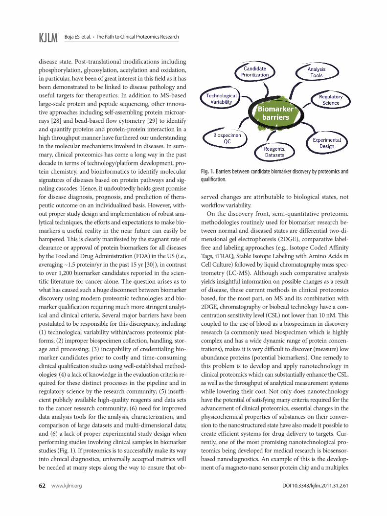
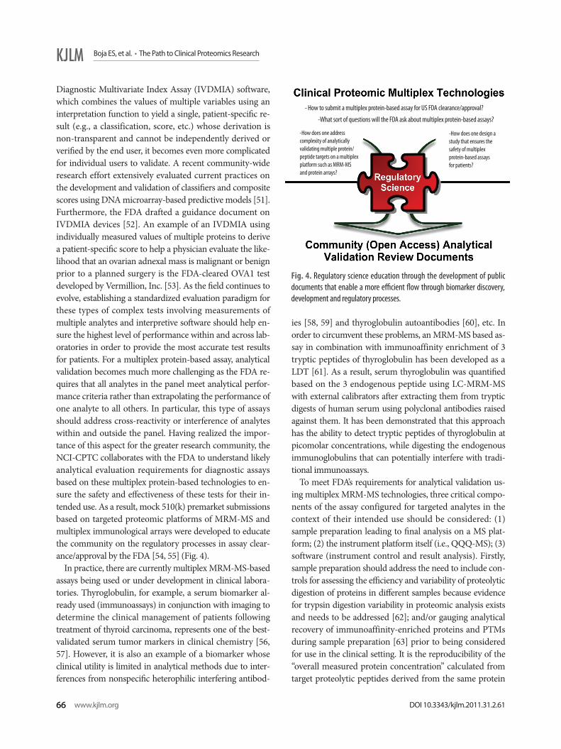
volves a discovery stage followed by a qualification stage (commonly known as biomarker validation) on large co-horts (Fig. 2A), prior to clinical implementation. Tradition-ally, the discovery stage is performed on a MS-based plat-form for global unbiased sampling of the proteome, while biomarker qualification and clinical implementation gener-ally involve the development of an antibody-based protocol, such as the widely used enzyme linked immunosorbent as-says (ELISAs). Although this process has the potential to deliver clinically important biomarkers, it is not the most ef-ficient as the latter is low-throughput, very costly and time-consuming. In many cases, affinity reagents for novel pro-tein candidates do not even exist and it is difficult to multi-plex targets without creating significant interferences and cross-reactivity. These limitations of immunoassays have in-centivized the development of alternative approaches. The
recent explosion in the advancement of proteomic technol-ogies centering on targeted MS and protein microarrays has provided great opportunities for researchers to use them as “bridging technologies” for clinical proteomic investigation of disease-relevant changes in tissues and biofluids.
To address many of the critical challenges facing the pro-tein biomarker community, the NCI launched the Clinical Proteomic Technologies for Cancer initiative (CPTC) in 2006 (http://proteomics.cancer.gov). The overall goals of CPTC during the first 5 yr were to focus on removing sev-eral of the major barriers in proteomics research to enable the accurate, efficient and reproducible identification, and quantification of meaningful numbers of proteins that could drive high value clinical biomarker qualification studies. Achieving this goal would provide a firm foundation for the field of discovery proteomics and enable the rational devel-opment of clinical biomarkers to address various needs in cancer drug development, diagnostics and clinical manage-ment.
Since its launch, CPTC has made significant progress in developing an accurate and quantitative biomarker assay workflow for proteomics, incorporating common technol-ogy standards, standard operating procedures (SOPs), data analysis standards, critically needed reagents (affinity and reference materials), and an open access proteomics data-base [32-37]. The new protein biomarker workflows devel-oped by CPTC that incorporate go/no-go decision points, address the variability of methods and technologies–which enable researchers to accurately, reliably and quantitatively
Fig. 2. The envisioned National Cancer Institute-Clinical Proteomic Technolo-gies for Cancer initiative (NCI-CPTC) development pipeline from discovery to qualification. (A) The gap in the current proteomics research pipeline. (B) The incorporation of verification into the NCI-CPTC pipeline between discovery and qualification.
Typical proteomics research pipeline
Discovery
Discovery
Gaps/barriers
ClinicalImplementation
ClinicalImplementation
> 10,000 analytes< 10 biospecimens
(blood)
> 10,000 analytes10s of biospecimens
(tissue or proximal fluid)
~10 analytes1000s of biospecimens
(blood)
~10 analytes1000s of biospecimens
(blood)
~100s of analytes100s of biospecimens
(blood)
·Many candidates·Prioritization uncertain·No mechanism for attrition· Few high-quality reagents·Need for redefined discovery and bridging techniques
Untargeted platforms~100s of candidate
biomarkers
Untargeted platforms~100s of candidate
biomarkers
Targeted platforms ~10s of verified
biomarkers
ImmunoassayPanel of qualified
biomarkers
ImmunoassayPanel of qualified
biomarkers
Qualification
QualificationVerification
NCI-CPTC proteomics research pipeline
A
B
64 www.kjlm.org
Boja ES, et al. • The Path to Clinical Proteomics Research
DOI 10.3343/kjlm.2011.31.2.61
KJLM
identify large numbers of proteins (see below). Conse-quently, CPTC has quickly evolved into a national (and in-ternational) community resource that links technologists with cancer biologists and clinicians to accelerate the devel-opment, improvement and standardization of proteomic technologies for the detection of cancer-relevant proteins/peptides in clinical biospecimens.
NCI-CPTC incorporated an intermediate “bridging” step called “biomarker verification” in its pipeline to efficiently translate proteomic discoveries into clinical qualification studies (Fig. 2B). In this context, biomarker verification is defined as the process of credentialing prioritized “bio-marker candidates” using analytically robust, reproducible and quantitative multiplex assays on statistically powered number of samples with clinical relevance. Credentialed proteins successfully passing this stage of the pipeline are considered verified biomarkers of high value for translating into large-scale clinical qualification studies.
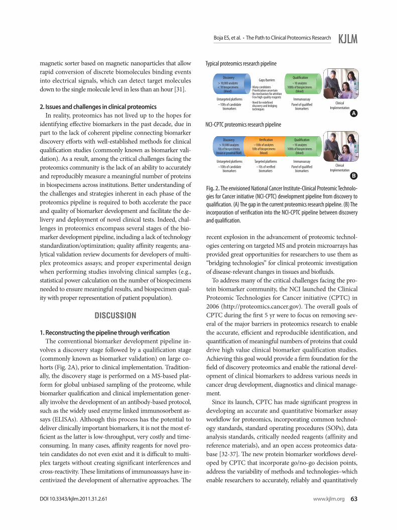
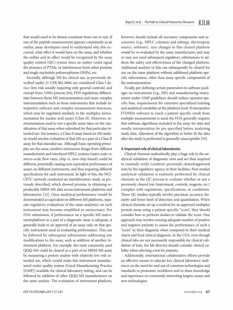
One of the main verification technologies currently being tested by CPTC is based on an existing technology, Multiple Reaction Monitoring Mass Spectrometry (MRM-MS), which has been used for decades in clinical reference labo-ratories to accurately measure small molecules in plasma, such as drug metabolites [38, 39]. MRM-MS has only re-cently proven suitable for use in pre-clinical studies to rap-idly screen and measure large numbers of candidate pro-teins in complex patient samples necessary for biomarker verification [40-43]. MRM-MS provides a rapid way to measure the abundance of a particular candidate(s) and de-termine whether changes in abundance correspond to the presence or stage of a disease. Prior to applying MRM-MS based assays on clinical biospecimen, precursor peptide ions are selected from previous experiments combining empiri-cal MS data, database searches of peptide libraries, and soft-ware predictions to develop the best peptide candidates to monitor. As illustrated in Fig. 3, the general components of MRM-MS based approaches for targeted protein quantita-tion encompass three quadrupoles (QQQ) for enhanced sensitivity and selectivity on a triple quadrupole mass spec-trometer. The first quadrupole (Q1) is designed to transmit only the selected precursor peptide ion (m/z) into the sec-ond quadrupole (Q2) where collisionally-induced dissocia-tion (CID) occurs to generate a signature fragment ion of particular m/z value or several fragment ions allowed to be admitted into the third quadrupole (Q3) for monitoring. Consequently, peptide quantitation is based on measuring the intensity of the product ion(s) selected from Q3. MRM-MS can precisely detect a wide variety of peptides and pro-teins via CID or MS/MS. When coupled with chemically
identical heavy isotope labeled internal standard peptides of known amounts (stable isotope dilution mass spectrome-try), this approach has the potential benefits for accurate quantitation of target peptides that make it a desirable alter-native to immunoassays including, but not limited to, shorter assay development timeline, lower cost, high multi-plexing capability, potential for high specificity, inclusion of internal standards, and particular advantages for PTMs, mutations and other variants of a protein. On the other hand, there are potential drawbacks of MRM-MS assays which may include: the complex selection process for deter-mining target precursor peptide ions to monitor, relatively low resolution of QQQ-MS (usually unit resolution) in re-solving components in complex biofluids, and the possibil-ity of interference from in-source fragmentation of abun-dant peptides that prevent analysis of desired precursor peptides. In terms of sensitivity, MRM-MS assays alone are at best in the range of μg per milliliter of biofluids. However, when coupled with immunoaffinity enrichment [i.e., Stable Isotope Standards and Capture by Anti-Peptide Antibodies (SISCAPA)] [44-46], it elevates the sensitivity of detection several orders of magnitude. With improvements in gener-ating monoclonal antibodies against target signature pep-tides, instrument designs and workflow automation, MRM-MS may potentially provide a very reliable go/no-go deci-sion point in the new CPTC biomarker development pipe-line. The complement of a LC-MRM-MS assay is iMALDI (immuno matrix-assisted laser desorption ionization), where the beads with the affinity-bound peptides attached are placed directly on a MALDI mass spectrometer, and the MALDI matrix solvent elutes the peptides from the beads [47, 48]. The presence of the peptide, and its peak height or
Fig. 3. Multiple Reaction Monitoring Mass Spectrometry (MRM-MS). A sche-matic of a triple quadrupole mass spectrometer (QQQ-MS) commonly used in MRM-MS analysis: Q1 and Q3 represent two mass filters for precursor and fragment ion selection while Q2 (collision cell) creates fragment ions via col-lisionally-induced dissociation (CID). In this case, one of the three peptide precursor ions (colored in blue) is selected in Q1, fragmented in Q2 and quantitated using one of its fragment ions (transition) selected in Q3 by the relative intensity of its peak area. An MRM-MS assay offers multiplexing ca-pability of many target analytes in a single HPLC run.
Boja ES, et al. • The Path to Clinical Proteomics Research
www.kjlm.org 65DOI 10.3343/kjlm.2011.31.2.61
KJLM
peak area, are then determined from an MS spectrum for quantitation in the MS mode, while peptide identities are confirmed with CID or MS/MS. In principle, iMALDI can be performed with only a MALDI-MS instrument, but it can also be used in the “MRM mode” on an MALDI-MS/MS mode.
2. Standardization and optimization of MRM-MS technologyTo determine the reliability and transferability of this
technology across instrument platforms and laboratories, the CPTC network conducted a first-of-its-kind seminal study to assess the reproducibility of this approach in 2009. CPTC data [34] shows that MRM-MS based platforms can consistently measure a number of candidate proteins across multiple laboratories sensitively and quantitatively in a high throughput fashion on instruments already deployed in clinical diagnostic laboratories. The collective results dem-onstrated the generation of reproducible MRM-MS data using 11 target peptides from 7 proteins (22 in total includ-ing their heavy isotope labeled internal standards) to con-struct linear concentration curves without affinity enrich-ment. The study has shown that even in the most complex scenario (study III) where multi-step sample preparation was individually performed at 8 sites, the highest CV was <23% using only a single transition of MRM (one fragment ion), except for one peptide (LEP-IND) whose CVs were consistently higher than others in all three studies. It is con-ceivable that analytical variability will further decrease with reduced or more streamlined sample preparation. Further-more, automation with robotics in conjunction with soft-ware development should eventually reduce labor-intensive workflow, variability between different instrumentation platforms, and the need for high level of expertise currently required to perform this type of assays, while assay specific-ity will be improved by monitoring multiple transitions for a single peptide and multiple peptides (an average of 3-5) from the same protein and highly specific antibodies. Nev-ertheless, this inter-laboratory study represents the first critical step toward potential widespread implementation of assays for the pre-clinical verification of candidate biomark-ers. Follow-up studies to this are ongoing which include targeting cancer-specific proteins, enhancing sensitivity, lowering the coefficients of variation (CV), increasing mul-tiplex level, and assessing reproducibility across a larger network of institutions on more instrument platforms.
3. Understanding regulatory scienceIn addition to restructuring the biomarker development
pipeline, it is critical to introduce regulatory science to the
proteomics research and clinical chemistry community in order for this technology to be translated from the laboratory to the clinic. Navigating the regulatory process is daunting by itself, and this intensifies when new technologies are brought under review as the kind of proteomic technologies being used for biomarkers are relatively new to the US FDA, which creates uncertainty both on the part of researchers in how their findings should be presented in a submission to the agency, and on the part of the FDA in evaluating the data.
In the USA, device clearance or approval rests on the abil-ity of the sponsor to provide analytical and clinical data that demonstrate that the device performance is adequate to meet its claimed intended use [49]. In case of novel mark-ers, simple analytical detection or quantification of an ana-lyte is inadequate. Significance of the measurement of novel markers for clinical management of the patient must also be demonstrated, either through clinical data, or in some cases, through sufficient credible published information that sup-ports clinical use. A premarket submission document in-cludes a device intended use/indication for use, a descrip-tion of the device covering both the instrument and re-agents, and analytical and clinical performance studies eval-uating performance of the device for its intended use. The analytical performance of the device (i.e., test or assay) is described in terms of precision, accuracy and performance around the cut-off point, along with other performance measurements such as specificity, sensitivity, linearity, limit of detection and limit of quantitation, as required for any assays. A detailed description of appropriate internal and external controls and calibrators used in the assay should also be included in the submission. To complicate these even more, laboratory-developed tests (LDTs or “home-brew tests”) currently exist for at least 96 protein analytes for which there is no FDA-approved test. LDTs have histori-cally been a general subject of FDA enforcement discretion. However, the FDA’s longstanding policy of enforcement dis-cretion may undergo some major changes as witnessed by the FDA’s public meeting on the oversight of LDTs [50] as proper clinical validation of multiplex proteomics-based tests used in a clinical setting might be beyond the scope of a single laboratory. Multiplex protein-based tests will likely require further simplification and analytical robustness in order to be used extensively in clinical applications.
Multiplex protein-based assays discussed here, defined as device/test systems where one or more protein/peptide tar-gets are simultaneously detected via a common process of sample preparation, measurement, and interpretation, are intrinsically very complex. If multiple results from multiple measurements are subsequently interpreted via an in vitro
66 www.kjlm.org
Boja ES, et al. • The Path to Clinical Proteomics Research
DOI 10.3343/kjlm.2011.31.2.61
KJLM
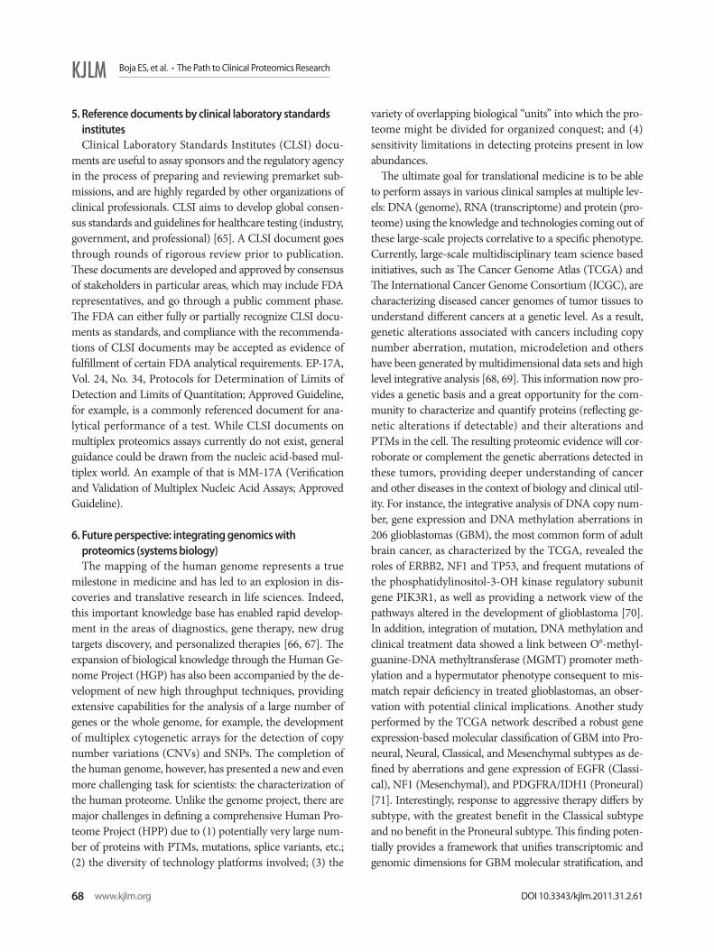
Diagnostic Multivariate Index Assay (IVDMIA) software, which combines the values of multiple variables using an interpretation function to yield a single, patient-specific re-sult (e.g., a classification, score, etc.) whose derivation is non-transparent and cannot be independently derived or verified by the end user, it becomes even more complicated for individual users to validate. A recent community-wide research effort extensively evaluated current practices on the development and validation of classifiers and composite scores using DNA microarray-based predictive models [51]. Furthermore, the FDA drafted a guidance document on IVDMIA devices [52]. An example of an IVDMIA using individually measured values of multiple proteins to derive a patient-specific score to help a physician evaluate the like-lihood that an ovarian adnexal mass is malignant or benign prior to a planned surgery is the FDA-cleared OVA1 test developed by Vermillion, Inc. [53]. As the field continues to evolve, establishing a standardized evaluation paradigm for these types of complex tests involving measurements of multiple analytes and interpretive software should help en-sure the highest level of performance within and across lab-oratories in order to provide the most accurate test results for patients. For a multiplex protein-based assay, analytical validation becomes much more challenging as the FDA re-quires that all analytes in the panel meet analytical perfor-mance criteria rather than extrapolating the performance of one analyte to all others. In particular, this type of assays should address cross-reactivity or interference of analytes within and outside the panel. Having realized the impor-tance of this aspect for the greater research community, the NCI-CPTC collaborates with the FDA to understand likely analytical evaluation requirements for diagnostic assays based on these multiplex protein-based technologies to en-sure the safety and effectiveness of these tests for their in-tended use. As a result, mock 510(k) premarket submissions based on targeted proteomic platforms of MRM-MS and multiplex immunological arrays were developed to educate the community on the regulatory processes in assay clear-ance/approval by the FDA [54, 55] (Fig. 4).
In practice, there are currently multiplex MRM-MS-based assays being used or under development in clinical labora-tories. Thyroglobulin, for example, a serum biomarker al-ready used (immunoassays) in conjunction with imaging to determine the clinical management of patients following treatment of thyroid carcinoma, represents one of the best-validated serum tumor markers in clinical chemistry [56, 57]. However, it is also an example of a biomarker whose clinical utility is limited in analytical methods due to inter-ferences from nonspecific heterophilic interfering antibod-
ies [58, 59] and thyroglobulin autoantibodies [60], etc. In order to circumvent these problems, an MRM-MS based as-say in combination with immunoaffinity enrichment of 3 tryptic peptides of thyroglobulin has been developed as a LDT [61]. As a result, serum thyroglobulin was quantified based on the 3 endogenous peptide using LC-MRM-MS with external calibrators after extracting them from tryptic digests of human serum using polyclonal antibodies raised against them. It has been demonstrated that this approach has the ability to detect tryptic peptides of thyroglobulin at picomolar concentrations, while digesting the endogenous immunoglobulins that can potentially interfere with tradi-tional immunoassays.
To meet FDA’s requirements for analytical validation us-ing multiplex MRM-MS technologies, three critical compo-nents of the assay configured for targeted analytes in the context of their intended use should be considered: (1) sample preparation leading to final analysis on a MS plat-form; (2) the instrument platform itself (i.e., QQQ-MS); (3) software (instrument control and result analysis). Firstly, sample preparation should address the need to include con-trols for assessing the efficiency and variability of proteolytic digestion of proteins in different samples because evidence for trypsin digestion variability in proteomic analysis exists and needs to be addressed [62]; and/or gauging analytical recovery of immunoaffinity-enriched proteins and PTMs during sample preparation [63] prior to being considered for use in the clinical setting. It is the reproducibility of the “overall measured protein concentration” calculated from target proteolytic peptides derived from the same protein
Fig. 4. Regulatory science education through the development of public documents that enable a more efficient flow through biomarker discovery, development and regulatory processes.
- How to submit a multiplex protein-based assay for US FDA clearance/approval?
-What sort of questions will the FDA ask about multiplex protein-based assays?
-How does one address complexity of analytically validating multiple protein/ peptide targets on a multiplex platform such as MRM-MS and protein arrays?
-How does one design a study that ensures the safety of multiplex protein-based assays for patients?
Boja ES, et al. • The Path to Clinical Proteomics Research
www.kjlm.org 67DOI 10.3343/kjlm.2011.31.2.61
KJLM
that would need to be shown consistent from run to run. If one of the peptide measurements appears consistently as an outlier, assay developers need to understand why this oc-curred, what effect it would have on the assay, and whether the outlier and its effect would be recognized by the assay quality control (QC) system since an outlier could signal the presence of PTMs, or interferences from other proteins and single nucleotide polymorphisms (SNPs), etc.
Secondly, although MS for clinical use, as previously de-scribed under 21 CFR 862.2860, are considered Class I de-vice (low risk usually requiring only general controls) and exempt from 510(k) process [64], FDA regulations differen-tiate between those MS instrumentation and more complex instrumentation such as those instruments that include in-terpretive software and complex measurement functions, which may be regulated similarly to the multiplex instru-mentation for nucleic acid assays (Class II). Moreover, in-strumentation used to run a specific assay takes on the clas-sification of that assay when submitted for that particular in-tended use. For instance, a Class II assay based on MS analy-sis would involve evaluation of that MS as a part of a Class II assay for that intended use. Although basic operating princi-ples are the same, modern instrument design from different manufacturers and interfaced HPLC systems (nano-scale vs. micro-scale flow rates, chip vs. non-chip-based) could be different, potentially causing non-equivalent performance of assays on different instruments, and thus requiring different specifications for each instrument. In light of this, the NCI-CPTC network conducted an interlaboratory study, as pre-viously described, which showed promise in obtaining re-producible MRM-MS data across instrument platforms and laboratories [32]. Once analytical performance would be demonstrated as equivalent on different MS platforms, sepa-rate regulatory evaluations of the same analyte(s) on each instrument may become simplified or unnecessary. For FDA submission, if performance on a specific MS instru-ment/platform as a part of a diagnostic assay is adequate, it generally leads to an approval of an assay only on that spe-cific instrument used in evaluating performance. This can be followed by subsequent submissions addressing any modifications to the assay, such as addition of another in-strument platform. For example, the most commonly used QQQ-MS could be cleared as a part of an MRM-MS assay by measuring a protein analyte with relatively low-risk in-tended use, which would make this instrument manufac-tured under quality system (Good Manufacturing Practice [GMP]) available for clinical laboratory testing, and can be followed by addition of other QQQ-MS manufacturers on the same analyte. The evaluation of instrument platform,
however, should include all necessary components and ac-cessories (e.g., HPLC columns and tubings, electrospray source, software). Any changes to this cleared platform would be re-evaluated by the assay manufacturer, and may or may not need subsequent regulatory submissions to ad-dress the safety and effectiveness of the changed platform. Additional analytes or kits can subsequently be cleared for use on the same platform without additional platform-spe-cific information, other than assay-specific components of the instrumentation.
Finally, pre-defining certain parameters in software pack-ages on instruments (e.g., MS) and manufacturing instru-ments under GMP guidelines should reduce operator-spe-cific bias, requirements for extensive specialized training and analytical variability at the platform level. If interpretive IVDMIA software to reach a patient-specific result from multiple measurements is used, the FDA generally requires that software algorithms included in the assay for data and results interpretation be pre-specified before analyzing study data. Alteration of the algorithm to better fit the data after the study is performed is generally unacceptable [54].
4. Important role of clinical laboratoriesClinical chemists undoubtedly play a huge role in the an-
alytical validation of diagnostic tests and are thus required to routinely verify (confirm) previously cleared/approved tests by the regulatory agency in their facilities. Post-market analytical validation is routinely performed by clinical chemists as the QC process to evaluate whether or not a previously cleared test (instrument, controls, reagents, etc.) complies with regulations, specifications, or conditions. These QC studies typically involve precision, accuracy, lin-earity and lower limit of detection and quantitation. When clinical chemists set up a method for an approved multiplex protein assay using a patient-specific “score”, they should consider how to perform studies to validate the score. One approach may involve running adequate number of positive and negative patients to assess the performance of such a “score” in their diagnosis when compared to their medical charts and final clinical diagnosis. In the USA, even though clinical labs are not necessarily responsible for clinical vali-dation of tests, the lab director should consider clinical va-lidity when selecting a test for patients.
Additionally, international collaborative efforts provide an effective means to educate key clinical laboratory audi-ences on the need for and use of common technologies and standards in proteomic workflows and to share knowledge and experience on commonly interesting targets, assays and new technologies.
68 www.kjlm.org
Boja ES, et al. • The Path to Clinical Proteomics Research
DOI 10.3343/kjlm.2011.31.2.61
KJLM
5. Reference documents by clinical laboratory standards institutes
Clinical Laboratory Standards Institutes (CLSI) docu-ments are useful to assay sponsors and the regulatory agency in the process of preparing and reviewing premarket sub-missions, and are highly regarded by other organizations of clinical professionals. CLSI aims to develop global consen-sus standards and guidelines for healthcare testing (industry, government, and professional) [65]. A CLSI document goes through rounds of rigorous review prior to publication. These documents are developed and approved by consensus of stakeholders in particular areas, which may include FDA representatives, and go through a public comment phase. The FDA can either fully or partially recognize CLSI docu-ments as standards, and compliance with the recommenda-tions of CLSI documents may be accepted as evidence of fulfillment of certain FDA analytical requirements. EP-17A, Vol. 24, No. 34, Protocols for Determination of Limits of Detection and Limits of Quantitation; Approved Guideline, for example, is a commonly referenced document for ana-lytical performance of a test. While CLSI documents on multiplex proteomics assays currently do not exist, general guidance could be drawn from the nucleic acid-based mul-tiplex world. An example of that is MM-17A (Verification and Validation of Multiplex Nucleic Acid Assays; Approved Guideline).
6. Future perspective: integrating genomics with proteomics (systems biology)
The mapping of the human genome represents a true milestone in medicine and has led to an explosion in dis-coveries and translative research in life sciences. Indeed, this important knowledge base has enabled rapid develop-ment in the areas of diagnostics, gene therapy, new drug targets discovery, and personalized therapies [66, 67]. The expansion of biological knowledge through the Human Ge-nome Project (HGP) has also been accompanied by the de-velopment of new high throughput techniques, providing extensive capabilities for the analysis of a large number of genes or the whole genome, for example, the development of multiplex cytogenetic arrays for the detection of copy number variations (CNVs) and SNPs. The completion of the human genome, however, has presented a new and even more challenging task for scientists: the characterization of the human proteome. Unlike the genome project, there are major challenges in defining a comprehensive Human Pro-teome Project (HPP) due to (1) potentially very large num-ber of proteins with PTMs, mutations, splice variants, etc.; (2) the diversity of technology platforms involved; (3) the
variety of overlapping biological “units” into which the pro-teome might be divided for organized conquest; and (4) sensitivity limitations in detecting proteins present in low abundances.
The ultimate goal for translational medicine is to be able to perform assays in various clinical samples at multiple lev-els: DNA (genome), RNA (transcriptome) and protein (pro-teome) using the knowledge and technologies coming out of these large-scale projects correlative to a specific phenotype. Currently, large-scale multidisciplinary team science based initiatives, such as The Cancer Genome Atlas (TCGA) and The International Cancer Genome Consortium (ICGC), are characterizing diseased cancer genomes of tumor tissues to understand different cancers at a genetic level. As a result, genetic alterations associated with cancers including copy number aberration, mutation, microdeletion and others have been generated by multidimensional data sets and high level integrative analysis [68, 69]. This information now pro-vides a genetic basis and a great opportunity for the com-munity to characterize and quantify proteins (reflecting ge-netic alterations if detectable) and their alterations and PTMs in the cell. The resulting proteomic evidence will cor-roborate or complement the genetic aberrations detected in these tumors, providing deeper understanding of cancer and other diseases in the context of biology and clinical util-ity. For instance, the integrative analysis of DNA copy num-ber, gene expression and DNA methylation aberrations in 206 glioblastomas (GBM), the most common form of adult brain cancer, as characterized by the TCGA, revealed the roles of ERBB2, NF1 and TP53, and frequent mutations of the phosphatidylinositol-3-OH kinase regulatory subunit gene PIK3R1, as well as providing a network view of the pathways altered in the development of glioblastoma [70]. In addition, integration of mutation, DNA methylation and clinical treatment data showed a link between O6-methyl-guanine-DNA methyltransferase (MGMT) promoter meth-ylation and a hypermutator phenotype consequent to mis-match repair deficiency in treated glioblastomas, an obser-vation with potential clinical implications. Another study performed by the TCGA network described a robust gene expression-based molecular classification of GBM into Pro-neural, Neural, Classical, and Mesenchymal subtypes as de-fined by aberrations and gene expression of EGFR (Classi-cal), NF1 (Mesenchymal), and PDGFRA/IDH1 (Proneural) [71]. Interestingly, response to aggressive therapy differs by subtype, with the greatest benefit in the Classical subtype and no benefit in the Proneural subtype. This finding poten-tially provides a framework that unifies transcriptomic and genomic dimensions for GBM molecular stratification, and
Boja ES, et al. • The Path to Clinical Proteomics Research
www.kjlm.org 69DOI 10.3343/kjlm.2011.31.2.61
KJLM
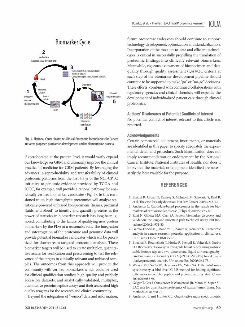
if corroborated at the protein level, it would vastly expand our knowledge on GBM and ultimately improve the clinical practice of medicine for GBM patients. By leveraging the advances in reproducibility and transferability of clinical proteomic platforms from the first 4.5 yr of the NCI-CPTC initiative to genomic evidence provided by TCGA and ICGC, for example, will provide a rational pathway for ana-lytically verified biomarker candidates (Fig. 5). In this envi-sioned route, high throughput proteomics will analyze sta-tistically-powered unbiased biospecimens (tissues, proximal fluids, and blood) to identify and quantify proteins as the power of statistics in biomarker research has long been ig-nored, contributing to the failure of qualifying new protein biomarkers by the FDA at a reasonable rate. The integration and interrogation of the proteomic and genomic data will provide potential biomarker candidates which will be priori-tized for downstream targeted proteomic analysis. These biomarker targets will be used to create multiplex, quantita-tive assays for verification and prescreening to test the rele-vance of the targets in clinically relevant and unbiased sam-ples. The outcomes from this approach will provide the community with verified biomarkers which could be used for clinical qualification studies; high quality and publicly accessible datasets; and analytically validated, multiplex, quantitative protein/peptide assays and their associated high quality reagents for the research and clinical community.
Beyond the integration of “-omics” data and information,
future proteomic endeavors should continue to support technology development, optimization and standardization. Incorporation of the most up-to-date and efficient technol-ogies is critical in successfully propelling the translation of proteomic findings into clinically relevant biomarkers. Meanwhile, rigorous assessment of biospecimen and data quality through quality assessment (QA)/QC criteria at each step of the biomarker development pipeline should continue to be supported to make “go” or “no-go” decisions. These efforts, combined with continued collaborations with regulatory agencies and clinical chemists, will expedite the development of individualized patient care through clinical proteomics.
Authors’ Disclosures of Potential Conflicts of InterestNo potential conflict of interest relevant to this article was reported.
AcknowledgementsCertain commercial equipment, instruments, or materials are identified in this paper to specify adequately the experi-mental detail and procedure. Such identification does not imply recommendation or endorsement by the National Cancer Institute, National Institutes of Health, nor does it imply that the materials or equipment identified are neces-sarily the best available for the purpose.
REFERENCES
1. Etzioni R, Urban N, Ramsey S, McIntosh M, Schwartz S, Reid B, et al. The case for early detection. Nat Rev Cancer 2003;3:243-52.
2. Anderson L. Candidate-based proteomics in the search for bio-markers of cardiovascular disease. J Physiol 2005;563:23-60.
3. Rifai N, Gillette MA, Carr SA. Protein biomarker discovery and validation: the long and uncertain path to clinical utility. Nat Bio-technol 2006;24:971-83.
4. García-Foncillas J, Bandrés E, Zárate R, Remírez N. Proteomic analysis in cancer research: potential application in clinical use. Clin Transl Oncol 2006;8:250-61.
5. Bouchal P, Roumeliotis T, Hrstka R, Nenutil R, Vojtesek B, Garbis SD. Biomarker discovery in low-grade breast cancer using isobaric stable isotope tags and two-dimensional liquid chromatography-tandem mass spectrometry (iTRAQ-2DLC-MS/MS) based quan-titative proteomic analysis. J Proteome Res 2009;8:362-73.
6. Wiener MC, Sachs JR, Deyanova EG, Yates NA. Differential mass spectrometry: a label-free LC-MS method for finding significant differences in complex peptide and protein mixtures. Anal Chem 2004;76:6085-96.
7. Geiger T, Cox J, Ostasiewicz P, Wisniewski JR, Mann M. Super-SI-LAC mix for quantitative proteomics of human tumor tissue. Nat Methods 2010;7:383-5.
8. Anderson L and Hunter CL. Quantitative mass spectrometric
Fig. 5. National Cancer Institute-Clinical Proteomic Technologies for Cancer initiative proposed proteomics development and implementation process.
Verification(targeted assay development)
Discovery(sample characterization)
Clinicalimplementation
Biomarkerqualification
Knowledge
- Highly credentialed protein candidates- Reference datasets- Library of multiplex assays, reagents and SOPs
Tissue
Blood
Genomicanalysis
Proteomicanalysis
Biomarker Cycle
70 www.kjlm.org
Boja ES, et al. • The Path to Clinical Proteomics Research
DOI 10.3343/kjlm.2011.31.2.61
KJLM
multiple reaction monitoring assays for major plasma proteins. Mol Cell Proteomics 2006;5:573-88.
9. Wang H, Wong CH, Chin A, Kennedy J, Zhang Q, Hanash S. Quantitative serum proteomics using dual stable isotope coding and nano LC-MS/MSMS. J Proteome Res 2009;8:5412-22.
10. Lee J, Soper SA, Murray KK. Microfluidic chips for mass spec-trometry-based proteomics. J Mass Spectrom 2009;44:579-93.
11. Pierobon M, Calvert V, Belluco C, Garaci E, Deng J, Lise M, et al. Multiplexed cell signaling analysis of metastatic and nonmetastatic colorectal cancer reveals COX2-EGFR signaling activation as a potential prognostic pathway biomarker. Clin Colorectal Cancer 2009;8:110-7.
12. Ramachandran N, Raphael JV, Hainsworth E, Demirkan G, Fuen-tes MG, Rolfs A, et al. Next-generation high-density self-assem-bling functional protein arrays. Nat Methods 2008;5:535-8.
13. Beirne P, Pantelidis P, Charles P, Wells AU, Abraham DJ, Denton CP, et al. Multiplex immune serum biomarker profiling in sarcoid-osis and systemic sclerosis. Eur Respir J 2009;34:1376-82.
14. Kelleher MT, Fruhwirth G, Patel G, Ofo E, Festy F, Barber PR, et al. The potential of optical proteomic technologies to individualize prognosis and guide rational treatment for cancer patients. Target Oncol 2009;4:235-52.
15. Wang P, Whiteaker JR, Paulovich AG. The evolving role of mass spectrometry in cancer biomarker discovery. Cancer Biol Ther 2009;8:1083-94.
16. Whiteaker JR, Zhang H, Eng JK, Fang R, Piening BD, Feng LC, et al. Head-to-head comparison of serum fractionation techniques. J Proteome Res 2007;6:828-36.
17. Ernoult E, Bourreau A, Gamelin E, Guette C. A proteomic ap-proach for plasma biomarker discovery with iTRAQ labeling and OFFGEL fractionation. J Biomed Biotechnol 2010;2010:927917.
18. Nirmalan NJ, Hughes C, Peng J, McKenna T, Langridge J, Cairns DA, et al. Initial development and validation of a novel extraction method for quantitative mining of the formalin-fixed, paraffin-embedded tissue proteome for biomarker investigations. J Pro-teome Res 2010;10:896-906.
19. Krishhan VV, Khan IH, Luciw PA. Multiplexed microbead immu-noassays by flow cytometry for molecular profiling: basic concepts and proteomics applications. Crit Rev Biotechnol 2009;29:29-43.
20. Cha S, Imielinski MB, Rejtar T, Richardson EA, Thakur D, Sgroi DC, et al. In situ proteomic analysis of human breast cancer epi-thelial cells using laser capture microdissection: annotation by protein set enrichment analysis and gene ontology. Mol Cell Pro-teomics 2010;9:2529-44.
21. Anderson KS, Sibani S, Wallstrom G, Qiu J, Mendoza EA, Raphael J, et al. Protein microarray signature of autoantibody biomarkers for the early detection of breast cancer. J Proteome Res 2011;10:85-96.
22. Bateman NW, Sun M, Hood BL, Flint MS, Conrads TP. Defining central themes in breast cancer biology by differential proteomics: conserved regulation of cell spreading and focal adhesion kinase. J Proteome Res 2010;9:5311-24.
23. Kristiansen TZ, Harsha HC, Grønborg M, Maitra A, Pandey A. Dif-ferential membrane proteomics using 18O-labeling to identify bio-markers for cholangiocarcinoma. J Proteome Res 2008;7:4670-7.
24. An HJ and Lebrilla CB. A glycomics approach to the discovery of potential cancer biomarkers. Methods Mol Biol 2010;600:199-213.
25. Choudhary C and Mann M. Decoding signalling networks by
mass spectrometry-based proteomics. Nat Rev Mol Cell Biol 2010;11:427-39.
26. Madian AG and Regnier FE. Profiling carbonylated proteins in human plasma. J Proteome Res 2010;9:1330-43.
27. Iwabata H, Yoshida M, Komatsu Y. Proteomic analysis of organ-specific post-translational lysine-acetylation and -methylation in mice by use of anti-acetyllysine and -methyllysine mouse mono-clonal antibodies. Proteomics 2005;5:4653-64.
28. Ceroni A, Sibani S, Baiker A, Pothineni VR, Bailer SM, LaBaer J, et al. Systematic analysis of the IgG antibody immune response against varicella zoster virus (VZV) using a self-assembled protein microarray. Mol Biosyst 2010;6:1604-10.
29. Wong J, Sibani S, Lokko NN, LaBaer J, Anderson KS. Rapid detec-tion of antibodies in sera using multiplexed self-assembling bead arrays. J Immunol Methods 2009;350:171-82.
30. Anderson NL. The clinical plasma proteome: a survey of clinical assays for proteins in plasma and serum. Clin Chem 2010;56:177-85.
31. Osterfeld SJ, Yu H, Gaster RS, Caramuta S, Xu L, Han SJ, et al. Multiplex protein assays based on real-time magnetic nanotag sensing. Proc Natl Acad Sci USA 2008;105:20637-40.
32. Paulovich AG, Billheimer D, Ham AJ, Vega-Montoto L, Rudnick PA, Tabb DL, et al. Interlaboratory study characterizing a yeast performance standard for benchmarking LC-MS platform perfor-mance. Mol Cell Proteomics 2010;9:242-54.
33. Rudnick PA, Clauser KR, Kilpatrick LE, Tchekhovskoi DV, Neta P, Blonder N, et al. Performance metrics for liquid chromatography-tandem mass spectrometry systems in proteomics analyses. Mol Cell Proteomics 2010;9:225-41.
34. Addona TA, Abbatiello SE, Schilling B, Skates SJ, Mani DR, Bunk DM, et al. Multi-site assessment of the precision and reproducibil-ity of multiple reaction monitoring-based measurements of pro-teins in plasma. Nat Biotechnol 2009;27:633-41.
35. Tabb DL, Vega-Montoto L, Rudnick PA, Variyath AM, Ham AJ, Bunk DM, et al. Repeatability and reproducibility in proteomic identifications by liquid chromatography-tandem mass spectrom-etry. J Proteome Res 2010;9:761-76.
36. Rodriguez H, Snyder M, Uhlén M, Andrews P, Beavis R, Borchers C, et al. Recommendations from the 2008 international summit on proteomics data release and sharing policy: the amsterdam principles. J Proteome Res 2009;8:3689-92.
37. Gloriam DE, Orchard S, Bertinetti D, Björling E, Bongcam-Rudl-off E, Borrebaeck CA, et al. A community standard format for the representation of protein affinity reagents. Mol Cell Proteomics 2010;9:1-10.
38. Xu X, Zhang J, Zhang L, Liu W, Weisel CP. Selective detection of monohydroxy metabolites of polycyclic aromatic hydrocarbons in urine using liquid chromatography/triple quadrupole tandem mass spectrometry. Rapid Commun Mass Spectrom 2004;18:2299-308.
39. Xu X, Roman JM, Issaq HJ, Keefer LK, Veenstra TD, Ziegler RG. Quantitative measurement of endogenous estrogens and estrogen metabolites in human serum by liquid chromatography-tandem mass spectrometry. Anal Chem 2007;79:7813-21.
40. James A and Jorgensen C. Basic design of MRM assays for peptide quantification. Methods Mol Biol 2010;658:167-85.
41. Kiyonami R, Schoen A, Prakash A, Peterman S, Zabrouskov V, Picotti P, et al. Increased selectivity, analytical precision, and thro-ughput in targeted proteomics. Mol Cell Proteomics 2011;10:
Boja ES, et al. • The Path to Clinical Proteomics Research
www.kjlm.org 71DOI 10.3343/kjlm.2011.31.2.61
KJLM
M110.002931.42. Gerszten RE, Carr SA, Sabatine M. Integration of proteomic-based
tools for improved biomarkers of myocardial injury. Clin Chem 2010;56:194-201.
43. Kuhn E, Wu J, Karl J, Liao H, Zolg W, Guild B. Quantification of C-reactive protein in the serum of patients with rheumatoid ar-thritis using multiple reaction monitoring mass spectrometry and 13C-labeled peptide standards. Proteomics 2004;4:1175-86.
44. Kuhn E, Addona T, Keshishian H, Burgess M, Mani DR, Lee RT, et al. Developing multiplexed assays for troponin I and interleu-kin-33 in plasma by peptide immunoaffinity enrichment and tar-geted mass spectrometry. Clin Chem 2009;55:1108-17.
45. Anderson NL, Anderson NG, Haines LR, Hardie DB, Olafson RW, Pearson TW. Mass spectrometric quantitation of peptides and pro-teins using stable isotope standards and capture by anti-peptide antibodies (SISCAPA). J Proteome Res 2004;3:235-44.
46. Ahn YH, Lee JY, Lee JY, Kim YS, Ko JH, Yoo JS. Quantitative anal-ysis of an aberrant glycoform of TIMP1 from colon cancer serum by L-PHA-enrichment and SISCAPA with MRM mass spectrom-etry. J Proteome Res 2009;8:4216-24.
47. Reid JD, Holmes DT, Mason DR, Shah B, Borchers CH. Towards the development of an immuno MALDI (iMALDI) mass spec-trometry assay for the diagnosis of hypertension. J Am Soc Mass Spectrom 2010;21:1680-6.
48. Jiang J, Parker CE, Fuller JR, Kawula TH, Borchers CH. An im-munoaffinity tandem mass spectrometry (iMALDI) assay for de-tection of Francisella tularensis. Anal Chim Acta 2007;605:70-9.
49. Mansfield E, O’Leary TJ, Gutman SI. Food and drug administra-tion regulation of in vitro diagnostic devices. J Mol Diagn 2005; 7:2-7.
50. U.S. Food and Drug Administration (FDA). FDA/CDRH Public Meeting: oversight of Laboratory Developed Tests (LDTs), July 19-20, 2010. http://www.fda.gov/MedicalDevices/NewsEvents/Work-shopsConferences/ucm212830.htm (Updated on Sep 2010).
51. Shi L, Campbell G, Jones WD, Campagne F, Wen Z, Walker SJ, et al. The MicroArray Quality Control (MAQC)-II study of common practices for the development and validation of microarray-based predictive models. Nat Biotechnol 2010;28:827-38.
52. Center for Devices and Radiological Health (CDRH). Draft guid-ance for industry, clinical laboratories, and FDA staff: in vitro diag-nostic multivariate index assays. http://www.fda.gov/downloads/MedicalDevices/DeviceRegulationandGuidance/GuidanceDocu-ments/ucm071455.pdf (Updated on Jul 2007).
53. http://vermillion.com/the-ova1atrade-test (more information is available at http://www.accessdata.fda.gov/cdrh_docs/reviews/K081754.pdf).
54. Rodriguez H, Tezak Z, Mesri M, Carr SA, Liebler DC, Fisher SJ, et al. Analytical validation of protein-based multiplex assays: a work-shop report by the NCI-FDA interagency oncology task force on molecular diagnostics. Clin Chem 2010;56:237-43.
55. Regnier FE, Skates SJ, Mesri M, Rodriguez H, Tezak Z, Kondrato-vich MV, et al. Protein-based multiplex assays: mock presubmis-
sions to the US food and drug administration. Clin Chem 2010;56: 165-71.
56. Pacini F. Follow-up of differentiated thyroid cancer. Eur J Nucl Med Mol Imaging 2002;29(S2):S492-6.
57. Saghari M, Gholamrezanezhad A, Mirpour S, Eftekhari M, Taka-var A, Fard-Esfahani A, et al. Efficacy of radioiodine therapy in the treatment of elevated serum thyroglobulin in patients with dif-ferentiated thyroid carcinoma and negative whole-body iodine scan. Nucl Med Commun 2006;27:567-72.
58. Spencer CA. Recoveries cannot be used to authenticate thyroglob-ulin (Tg) measurements when sera contain Tg autoantibodies. Clin Chem 1996;42:661-3.
59. Spencer CA. Challenges of serum thyroglobulin (Tg) measurement in the presence of Tg autoantibodies. J Clin Endocrinol Metab 2004; 89:3702-4.
60. Preissner CM, O’Kane DJ, Singh RJ, Morris JC, Grebe SK. Phan-toms in the assay tube: heterophile antibody interferences in se-rum thyroglobulin assays. J Clin Endocrinol Metab 2003;88:3069-74.
61. Hoofnagle AN, Becker JO, Wener MH, Heinecke JW. Quantifica-tion of thyroglobulin, a low-abundance serum protein, by immu-noaffinity peptide enrichment and tandem mass spectrometry. Clin Chem 2008;54:1796-804.
62. Hoofnagle AN. Peptide lost and found: internal standards and the mass spectrometric quantification of peptides. Clin Chem 2010; 56:1515-7.
63. Ciccimaro E, Hanks SK, Yu KH, Blair IA. Absolute quantification of phosphorylation on the kinase activation loop of cellular focal adhesion kinase by stable isotope dilution liquid chromatography/mass spectrometry. Anal Chem 2009;81:3304-13.
64. U.S. Food and Drug Administration (FDA). CFR-Code federal regulations title 21. http://www.accessdata.fda.gov/scripts/cdrh/cf-docs/cfcfr/CFRSearch.cfm?fr=862.2860 (Updated on Apr 2010).
65. Clinical and Laboratory Standards Institute (CLSI). http://www.clsi.org (Updated on Feb 2011).
66. Spitz MR and Bondy ML. The evolving discipline of molecular ep-idemiology of cancer. Carcinogenesis 2010;31:127-34.
67. Rosa DD, Ismael G, Lago LD, Awada A. Molecular-targeted thera-pies: lessons from years of clinical development. Cancer Treat Rev 2008;34:61-80.
68. Bredel M, Scholtens DM, Harsh GR, Bredel C, Chandler JP, Ren-frow JJ, et al. A network model of a cooperative genetic landscape in brain tumors. JAMA 2009;302:261-75.
69. International Cancer Genome Consortium. International network of cancer genome projects. Nature 2010;464:993-8.
70. Cancer Genome Atlas Research Network. Comprehensive genomic characterization defines human glioblastoma genes and core path-ways. Nature 2008;455:1061-8.
71. Verhaak RG, Hoadley KA, Purdom E, Wang V, Qi Y, Wilkerson MD, et al. Integrated genomic analysis identifies clinically relevant subtypes of glioblastoma characterized by abnormalities in PDG-FRA, IDH1, EGFR, and NF1. Cancer Cell 2010;17:98-110.

![ORIGINAL aaps ARTICLE - KoreaMed Synapse€¦ · · 2014-07-16ORIGINAL ARTICLE ... poor functional or aesthetic outcomes and are the major short comings of this procedure [2]. ...](https://static.fdocuments.us/doc/165x107/5ac82f557f8b9a6b578bdb8f/original-aaps-article-koreamed-synapse-2014-07-16original-article-poor-functional.jpg)


![Review Article - KoreaMed › Synapse › Data › PDFData › 2020... · 2016-12-08 · world for viewing and diagnosing of a wide range of intra-abdominal pathology [1]. The technique](https://static.fdocuments.us/doc/165x107/5f03ae707e708231d40a3fd3/review-article-koreamed-a-synapse-a-data-a-pdfdata-a-2020-2016-12-08.jpg)


