Pathological mechanisms and therapeutic outlooks for ...
24
REVIEW ARTICLE OPEN Pathological mechanisms and therapeutic outlooks for arthrofibrosis Kayley M. Usher 1 , Sipin Zhu 2 , Georgios Mavropalias 3 , John A. Carrino 4 , Jinmin Zhao 5,6 and Jiake Xu 1,5 Arthrofibrosis is a fibrotic joint disorder that begins with an inflammatory reaction to insults such as injury, surgery and infection. Excessive extracellular matrix and adhesions contract pouches, bursae and tendons, cause pain and prevent a normal range of joint motion, with devastating consequences for patient quality of life. Arthrofibrosis affects people of all ages, with published rates varying. The risk factors and best management strategies are largely unknown due to a poor understanding of the pathology and lack of diagnostic biomarkers. However, current research into the pathogenesis of fibrosis in organs now informs the understanding of arthrofibrosis. The process begins when stress signals stimulate immune cells. The resulting cascade of cytokines and mediators drives fibroblasts to differentiate into myofibroblasts, which secrete fibrillar collagens and transforming growth factor-β (TGF-β). Positive feedback networks then dysregulate processes that normally terminate healing processes. We propose two subtypes of arthrofibrosis occur: active arthrofibrosis and residual arthrofibrosis. In the latter the fibrogenic processes have resolved but the joint remains stiff. The best therapeutic approach for each subtype may differ significantly. Treatment typically involves surgery, however, a pharmacological approach to correct dysregulated cell signalling could be more effective. Recent research shows that myofibroblasts are capable of reversing differentiation, and understanding the mechanisms of pathogenesis and resolution will be essential for the development of cell-based treatments. Therapies with significant promise are currently available, with more in development, including those that inhibit TGF-β signalling and epigenetic modifications. This review focuses on pathogenesis of sterile arthrofibrosis and therapeutic treatments. Bone Research (2019)7:9 ; https://doi.org/10.1038/s41413-019-0047-x INTRODUCTION Arthrofibrosis is a fibrotic joint disorder characterised by excessive collagen production and adhesions that result in restricted joint motion and pain. It can occur in most joints, 1 and is referred to by a number of names including frozen shoulder, adhesive capsulitis, joint contracture, stiff knee and stiff elbow. Sterile arthrofibrosis is typically caused by chronic or repetitive injury or surgery that leads to a dysregulated immune reaction and fibrosis in and/or around a joint 2 to varying degrees. The fibrotic scar tissue that forms in the joint is known as extracellular matrix (ECM), and is primarily composed of collagen. Although the term ECM includes a wide variety of biological components we use this established terminology when discussing fibrotic scar tissue. This forms adhesions within joint capsules and contracts tendons and bursa around the joint, 3 causing the loss of joint flexion and/or extension. In addition, scarred bursa may impinge into the joint causing more inflammation. Together with reduced range of motion (ROM), pain and varying amounts of swelling are commonly reported by patients. Arthrofibrosis affects people of all ages, although it is rare in children. 4 Arthrofibrosis frequently causes signi ficant disability; how- ever, the nature of the disability depends on the joint affected and disease severity. When arthrofibrosis affects the knee symptoms become intensified during walking and standing, and the condition is frequently more debilitating than the original injury or degenerative condition. 5 Even a small loss of knee extension of 5° creates difficulties in walking while a loss of flexion creates problems with stair climbing, sitting, getting in and out of chairs 6 and cars and driving. Papers sometimes state that arthrofibrosis is a “frustrating” or “disappointing” problem for both surgeon and patient, 7–11 however, these descriptions do not adequately describe the effects that arthrofibrosis has on patients’ lives. Patients frequently suffer constant pain, severe limitations on physical activity and difficulty sleeping, sitting and weight bearing. 12 These symp- toms may lead to the loss of job/career and difficulty socialising and performing daily living tasks, negatively impacting physical and emotional well-being. On a cellular level arthrofibrosis is characterised by upregulated myofibroblast proliferation with reduced apoptosis, adhesions, aggressive synthesis of ECM that can fill and contract joint pouches and tissues and often also heterotrophic ossifica- tion. 1,13,14 Although ECM is necessary for healing and wound repair, dysregulation of production and degradation leads to pathologic fibrosis. 1,15 While there are relatively few studies into Received: 27 July 2018 Revised: 17 February 2019 Accepted: 26 February 2019 1 School of Biomedical Sciences, University of Western Australia, Crawley, Western Australia, Australia; 2 Department of Orthopaedics, The Second Affiliated Hospital and Yuying Children’s Hospital of Wenzhou Medical University, Wenzhou, Zhejiang, China; 3 School of Medical and Health Sciences, Edith Cowan University, Joondalup, Western Australia, Australia; 4 Hospital for Special Surgery, New York, NY, USA; 5 Guangxi Key Laboratory of Regenerative Medicine, Guangxi Medical University, Nanning, Guangxi, China and 6 Department of Orthopaedic Surgery, The First Affiliated Hospital of Guangxi Medical University, Nanning, Guangxi, China Correspondence: Kayley M. Usher ([email protected]) or Jiake Xu ([email protected]) www.nature.com/boneres Bone Research © The Author(s) 2019
Transcript of Pathological mechanisms and therapeutic outlooks for ...

REVIEW ARTICLE OPEN
Pathological mechanisms and therapeutic outlooks forarthrofibrosisKayley M. Usher1, Sipin Zhu2, Georgios Mavropalias3, John A. Carrino4, Jinmin Zhao5,6 and Jiake Xu1,5
Arthrofibrosis is a fibrotic joint disorder that begins with an inflammatory reaction to insults such as injury, surgery and infection.Excessive extracellular matrix and adhesions contract pouches, bursae and tendons, cause pain and prevent a normal range of jointmotion, with devastating consequences for patient quality of life. Arthrofibrosis affects people of all ages, with published ratesvarying. The risk factors and best management strategies are largely unknown due to a poor understanding of the pathology andlack of diagnostic biomarkers. However, current research into the pathogenesis of fibrosis in organs now informs the understandingof arthrofibrosis. The process begins when stress signals stimulate immune cells. The resulting cascade of cytokines and mediatorsdrives fibroblasts to differentiate into myofibroblasts, which secrete fibrillar collagens and transforming growth factor-β (TGF-β).Positive feedback networks then dysregulate processes that normally terminate healing processes. We propose two subtypes ofarthrofibrosis occur: active arthrofibrosis and residual arthrofibrosis. In the latter the fibrogenic processes have resolved but thejoint remains stiff. The best therapeutic approach for each subtype may differ significantly. Treatment typically involves surgery,however, a pharmacological approach to correct dysregulated cell signalling could be more effective. Recent research shows thatmyofibroblasts are capable of reversing differentiation, and understanding the mechanisms of pathogenesis and resolution will beessential for the development of cell-based treatments. Therapies with significant promise are currently available, with more indevelopment, including those that inhibit TGF-β signalling and epigenetic modifications. This review focuses on pathogenesis ofsterile arthrofibrosis and therapeutic treatments.
Bone Research (2019) 7:9 ; https://doi.org/10.1038/s41413-019-0047-x
INTRODUCTIONArthrofibrosis is a fibrotic joint disorder characterised byexcessive collagen production and adhesions that result inrestricted joint motion and pain. It can occur in most joints,1 andis referred to by a number of names including frozen shoulder,adhesive capsulitis, joint contracture, stiff knee and stiff elbow.Sterile arthrofibrosis is typically caused by chronic or repetitiveinjury or surgery that leads to a dysregulated immune reactionand fibrosis in and/or around a joint2 to varying degrees.The fibrotic scar tissue that forms in the joint is knownas extracellular matrix (ECM), and is primarily composed ofcollagen. Although the term ECM includes a wide variety ofbiological components we use this established terminologywhen discussing fibrotic scar tissue. This forms adhesions withinjoint capsules and contracts tendons and bursa around thejoint,3 causing the loss of joint flexion and/or extension. Inaddition, scarred bursa may impinge into the joint causing moreinflammation. Together with reduced range of motion (ROM),pain and varying amounts of swelling are commonly reportedby patients. Arthrofibrosis affects people of all ages, although itis rare in children.4
Arthrofibrosis frequently causes significant disability; how-ever, the nature of the disability depends on the joint affected
and disease severity. When arthrofibrosis affects the kneesymptoms become intensified during walking and standing,and the condition is frequently more debilitating than theoriginal injury or degenerative condition.5 Even a small loss ofknee extension of 5° creates difficulties in walking while a lossof flexion creates problems with stair climbing, sitting, gettingin and out of chairs6 and cars and driving. Papers sometimesstate that arthrofibrosis is a “frustrating” or “disappointing”problem for both surgeon and patient,7–11 however, thesedescriptions do not adequately describe the effects thatarthrofibrosis has on patients’ lives. Patients frequently sufferconstant pain, severe limitations on physical activity anddifficulty sleeping, sitting and weight bearing.12 These symp-toms may lead to the loss of job/career and difficulty socialisingand performing daily living tasks, negatively impacting physicaland emotional well-being.On a cellular level arthrofibrosis is characterised by upregulated
myofibroblast proliferation with reduced apoptosis, adhesions,aggressive synthesis of ECM that can fill and contract jointpouches and tissues and often also heterotrophic ossifica-tion.1,13,14 Although ECM is necessary for healing and woundrepair, dysregulation of production and degradation leads topathologic fibrosis.1,15 While there are relatively few studies into
Received: 27 July 2018 Revised: 17 February 2019 Accepted: 26 February 2019
1School of Biomedical Sciences, University of Western Australia, Crawley, Western Australia, Australia; 2Department of Orthopaedics, The Second Affiliated Hospital and YuyingChildren’s Hospital of Wenzhou Medical University, Wenzhou, Zhejiang, China; 3School of Medical and Health Sciences, Edith Cowan University, Joondalup, Western Australia,Australia; 4Hospital for Special Surgery, New York, NY, USA; 5Guangxi Key Laboratory of Regenerative Medicine, Guangxi Medical University, Nanning, Guangxi, China and6Department of Orthopaedic Surgery, The First Affiliated Hospital of Guangxi Medical University, Nanning, Guangxi, ChinaCorrespondence: Kayley M. Usher ([email protected]) or Jiake Xu ([email protected])
www.nature.com/boneresBone Research
© The Author(s) 2019
the pathogenesis and molecular biology of arthrofibrosis com-pared to other fibrotic diseases,1 there are common pathogenicpathways.16–18
This review highlights current progress in understandingthe pathogenesis of sterile arthrofibrosis, focusing on arthrofi-brosis of the knee to illustrate the condition. The regulation ofinflammation, myofibroblast proliferation and survival and ECMproduction involves a highly complex array of mediators, celltypes, receptors and interactions. A detailed explanation of all ofthese factors is beyond the scope of this review; therefore, wepresent a summary of the important cytokines and mediatorsinvolved in the condition. In addition this review examinescurrently available medications and developing pharmacologi-cal therapies that hold significant promise in the treatment ofarthrofibrosis.
CHARACTERISATION AND CLASSIFICATION OFARTHROFIBROSISAlthough arthrofibrosis is often attributed to surgery, it can becaused by injury alone.19 This may be particularly true for shoulderarthrofibrosis (frozen shoulder), where the cause is often notknown,20 but which may result from repeated small injuries overtime, or damaged structures that place ongoing stress on the joint.21
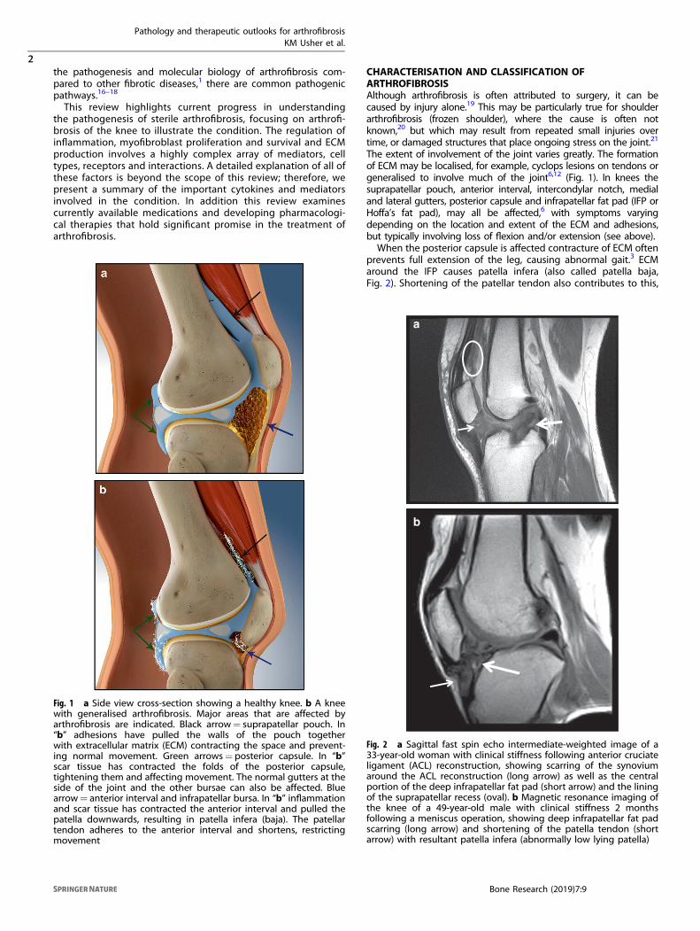
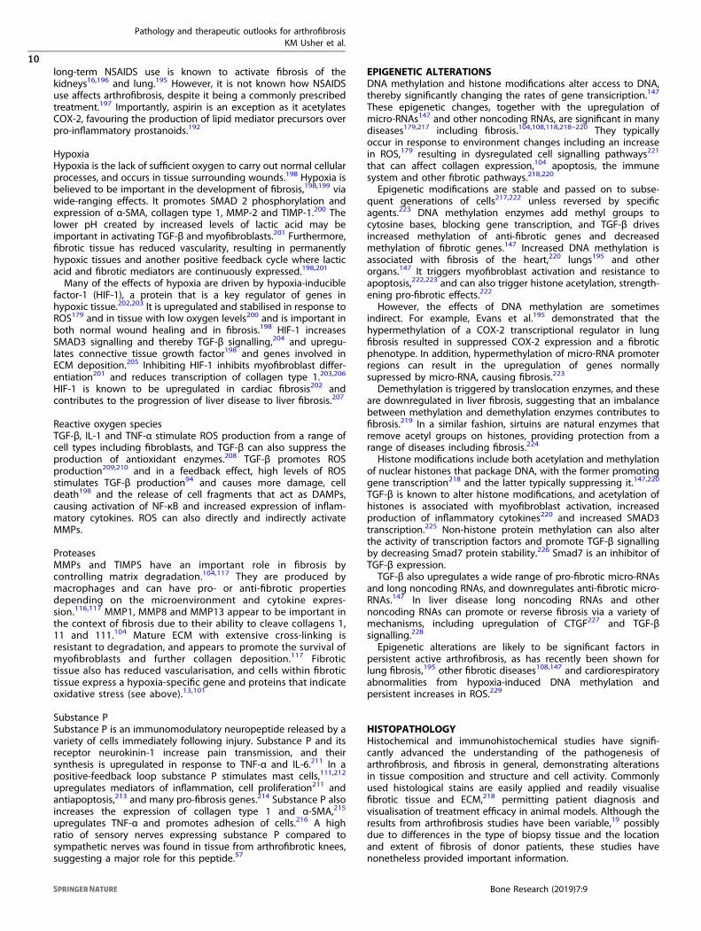
The extent of involvement of the joint varies greatly. The formationof ECM may be localised, for example, cyclops lesions on tendons orgeneralised to involve much of the joint6,12 (Fig. 1). In knees thesuprapatellar pouch, anterior interval, intercondylar notch, medialand lateral gutters, posterior capsule and infrapatellar fat pad (IFP orHoffa’s fat pad), may all be affected,6 with symptoms varyingdepending on the location and extent of the ECM and adhesions,but typically involving loss of flexion and/or extension (see above).When the posterior capsule is affected contracture of ECM often
prevents full extension of the leg, causing abnormal gait.3 ECMaround the IFP causes patella infera (also called patella baja,Fig. 2). Shortening of the patellar tendon also contributes to this,
Fig. 1 a Side view cross-section showing a healthy knee. b A kneewith generalised arthrofibrosis. Major areas that are affected byarthrofibrosis are indicated. Black arrow= suprapatellar pouch. In“b” adhesions have pulled the walls of the pouch togetherwith extracellular matrix (ECM) contracting the space and prevent-ing normal movement. Green arrows= posterior capsule. In “b”scar tissue has contracted the folds of the posterior capsule,tightening them and affecting movement. The normal gutters at theside of the joint and the other bursae can also be affected. Bluearrow= anterior interval and infrapatellar bursa. In “b” inflammationand scar tissue has contracted the anterior interval and pulled thepatella downwards, resulting in patella infera (baja). The patellartendon adheres to the anterior interval and shortens, restrictingmovement
a
b
Fig. 2 a Sagittal fast spin echo intermediate-weighted image of a33-year-old woman with clinical stiffness following anterior cruciateligament (ACL) reconstruction, showing scarring of the synoviumaround the ACL reconstruction (long arrow) as well as the centralportion of the deep infrapatellar fat pad (short arrow) and the liningof the suprapatellar recess (oval). b Magnetic resonance imaging ofthe knee of a 49-year-old male with clinical stiffness 2 monthsfollowing a meniscus operation, showing deep infrapatellar fat padscarring (long arrow) and shortening of the patella tendon (shortarrow) with resultant patella infera (abnormally low lying patella)
Pathology and therapeutic outlooks for arthrofibrosisKM Usher et al.
2
Bone Research (2019) 7:9
1234567890();,:

leading to patellofemoral pain22,23 and often osteoarthritis (OA) ata later stage. The IFP may become fibrotic and impinge in the jointwhen the knee is flexed, creating further inflammation andfibrosis, loss of flexion and pain.24 The IFP is a store of immunecells that secrete inflammatory cytokines under stressful condi-tions25 (see “Risk assessment”), and can fill with ECM whenadipose cells transform into fibrous tissue.26
The causes of arthrofibrosis are poorly understood,27 andexplanations frequently depend on the training of authors. Shoulderarthrofibrosis has been recognised as an inflammatory condition forsome time,28 however, orthopaedic surgeons specialising in kneeshave traditionally cited physical/mechanical causes such as poorsurgical technique and non-compliance of patients in rehabilitation(for example,7,9,29,30). Nonetheless, the role that inflammation playsin arthrofibrosis is increasingly being recognised by the surgicalcommunity.6,12,31 Studies by immunologists and rheumatologistsdemonstrate that dysregulation of the immune system and woundhealing processes, including inflammatory chemokines, cytokinesand proteins, leads to fibrosis18 following an insult such as surgery.Indeed, surgery to treat anterior cruciate ligament (ACL) injury hasbeen associated with a significantly higher risk of arthrofibrosis thanconservative management.29 Immobilisation is also frequently citedas a contributing factor.14,19
Understanding arthrofibrosis, its causes, rates of occurrence andthe success or failure of treatments has been complicated becausethe condition was poorly defined.32 Definitions have varied widelyand are sometimes subjective, as are measures of treatmentoutcomes.33 Recently, an international panel of experts frommultiple medical disciplines developed a consensus definition andclassification of knee arthrofibrosis, which stated “post-operativefibrosis of the knee was defined as a limited ROM in extensionand/or flexion”, measured by active flexion and extension, whichwas not caused by infection of other specific causes.32 Mild,moderate and severe arthrofibrosis was classified as flexion rangeof 90°–100°, 70°–89°, and less than 70°, respectively, and/or a lossof extension of 5°–10°, 11°–20° and more than 20o, respectively.32
The presence of pain was acknowledged as being an importantaspect of the condition. This consensus definition should assistarthrofibrosis research and should be widely applied.The Shelbourne classification34 has been widely used for knee
arthrofibrosis in the past, but was developed from patients witharthrofibrosis arising from ACL reconstruction. Using these criteriaa diagnosis of arthrofibrosis requires a loss of extension, excludingmany patients with debilitating arthrofibrosis that have pain and aloss of flexion but not a loss of extension. For example, a recentcase report identified a young woman with arthrofibrosis who hadonly minimal loss of ROM, but considerable pain, inflammationand disability.12 The presence of excessive ECM was confirmed byarthroscopy.It is sometimes stated that arthrofibrosis is a rare complication
of surgery29; however, some authors describe the condition as acommon complication of total knee replacement (TKR) and ACLreconstruction surgeries.8,33,35–37 Estimates of the rates ofarthrofibrosis following ACL reconstruction range from 2% to35%,22,29 and after TKR between 0.2% and 10%38 with othersreporting rates up to 15% (ref.32 and references within).39 Onelarge study of TKRs in more than 64 000 patients in the US foundthat rates of arthrofibrosis for which revision surgery wasperformed was 0.2%.38 However, Abdul et al. reported post-TKRrates of arthrofibrosis of between 3% and 10%,40 and rates of 4%41
and 12%42 have been reported, with one review paper citing ratesof stiffness from 8% to 60% following a TKR.36
In a study by Werner et al.,5 all surgeries in a national sample ofspecific cohorts for non-TKR knee surgeries were investigated.Rates of arthrofibrosis requiring a manipulation under anaesthesia(MUA) or arthroscopy within 6 months of the initial surgery wereup to 8%. This study showed that rates of arthrofibrosis requiringsurgical treatment were significantly higher for ACL reconstruction
compared to meniscectomy and microfracture.5 However, evenexploratory arthroscopies are capable of causing arthrofibrosis.12
While some of the confusion about the rates of post-operativearthrofibrosis are due to the lack of an agreed definition,32 otherfactors most likely come into play too. Papers may not reflect thetrue rates of arthrofibrosis29 due to reporting bias. Actual rates ofarthrofibrosis following surgery are likely to be higher than thereported rates, since patients may not be treated surgically.5
Registries of joint replacement outcomes do not includearthrofibrosis unless the patient undergoes a surgical procedureto exchange or remove prostheses,32 and the incidence ofuntreated arthrofibrosis is unknown.Arthrofibrosis is a form of fibrosis43 and common pathogenic
pathways occur in fibrosis of organs and tissues.15,17,44,45 However,specialised cell types in some organs may have organ-specificinfluences.43 In fibrosis myofibroblasts are activated and dysregu-lated as a result of inflammation,46 and inflammatory cytokines areknown to upregulate the factors that induce arthrofibrosis.43
Despite the increasing use of preventative measures aftersurgery, it appears that arthrofibrosis rates have remainedrelatively constant.29 A lack of an understanding of the role thatinflammation plays in arthrofibrosis can lead to overly aggressivephysical therapy programmes, with papers frequently recom-mending “aggressive” physical therapy as soon as possible aftersurgery.7,42,47,48 However, aggressive exercise can initiate orworsen arthrofibrosis32,48 because exercise triggers an inflamma-tory response49 including an increase in inflammatory cytokines,collagen production and TGF-β,50,51 factors that are dysregulatedin fibrosis (see below). Some patients on international knee forumsreport that their symptoms either began or became significantlyworse after they were instructed to “push through the pain”during rehabilitation, or performed more strenuous exercise.
TWO “TYPES” OF ARTHROFIBROSIS?Pain and some degree of inflammation are recognised symptomsof arthrofibrosis,32 yet some papers on knee arthrofibrosis onlydiscuss “stiffness” as a symptom, for example,8,11,52 and eitherspecify a painless joint,7 or do not mention pain and inflammationat all. We suggest that what is termed “arthrofibrosis” may be twodifferent conditions, (1) an active condition in which ECMformation and inflammation are continuous processes driven bypositive feedback loops and (2) residual arthrofibrosis, in whichthe joint has limited ROM due to existing ECM, but the activeinflammatory and ECM deposition phases have resolved. Thepresence of the inflammatory cytokines tumour necrosis factoralpha (TNF-α) and interleukin-1β (IL-1β) in acute pulmonaryfibrotic tissue, but not in older fibrotic tissue,53 suggests oneway in which active and residual fibrosis may differ, and anexplanation in part for differing pain levels between the twoarthrofibrosis groups, but research is lacking.Misdiagnoses may complicate the understanding of arthrofi-
brosis. For example, Pujol et al.35 describe two types of patientswith arthrofibrosis, those with swelling and pain in addition to lossof ROM, and those with primarily a loss of ROM. The first group ofpatients is described as having complex regional pain syndrome(CRPS), a type of neuropathic pain caused by nerve damage, andthe authors recognise that this group of patients should not beoperated on. However, there are no specific diagnostic tests forCRPS, and no clinical features that identify it.54,55 Consequently,the diagnosis of CRPS is made in the absence of otherexplanations for pain and swelling, and it remains a controversialdiagnosis.54,55
Without publically available blood tests for arthrofibrosis, itseems likely that many patients that have been diagnosed withCRPS do in fact have active arthrofibrosis and a dysregulatedinflammatory response. Indeed, a significant majority of patientsdiagnosed with CPRS type 1 have muscle weakness or limited
Pathology and therapeutic outlooks for arthrofibrosisKM Usher et al.
3
Bone Research (2019) 7:9

ROM (ref.55 and references within). It is nonetheless worthrecognising that inflammatory cytokines sensitise the peripheraland central nervous system leading to persistent pain in thepresence of chronic low-grade inflammation.56
Indeed, under these conditions it is thought that persistentsynthesis of substance P, a known pain sensitiser and activator ofmast cells and fibroblasts, occurs, and creates a positive feedbackloop.14 In support of this, an increased ratio of sensory nerves(expressing substance P) to sympathetic nerves was found intissue from arthrofibrotic knees.57 Also of note is the fact thatchronic low grade inflammation frequently does not have obviousphysical signs or markers in the blood,56 but can nonetheless playa role in active arthrofibrosis.More research is needed to understand the difference between
active and residual arthrofibrosis, as the response of patientswithin these groups to surgery and exercise may be significantlydifferent. In support of this, Panni et al.7 report that painful stiffknees do not respond well to arthroscopic surgery to lyseadhesions, and Babis et al.27 report that surgery to treatarthrofibrosis in TKR patients resulted in worse outcomes for painin all patients, with some also losing flexion. Surgical lysis offibrotic material is the standard treatment for arthrofibrosis,however, surgery stimulates wound healing processes, includingECM proliferation, and is associated with increased inflamma-tion.58 In addition, immune system memory and/or feedbackprocesses that may be occurring in a patient with activearthrofibrosis may be further stimulated by surgery. It is knownthat re-occurrence is frequent after the removal of ECM in someconditions.15
Possible parallels with active and residual knee arthrofibrosiscan be found in shoulder arthrofibrosis, in which pain may resolvewith time or remain together with ROM limitations,28 and in otherfibrotic diseases. There are several fibrotic diseases of the lungs,including simple pneumoconiosis, in which fibrosis begins andstops, and progressive massive fibrosis, in which extensive fibrosisprogresses until fatal.59 Simple pneumoconiosis can turn intoprogressive massive fibrosis if exposure to dust and inflammationcontinues. Liver fibrosis is another possible parallel, as it cansometimes be stopped and even reversed60 using anti-inflammatory or anti-viral medications, but can turn into active,progressive fibrosis.61 Active fibrosis results from a switch from aninitial Th1 inflammatory cell response to a Th2 cell response withprolonged exposure to an inflammatory stimulus. While thisswitch helps to control the damage caused by immune cells andpromotes healing, it also activates collagen deposition andfibrosis.62
GENDERS DIFFERENCES IN RATES OF ARTHROFIBROSISWomen have been reported to be more likely to developarthrofibrosis than men,21,63 with studies citing rates 2.5–2.8 timeshigher,29,64 although others have not found a gender differ-ence.33,38 It has been suggested that the higher rates ofarthrofibrosis in women may be due to psychological differencesbetween the genders and that women may be less active post-operatively, may not perform rehabilitation as well as men, mayseek more medical interventions, and have “different” paintolerance than men.29 But Hemsley65 found no differences inpain perception or pain reflex between patients at 6 weeks post-ACL reconstruction surgery, almost half of whom did not recoverfull ROM.However, it is well established that the genders differ in their
immunological responses, with 80% of autoimmune diseaseoccurring in women.66 Being female is also a risk factor forOA,38,67 with more women undergoing TKR than men, despitewomen having a greater unmet need for this surgery.68 Recentresearch shows that OA is initiated and progressed by inflamma-tion (see below in Risk factors), and that patients with OA havehigh levels of inflammatory cytokines in the knee.58
The gender difference in inflammatory responses is due to bothgenes and hormones. Women have stronger innate and adaptiveimmune responses than men, leading to increased rates ofinflammatory and autoimmune diseases.66 The corollary is thatwomen have around half the risk of serious post-surgical septicinfection,69 possibly because oestrogen upregulates pro-inflammatory cytokines including IL-1 and IL-6.70 Transforminggrowth factor β (TGF-β), the primary driver of fibrosis, is alsoupregulated and activated by progesterone and oestrogen,71
driving an increase in Treg cells at ovulation.72 Because immunesystem dysfunction and acute inflammation cause fibrosis,2 thehigher rates of arthrofibrosis in women is likely due to theseimmunological differences between the genders.
RISK FACTORS FOR ARTHROFIBROSISThere are no established methods for determining the risk ofdeveloping arthrofibrosis following surgery. However, by under-standing the pathology of the condition, it may be possible toprevent or successfully treat arthrofibrosis,13,42 and a number offactors are known to be involved (Table 1). Early onset OA may bea risk factor/indicator for developing arthrofibrosis after injury orsurgery. OA is associated with inflammation,73–76 and theinflammatory cytokines IL-6 and TNF-α are upregulated in OAsynovial fluid.67,74 Importantly, in a study by Remst et al. over half
Table 1. The stages of pathogenesis of sterile arthrofibrosis of the knee with corresponding clinical features, risk factors and current managements
Pathogenesis Clinical features Risk factors Current management
Inflammatory response, upregulated TGF-β Pain, redness and swelling Surgery or injury •Elevation and icing•Corticosteroids•Aspirin
Proliferation of myofibroblasts and ECMproduction
Stiffness and restricted range of motion Surgery or injury
Dysregulation of inflammation and TGF-βsignalling, excessive ECM in and around joint,adhesions and contractions. Epigeneticalterations
Persistent pain and restricted ROM, withtypically mild swelling. Further ECMproduction and contractions of soft tissues,abnormal gait
•Previous surgeries•Mutations causingexcessive TGF-β orinflammation•Female gender?•Early onset OA•Inflammatory andautoimmune diseases
•Daily CPM•Exercise rehabilitation•Control of inflammation•MUA•Surgery to lyseadhesions and debrideECM
ECM extracellular matrix, TGF-β transforming growth factor β, ROM range of motion, OA osteoarthritis, CPM continuous passive motion machine, MUAmanipulation under anaesthesia
Pathology and therapeutic outlooks for arthrofibrosisKM Usher et al.
4
Bone Research (2019) 7:9

of patients with OA were found to have fibrosis of the synovium,43
and other studies have also found an association between OA andfibrosis.75,76
This link with arthrofibrosis is likely due to over-expression ofTGF-β, a well-known initiator of fibrosis (see below) that is alsoimplicated in the development of OA when expressed at highlevels in subchondral bone and synovial cells.77 TGF-β levels werehigher in subchondral bone of patients with OA compared tohealthy controls, and appeared to lead to increased blood vesselformation, bone resorption and stress on articular cartilage.78 Insupport of this, high levels of TGF-β induced in rats and mice haveled to OA-like lesions.78,79
This suggests that a pro-inflammatory, pro-fibrosis scene existsfor patients with early onset OA. The high numbers of fibroblastsin knee synovium can drive inflammation67 and become furtheractivated following surgery. In addition, patients with OA have amore pro-inflammatory lipid profile in the IFP than individualswith healthy joints.25The bursa around the knee, particularly theIFP, produce and store inflammatory cytokines26,58 and immunecells, including macrophages, T cells, B cells and mast cells thatcan be locally activated by an insult to secrete inflammatorycytokines, particularly TNF-α and IL-6.25,80 Macrophages have beendetected in the IFP at 20 weeks post-ACL reconstruction surgery,58
and are known to play a key role in all stages arthrofibrosis.81
Injury prior to surgery is also a risk factor for arthrofibrosis. ACLtears have been demonstrated to increase the levels of IL-1β andTNF-α in synovial fluid, with levels increasing with the degree ofdamage and with time since injury.82 It has been suggested thathigher levels of these cytokines are responsible for the laterdevelopment of OA.82 TGF-β is also upregulated in the IFP at2 weeks post-ACL reconstruction surgery,58 potentially contribut-ing to the high rates of arthrofibrosis after this type of surgery.More than two previous surgeries are also a risk factor for post-operative arthrofibrosis,11 indicating that there is a potentiation or“memory” of each insult, as demonstrated in other fibroticdiseases.In other surgery, such as TKR and reconstructive surgery using
artificial ligaments, the implantation of a prosthesis triggers theformation of fibrotic tissue as the body attempts to encapsulatethe foreign material.83 Implants such as screws that impinge ontissues also cause an inflammatory reaction,84 and may promotearthrofibrosis of TKRs that are not well fitted.Other factors can also come into play. Childhood adversity such
as neglect or abuse is associated with disease and disability laterin life,85 causing higher Th17 cell numbers, a higher IL-6 responseto stress, and autoimmune and inflammatory diseases.86 Depres-sion and associated poor rehabilitation compliance are sometimescited as causative factors for arthrofibrosis,7 however, it isinteresting to note that depression is strongly associated withinflammation, and inflammation can cause depression.87,88 There-fore, it seems likely that the inflammatory processes associatedwith active arthrofibrosis cause depression.Other risk factors include pre-existing inflammatory or auto-
immune diseases, including type II diabetes,20 ankylosingspondylitis and rheumatoid arthritis.7 One study found thatpatients with diabetes mellitus had increased rates of arthrofi-brosis after a TKR,38 possibly due to a pro-inflammatoryphysiology.Biomarkers to assess the risk of developing post-surgical
arthrofibrosis are urgently needed. In addition to pre-surgeryapplications, biomarkers could also be used post-operatively for alljoint surgeries to monitor potential for developing arthrofibrosis,and following a diagnosis, to monitor the condition and itsresolution. Such biomarkers will be essential for the developmentand testing of therapies.89 Ideally tests should be minimallyinvasive, for example, serum parameters and imaging, andapplicable before surgery and during treatment to followprogress.90
GENETIC RISK FACTORSSome patients may have a genetic predisposition for developingfibrosis,91 with a twin study finding there was a geneticcomponent to shoulder arthrofibrosis.92 Because multiple biolo-gical pathways impact on the pathology of arthrofibrosis, it islikely that there are many types of mutations that can affect therisk of developing it, including mutations in the immune system,TGF-β signalling and genes involved in the synthesis ordegradation of collagen. Skutek et al.93 found a possible linkbetween some varieties of human leucocyte antigen and the riskof arthrofibrosis. The human leucocyte antigen complex isinvolved in immune system functioning.People with mutations involving TGF-β production or signalling,
which can result in excessive ECM formation,94 may be atparticular risk of developing arthrofibrosis. One candidate condi-tion is Aneurysms-OA Syndrome, now included under the nameLoeys–Dietz syndrome, in which upregulation of TGF-β signallingcauses early onset OA.95–97
PATHOGENESIS OF FIBROSISThere is little research into the cell biology and pathogenesis ofarthrofibrosis. However, a wealth of organ fibrosis researchprovides important insights into the processes involved inarthrofibrosis, and is reviewed here. Fibrosis results from acomplex dysregulation of innate and adaptive immunity that isinvolved in most chronic inflammatory diseases,15,45,46 and is aleading cause of mortality.62 Injury causes oxidative stress andan inflammatory response, inducing pro-inflammatory cyto-kines98–100 and TGF-β (Figs. 3 and 4).101 This leads to an increasein mast cells, macrophages and lymphocytes that promotefibroblast proliferation and reduced vascularisation.13,62
A lack of apoptosis and autophagy within fibrotic tissues havealso been implicated in a number of fibrotic conditions, and maycontribute to fibrotic tissue formation.13 Reduced autophagy leadsto a build-up of defective mitochondria and oxidative stress.102
Immune cell signalling also stimulates an increase in reactiveoxygen and nitrogen species (RONS)13 and positive feedbackbetween macrophages and lymphocytes, leading to immune celldysregulation.62 However, the severity of fibrosis is often not wellcorrelated with the degree of inflammation,62 and low-levelinflammation that persists over long periods also causes fibrosis.46
Pro-fibrotic cytokines are thought to cause an imbalancebetween ECM production and degradation, leading to excessivedeposition of matrix proteins, which are both collagenous andnoncollagenous.15,45 Collagen type I is the main constituent ofECM. It has high-tensile strength that prevents normal stretching,and in fibrosis there is a higher ratio of collagen type I to stretchyelastin, compared to healthy tissues.103 In addition to alteredcomposition, fibrotic ECM has extensive cross-linking that makes itvery difficult to degrade.89,104 In particular, levels of hydroxyally-sine cross-linking is increased, and appears to lead to irreversiblecollagen accumulation105 together with other effects on cellsignalling and ECM synthesis.89
The ECM that forms in fibrosis is largely cell-free, and serves as aconduit for immune cells, fibroblasts, nutrients and endothelialcells during angiogenesis. In addition to proteins the groundsubstance of the ECM is comprised of proteoglycans, and thesebind and inhibit or enhance a range of growth factors, proteases,protease inhibitors and TGF-β (for review see ref. 103).The inflammatory cytokines and mediators that trigger fibrosis,
together with the cells that express them (see below), areessential components of a healthy immune system. Typically,inflammatory cytokines are downregulated after a period of time,but the continued presence of inflammatory cytokines andmediators can cause tissue to become pro-inflammatory andfibrosis may develop. The presence of one inflammatory cytokinecauses the receptors for other cytokines to be made, sensitising
Pathology and therapeutic outlooks for arthrofibrosisKM Usher et al.
5
Bone Research (2019) 7:9
cells to respond strongly.106 Repeated trauma and/or long-terminflammation can trigger epigenetic modifications and activationof myofibroblasts and matrix-related genes.46,107,108 Chronicinflammation may also result from a lack of bioactive lipidmediators (LMs) that causes deficient or non-existent resolution
(see “resolvins”), or LMs that don’t have the required regulatoryeffects.109
Almost all types of immune cells are involved in fibrosis110
and the pathways are extremely complex. Consequently,a detailed discussion is beyond the scope of this review,
ROS, PDGF, TGF-β1, Inflammatory cytokines
MacrophagesTh2 cells,Lymphocytes,Mast cells
Fibroblasts
Myofibroblasts
Scar tissue,Adhesions
Chemokines, NF-κB, TGF-β1, Infammatory cytokines, ROS
PDGF, ROS, TGF-β1, Infammatory cytokines
Mechanical stress
Focal adhesion
Tissue
Tissue
Contraction
Cross linkagesforming
α- SMA stressfibers
Wound or injury: Hypoxia,Substance P,Inflammasome activation
Extracellular matrix proteins
Extracellular matrix
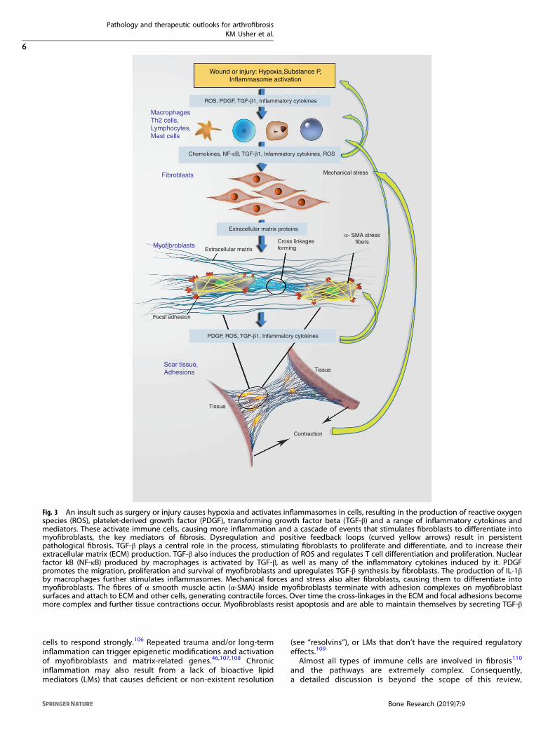
Fig. 3 An insult such as surgery or injury causes hypoxia and activates inflammasomes in cells, resulting in the production of reactive oxygenspecies (ROS), platelet-derived growth factor (PDGF), transforming growth factor beta (TGF-β) and a range of inflammatory cytokines andmediators. These activate immune cells, causing more inflammation and a cascade of events that stimulates fibroblasts to differentiate intomyofibroblasts, the key mediators of fibrosis. Dysregulation and positive feedback loops (curved yellow arrows) result in persistentpathological fibrosis. TGF-β plays a central role in the process, stimulating fibroblasts to proliferate and differentiate, and to increase theirextracellular matrix (ECM) production. TGF-β also induces the production of ROS and regulates T cell differentiation and proliferation. Nuclearfactor kB (NF-κB) produced by macrophages is activated by TGF-β, as well as many of the inflammatory cytokines induced by it. PDGFpromotes the migration, proliferation and survival of myofibroblasts and upregulates TGF-β synthesis by fibroblasts. The production of IL-1βby macrophages further stimulates inflammasomes. Mechanical forces and stress also alter fibroblasts, causing them to differentiate intomyofibroblasts. The fibres of α smooth muscle actin (α-SMA) inside myofibroblasts terminate with adhesion complexes on myofibroblastsurfaces and attach to ECM and other cells, generating contractile forces. Over time the cross-linkages in the ECM and focal adhesions becomemore complex and further tissue contractions occur. Myofibroblasts resist apoptosis and are able to maintain themselves by secreting TGF-β
Pathology and therapeutic outlooks for arthrofibrosisKM Usher et al.
6
Bone Research (2019) 7:9

however, we explore the major cell types and cytokinesinvolved below.
MyofibroblastsMyofibroblasts are the key effector cells of fibrosis,46,111 remodel-ling the ECM, and depositing dense fibrotic collagen.15,44,112,113
Myofibroblasts also form cell-to-cell connections and connectionsbetween cells and ECM, creating contractile units and causing thecontraction of surrounding tissues.103,114–116 In the presence ofTGF-β myofibroblasts produce fibres of α-smooth muscle actin (α-SMA) together with collagen type 1 (for review see115,117). Overtime the focal adhesions become more complex and further tissuecontractions occur, together with extensive collagen cross-linking.108
Myofibroblasts are important in wound healing, however, theyare not usually found in healthy tissue.103 They are derived fromfibroblasts115,116 and a range of other cells107,113 that havedifferentiated in response to inflammatory cytokines such asTGF-β, IL-1β and IL-6. However, myofibroblasts also produce TGF-β, IL-1β, IL-6 and platelet-derived growth factor (PDGF), in additionto reactive oxygen species (ROS) and a range of paracrine signalsthat further activate a fibrotic response (for reveiw see ref. 103).Thus myofibroblasts resist apoptosis and are able to maintainthemselves by secreting TGF-β15 and inflammatory cytokines,activating immune cells and further fibrosis. In addition, mechan-ical forces also alter the biochemical actions of fibroblasts, causingthem to differentiate into myofibroblasts.114
During normal wound healing and resolution of inflammationsome myofibroblasts become apoptotic, while others revert to theoriginal cell type, however, the processes by which this occurs arenot yet understood.90 In fibrosis epigenetic alterations inmyofibroblasts increase the activity of inflammatory and pro-fibrotic genes118 (see below in Epigenetic alterations), and appearto serve as a type of memory of the insult.108 Myofibroblasts thathave reverted back to fibroblasts are more likely to become re-activated when exposed to further insult.60,90 This has implicationsfor repeated joint surgeries as fibrosis may resolve naturallyand unnoticed, but the presence of reverted fibroblasts that serveas a store of pre-fibrotic cells may leave the patient susceptibleto arthrofibrosis after subsequent surgeries, as discussed earlier. Itis not known if the formation of ECM is common followingsurgery, only becoming apparent when normal function iscompromised.
INFLAMMATORY CELLS AND CELL STRUCTURESA number of cell types contribute to the initiation andmaintenance of chronic inflammation and fibrotic diseases,including macrophages, myofibroblasts and Th2 cells.62 Inaddition to these factors, protein complexes within the cytoplasmof cells called inflammasomes produce inflammatory cytokines,and which serve as a type of “memory” of insult (see below).
MacrophagesMacrophages react to a diverse range of signals by secretingcytokines and chemokines, and are found in close association withmyofibroblasts.119 They can be activated by TGF-β and can beimportant in fibrosis.16 Classically activated macrophages (M1)secrete inflammatory cytokines, including TNF-α, IL-1 and IL-6.120
M1 also promote the differentiation of Th17 cells, which are alsopro-inflammatory (see below). However, M2 macrophages secreteanti-inflammatory cytokines, including IL-10 and IL-13, and areimportant in the resolution of inflammation.120
Recent research shows that distinct macrophage populationsmay control the initiation, maintenance and resolution offibrosis.81 Macrophages are an important source of the pro-fibrosis mediators TGF-β, IL-1β and PDGF.46 PDGF promotes themigration, proliferation and survival of myofibroblasts,121,122 andupregulates TGF-β synthesis by fibroblasts.123 In addition, theproduction of IL-1β by macrophages can stimulate inflamma-somes in the lung.46 Macrophages may be able to regulate ECMsynthesis independently of TGF-β,119,124 however, macrophagesare also involved in the resolution of fibrosis via multiplemechanisms, including the clearing of excess collagen fromdamaged tissues and the secretion of collagenases that degradeECM components.81
Mast cellsMast cells initiate and maintain inflammation.111 They may play animportant part in the development of fibrosis125 and appear to beable to maintain a pro-fibrotic response, producing and storingmany of the cytokines that promote fibrosis14,111 (see below underCytokines), including TNF-α, IL-17 and TGF-β.125,126 Mast cellsnumbers are increased in fibrotic organs including the lung,127
heart and kidneys. Trautmann et al.128 demonstrated that mastcells stimulate fibroblast proliferation after attaching and directlyreleasing cytokines into their cytoplasm, suggesting an importantmechanism by which fibrosis is promoted and maintained.
IRAK
G-protein
InactivatedSmad
TNF-α TGF-βInterleukins Chemokines
TRAF
InactiveIKKK
ActivatedIKKK
Iκ-B NF-κB
Inflammasomes
NF-κB
Matrix production and fibrogenesis
Serine/ThreonineKinase domain
ActivatedSmad
InactivatedMAPK
ActivatedMAPK
αβ γα
GTP
p
ppp
p
p
Fig. 4 Four potential signal transduction pathways and their corresponding receptors associated with arthrofibrosis; including TNF-α,Interleukins (IL1, IL6, IL17, etc.), TGF-β and chemokines ligand-receptor superfamilies, which lead to activation of NF-κB, Smad, MAPK andmultiple downstream gene transcriptions responsible for matrix production and fibrogenesis
Pathology and therapeutic outlooks for arthrofibrosisKM Usher et al.
7
Bone Research (2019) 7:9

T cellsThe type of T cell response to inflammation controls themagnitude of fibrosis, with Th2 cells promoting the productionof ECM and fibrosis, while Th1 cells are typically suppressive.62,116
Th17 cells are a subset of T reg cells that differentiate in theperiphery in the presence of IL-1β, IL-6 and TGF-β.129 They secreteIL-17, a cytokine that is important for the activation and migrationof immune cells, inducing them to secrete inflammatory cytokinesand chemokines.129 Th17 cells are suppressed by the amino acidlimitation response, which also enhances autophagy.102
InflammasomesInflammasomes are intracellular protein complexes that activatean inflammatory cascade by upregulating the production andmaturation of inflammatory cytokines IL-1β and IL-18.2,120,130
Activated inflammasomes play a central role in fibrosis of organsincluding the liver,131,132 lungs133 and kidneys,130 upregulatingα-SMA, connective tissue growth factor and collagen type I131.Inflammasomes serve as an inflammatory memory, however, it isnot yet clear how they remain active in chronic fibroticdiseases.2
Inflammasomes are present in immune cells and a wide varietyof cells in tissues, including myofibroblasts and fibroblasts, and areactivated by an array of different signals from wounds andinfection. Sterile activators include nuclear factor kB (NF-κB)134
and stimuli generated by cell death or damage, referred to asdamage-associated molecular patterns (DAMPS), which signal theinflammasome via cell receptors. These diverse stimuli includeROS, adenosine triphosphate (ATP), mitochondrial DNA andproteins released from damaged ECM, such as hyaluronan,heparin sulphate and biglycan.2,120
Inflammasome activity is also regulated by secreted factors andby cell-to-cell interactions.2 In addition, some inflammatorycytokines that are released by dying cells, including TNF-α, IL-1αand IL-1β can act as DAMPS and activate inflammasomes.2,120
Intracellular proteins such as the chromatin associated proteinhigh-mobility group box 1 (HMGB1) are also released by necroticcells and act as DAMPS. Macrophages activated by TNF-α and TGF-β can also release HMGB1,2 activating inflammasomes andcreating crosstalk between the production of inflammatorycytokines and the TGF-β signalling, with potential feedback loopsand implications for fibrosis.Inflammasomes directly and indirectly activate matrix produc-
tion and fibrogenesis in tissue,98 and activate macrophages viaproduction of IL-1β.98 It is of interest that IL-1β can stimulate NF-kB and p38 MAPK pathways and the resulting transcription ofinflammatory cytokines including IL-6,120,135 perhaps leading toanother feedback loop between inflammasome activation, IL-1βsecretion and TGF-β production.The inflammasome component nucleotide-binding domain and
leucine-rich repeats containing pyrin domain 3 (NLRP3) is wellstudied. The NLRP3 inflammasome is a key player in sterileinflammation, and is associated with a range of auto-inflammatoryand autoimmune diseases.2 Tissue damage and the accumulationof damaged mitochondria increases mitochondrial ROS produc-tion, which, along with other signals activates NLRP3 andstimulates processing of IL-1β pre-cursers into the biologicallyactive form.2,136,137 NLRP3 also regulates ROS production bymitochondria.138 The activation of capase-1 by NLRP3 activates IL-1β and IL-18 precursors,2,137 and also causes the secretion of IL-1αand fibroblast growth factor 22.
CYTOKINESMany cytokines have been associated with fibrosis, the mostimportant being TGF-β. Other cytokines known to have involve-ment are TNF-α, IL-17, IL-1β and the anti-inflammatory IL-10.139 Acombination of inflammatory cytokines upregulates expression of
TGF-β receptors, and inflammation plays an important role in thedevelopment of fibrosis.140
Transforming growth factor betaTransforming growth factor beta (TGF-β) plays a central role inthe pathology of arthrofibrosis1 and all fibrotic diseases,141–144
causing activation and proliferation of myofibroblasts, inhibitionof collagen degradation, and an increase in ECM synthesis.144,145
TGF-β is produced by most cells, including inflammatory andeffector cells16,146 and regulates immunity.146 It is secreted ina latent state, and must be activated by cleavage.94 Fourisoforms are known and are involved in the regulation of cellproliferation, differentiation, adhesion, apoptosis, migration andfibrosis.94,101,147 TGF-β1 is the most abundant isoform, and isthought to be the most important in the pathology of fibrosis.148
Experimental induction of TGF-β causes excessive proliferationof fibroblasts in the knee joints of rats1 and stimulates theproduction of ECM, causing rat knee joints to becomecompletely encased in fibrous tissue.16 ECM also stores latentTGF-β,94 which is released and activated by the stress betweencell surfaces and ECM149 that occurs during the contraction ofmyofibroblasts.Production of activated TGF-β is stimulated by oxidative
stress,138 platelet degranulation144 and ROS released after injuryor surgical insult.94 While ROS activates TGF-β and results inapoptosis,150 TGF-β also induces the production of ROS,101,138,151
thus creating a positive feedback cycle. This cycle may beexaggerated by another effect of TGF-β, the inhibition of theexpression of antioxidant enzymes, including glutathione.101 Theresulting higher levels of mitochondrial ROS significantly upregu-lates inflammatory cytokines and the production of inflamma-somes.138 TGF-β also regulates T cell differentiation andproliferation and the activation and development of natural killercells.72
Following the binding of TGF-β to its receptor complex,cytoplasmic signal transducer proteins called Smads are phos-phorylated and promote the transcription of target genes in thenucleus.138,144 TGF-β also signals via non-Smad pathways includ-ing the extracellular signal-regulated kinase pathway (for reviewsee ref. 141). In addition, TGF-β induces epigenetic modifications147
(see Epigenetic alterations below) and upregulates matricellularproteins, which interact with cell surface receptors and the ECM.1
However, the specific DNA sequences that are upregulated byTGF-β signalling is dependent on cell-specific DNA-binding co-factors.94
TGF-β drives a shift from Th-1 cells to pro-inflammatory Th-17cells72 and upregulates the production of IL-11, a cytokine with asignificant involvement in the development of fibrosis, infibroblasts (see below). However, it can have different effectsdepending on the type of cells that secreted it, and the presenceof other cytokines. For example, TGF-β secreted by regulatoryT cells in the presence of IL-10 can inhibit inflammation andfibrosis,62 while TGF-β produced by macrophages is pro-fibrotic.81
Mitogen-activated protein kinases (MAPK) upregulate TGF-βexpression in the presence of inflammatory cytokines,147 andform another feedback loop.TGF-β is known to start a cascade of other downstream
regulatory effects including a reduction in ECM degradation viathe downregulation of a family of matrix metalloproteinases(MMPs),152 which include collagenases. Some MMPs are asso-ciated with the progression of fibrosis, however, some have aprotective effect.153,154 TGF-β also induces tissue inhibitors ofMMPs (TIMPS) that block ECM degradation and regulate MMPactivity.1,15,81 MMPs play a key role in regulating a number ofprocesses including ECM remodelling, proliferation, apoptosis andangiogenesis.155 MMPs are also induced by IL-17A, anothercytokine with a significant involvement in the development offibrosis15 (see below).
Pathology and therapeutic outlooks for arthrofibrosisKM Usher et al.
8
Bone Research (2019) 7:9

Interleukin-1IL-1β is believed to be an important mediator of fibrosis,98
influencing the migration of cells, adhesion, matrix metalloprotei-nase production and the expression of immune-modulatorygenes.156 It is a powerful inflammatory cytokine that inducesTGF-β133,145 and PDGF,103 driving the development of fibrosis116
following injury or infection. IL-1β is expressed in fibrotic tissues53
by a range of cell types, but is mainly produced by macro-phages.157 However, it has been demonstrated that in fibrosis ofthe lungs IL-1β acts via TGF-β induction and signalling.145 In auto-inflammatory diseases IL-1β sets up a feedback loop such that itstimulates its own production.158
Interleukin-6IL-6 is a family of cytokines that have been associated with lunginjury and the initiation of lung fibrosis,159 with fewer fibroticchanges seen in IL-6 deficient mice.160,161 Animal models showthat this cytokine increases the expression of TGF-β receptors andtheir signal transduction,162 demonstrating another link betweeninflammation and fibrosis. IL-6 is essential for host defence againstbacterial and viral infections, controlling T cell functions andsurvival. IL-6 also appears to be involved in the “memory” ofinflammation163 and the development of chronic fibrosis.161
Recently, Schafer et al. demonstrated that IL-11 is strongly pro-fibrotic, driving the synthesis of the proteins involved in ECMproduction, contraction and other processes active in fibrosis.143
Production of IL-11 is upregulated by TGF-β. Neutralisingantibodies to IL-11 and the deletion of IL-11 receptors inhibitedthe effects of TGF-β, suggesting new therapeutic targets forfibrosis.143 IL-11 is expressed by fibroblasts and other cells.164 It isa member of the IL-6 family of cytokines, and is also implicated intumour progression.165
Tumour necrosis factor alphaTNF-α is thought to be important in the pathogenesis offibrosis.46,116,166–168 It is a pleiotropic inflammatory cytokine169
that causes significant upregulation of TGF-β production168,170
and receptor expression140 and may stimulate fibroblast growthand collagen type I expression.171 TNF-α also causes fibroblast-likedifferentiation and inflammation,138 and PGE2 expression.172 TNF-α and IL-1 upregulate cyclooxygenase 2 (COX-2) synthesis inresponse to an insult.158 These cytokines also induce theexpression of intracellular adhesion molecule-1 (ICAM-1),158
expressed in vascular endothelium, macrophages and lympho-cytes, and associated with the development of fibrosis.59 Robertset al.152 reported that TNF-α and IL-1β upregulated MMPs in vitro,potentially providing some anti-fibrotic effects, however, thesecytokines also have pro-fibrotic effects. TNF-α may also beinvolved in the “memory” of insult, as TNF messenger RNA isable to remain elevated for more than 70 days.173
Interleukin-17IL-17 upregulates the production of TGF-β174 and inflammatorycytokines from chondrocytes and synovial fibroblasts,175 andpromotes the survival of fibroblasts.67 IL-17 can directly induce theproduction of collagen type 1139 and disrupt ECM homoeosta-sis,176 while promoting MMP production.176,177 It is secreted by anumber of cells types, primarily T-helper 17 (Th17), NK cells andmast and myeloid cells.67,164 The feedback loops between IL-17and IL-6, TNF-α and IL-1 are considered important drivers ofchronic inflammatory diseases,139,175 and suggest a mechanismfor the development of chronic fibrosis. IL-17 acts as a painsensitiser,67 induces monocyte migration and activates monocyte-derived macrophages to produce IL-1, TNF-α and PGE2.178
A number of other chemokines and cytokines including IL-13,IL-4 and IL-5 are associated with a higher risk of fibrosis, while IL-10 and IL-12 are protective.62,116,161 There is conflicting evidencefor the role of interferon-γ.161
OTHER PRO AND ANTI-FIBROGENIC MEDIATORSNF-κB is a family of proteins that occur in the cytoplasm of cells inan inactive form. NF-κB regulates genes and cells involved ininflammatory responses,179 including the activation, differentia-tion and function of inflammatory T cells and inflamma-somes.120,134 It directly and indirectly promotes Th17differentiation, and dysregulated production of NF-κB is asso-ciated with a range of autoimmune and inflammatory diseases.120
NF-κB upregulates the transcription of chemokines andinflammatory cytokines including TNF-α, IL-1β and IL-6 in a rangeof innate immune cells, inducing inflammation.120,157 NF-κB inmacrophages and fibroblasts is activated by TGF-β-activatedkinase 1, as well as many of the inflammatory cytokines inducedby it,120 leading to another feedback loop of inflammation andfibrosis. However, NF-κB is also necessary for inhibiting NLRPinflammasome activation in macrophages.134
The 5′-adenosine monophosphate-activated protein kinasepathwayAdenosine monophosphate-activated protein kinase (AMPK) is awidely expressed member of the serine/threonine kinase familythat is involved in energy regulation and the regulation of a rangeof genes involved in fibrosis.180 AMPK activation appears toregulate macrophages,181 limits ROS production,182 and isincreasingly recognised as playing an important role in suppres-sing fibrosis.180,181 In addition, AMPK also appears to inhibitdifferentiation and proliferation of myofibroblasts and suppresscollagen production.180 Stimulation of the AMPK pathway canoccur via caloric restriction, exercise or medication.181
Specialised pro-resolving lipid mediatorsThe discovery of resolvins, protectins, lipoxins and maresins hasrevolutionised the understanding of how inflammation is resolved.We now know that resolution is an active biochemical processmediated by these specialised pro-resolving LMs (SPMs), which actas a stop signal for inflammation and a return to homoeostasis.109
Specific SPMs have distinct anti-inflammatory, anti-microbial andpro-resolving effects.183,184 SPMs are derived from essential fattyacids, particularly omega 3 polyunsaturated fatty acids (ω-3 PUFA)found in fish oils and some plants, and are necessary in the humandiet.185 SPMs have synergistic effects on immune function,186
downregulating the production of TNF-α and IL-1β,184 reducingpain, inhibiting neutrophil migration and protecting againstuncontrolled inflammatory responses.185,187
Oral supplements of ω-3 PUFA result in biologically active levelsof SPMs in serum including the important subtypes RVD1 andRVD2,188 and in synovial fluid, where SPM levels were negativelycorrelated with pain.189 These and other SPMs are able to switchmacrophage phenotypes from pro-inflammatory to pro-resolving(ref.183 and references within), and reduce the expression ofinflammasomes.184 SPM profiles in patients correlate with out-comes, with a lack of them linked to delayed resolution ofinflammation.184
Importantly, SPMs were shown to be anti-fibrotic in organsincluding the kidney190 and liver.191 PDGF-induced myofibroblastproliferation is inhibited,190 along with the production ofinflammatory cytokines, and SPMs may represent an importantnew treatment for fibrosis.190 Although SPMs have a short half-lifein vivo, more stable synthetic analogues have been developed,192
and may become a useful therapy for a range of inflammatorydiseases and fibrosis.
Nonsteroidal anti-inflammatory drugsNonsteroidal anti-inflammatory drugs (NSAIDS) may prolongchronic inflammation if used for more than 48 h because theresulting inhibition of COX-2193 causes inhibition of resolvinproduction and other SPMs.56,194 COX-2 is an important anti-fibrotic enzyme.195 The chronic inflammation induced by
Pathology and therapeutic outlooks for arthrofibrosisKM Usher et al.
9
Bone Research (2019) 7:9
long-term NSAIDS use is known to activate fibrosis of thekidneys16,196 and lung.195 However, it is not known how NSAIDSuse affects arthrofibrosis, despite it being a commonly prescribedtreatment.197 Importantly, aspirin is an exception as it acetylatesCOX-2, favouring the production of lipid mediator precursors overpro-inflammatory prostanoids.192
HypoxiaHypoxia is the lack of sufficient oxygen to carry out normal cellularprocesses, and occurs in tissue surrounding wounds.198 Hypoxia isbelieved to be important in the development of fibrosis,198,199 viawide-ranging effects. It promotes SMAD 2 phosphorylation andexpression of α-SMA, collagen type 1, MMP-2 and TIMP-1.200 Thelower pH created by increased levels of lactic acid may beimportant in activating TGF-β and myofibroblasts.201 Furthermore,fibrotic tissue has reduced vascularity, resulting in permanentlyhypoxic tissues and another positive feedback cycle where lacticacid and fibrotic mediators are continuously expressed.198,201
Many of the effects of hypoxia are driven by hypoxia-induciblefactor-1 (HIF-1), a protein that is a key regulator of genes inhypoxic tissue.202,203 It is upregulated and stabilised in response toROS179 and in tissue with low oxygen levels200 and is important inboth normal wound healing and in fibrosis.198 HIF-1 increasesSMAD3 signalling and thereby TGF-β signalling,204 and upregu-lates connective tissue growth factor198 and genes involved inECM deposition.205 Inhibiting HIF-1 inhibits myofibroblast differ-entiation201 and reduces transcription of collagen type 1.203,206
HIF-1 is known to be upregulated in cardiac fibrosis202 andcontributes to the progression of liver disease to liver fibrosis.207
Reactive oxygen speciesTGF-β, IL-1 and TNF-α stimulate ROS production from a range ofcell types including fibroblasts, and TGF-β can also suppress theproduction of antioxidant enzymes.208 TGF-β promotes ROSproduction209,210 and in a feedback effect, high levels of ROSstimulates TGF-β production94 and causes more damage, celldeath198 and the release of cell fragments that act as DAMPs,causing activation of NF-κB and increased expression of inflam-matory cytokines. ROS can also directly and indirectly activateMMPs.
ProteasesMMPs and TIMPS have an important role in fibrosis bycontrolling matrix degradation.104,117 They are produced bymacrophages and can have pro- or anti-fibrotic propertiesdepending on the microenvironment and cytokine expres-sion.116,117 MMP1, MMP8 and MMP13 appear to be important inthe context of fibrosis due to their ability to cleave collagens 1,11 and 111.104 Mature ECM with extensive cross-linking isresistant to degradation, and appears to promote the survival ofmyofibroblasts and further collagen deposition.117 Fibrotictissue also has reduced vascularisation, and cells within fibrotictissue express a hypoxia-specific gene and proteins that indicateoxidative stress (see above).13,101
Substance PSubstance P is an immunomodulatory neuropeptide released by avariety of cells immediately following injury. Substance P and itsreceptor neurokinin-1 increase pain transmission, and theirsynthesis is upregulated in response to TNF-α and IL-6.211 In apositive-feedback loop substance P stimulates mast cells,111,212
upregulates mediators of inflammation, cell proliferation211 andantiapoptosis,213 and many pro-fibrosis genes.214 Substance P alsoincreases the expression of collagen type 1 and α-SMA,215
upregulates TNF-α and promotes adhesion of cells.216 A highratio of sensory nerves expressing substance P compared tosympathetic nerves was found in tissue from arthrofibrotic knees,suggesting a major role for this peptide.57
EPIGENETIC ALTERATIONSDNA methylation and histone modifications alter access to DNA,thereby significantly changing the rates of gene transicription.147
These epigenetic changes, together with the upregulation ofmicro-RNAs147 and other noncoding RNAs, are significant in manydiseases179,217 including fibrosis.104,108,118,218–220 They typicallyoccur in response to environment changes including an increasein ROS,179 resulting in dysregulated cell signalling pathways221
that can affect collagen expression,104 apoptosis, the immunesystem and other fibrotic pathways.218,220
Epigenetic modifications are stable and passed on to subse-quent generations of cells217,222 unless reversed by specificagents.223 DNA methylation enzymes add methyl groups tocytosine bases, blocking gene transcription, and TGF-β drivesincreased methylation of anti-fibrotic genes and decreasedmethylation of fibrotic genes.147 Increased DNA methylation isassociated with fibrosis of the heart,220 lungs195 and otherorgans.147 It triggers myofibroblast activation and resistance toapoptosis,222,223 and can also trigger histone acetylation, strength-ening pro-fibrotic effects.222
However, the effects of DNA methylation are sometimesindirect. For example, Evans et al.195 demonstrated that thehypermethylation of a COX-2 transcriptional regulator in lungfibrosis resulted in suppressed COX-2 expression and a fibroticphenotype. In addition, hypermethylation of micro-RNA promoterregions can result in the upregulation of genes normallysupressed by micro-RNA, causing fibrosis.223
Demethylation is triggered by translocation enzymes, and theseare downregulated in liver fibrosis, suggesting that an imbalancebetween methylation and demethylation enzymes contributes tofibrosis.219 In a similar fashion, sirtuins are natural enzymes thatremove acetyl groups on histones, providing protection from arange of diseases including fibrosis.224
Histone modifications include both acetylation and methylationof nuclear histones that package DNA, with the former promotinggene transcription218 and the latter typically suppressing it.147,220
TGF-β is known to alter histone modifications, and acetylation ofhistones is associated with myofibroblast activation, increasedproduction of inflammatory cytokines220 and increased SMAD3transcription.225 Non-histone protein methylation can also alterthe activity of transcription factors and promote TGF-β signallingby decreasing Smad7 protein stability.226 Smad7 is an inhibitor ofTGF-β expression.TGF-β also upregulates a wide range of pro-fibrotic micro-RNAs
and long noncoding RNAs, and downregulates anti-fibrotic micro-RNAs.147 In liver disease long noncoding RNAs and othernoncoding RNAs can promote or reverse fibrosis via a variety ofmechanisms, including upregulation of CTGF227 and TGF-βsignalling.228
Epigenetic alterations are likely to be significant factors inpersistent active arthrofibrosis, as has recently been shown forlung fibrosis,195 other fibrotic diseases108,147 and cardiorespiratoryabnormalities from hypoxia-induced DNA methylation andpersistent increases in ROS.229
HISTOPATHOLOGYHistochemical and immunohistochemical studies have signifi-cantly advanced the understanding of the pathogenesis ofarthrofibrosis, and fibrosis in general, demonstrating alterationsin tissue composition and structure and cell activity. Commonlyused histological stains are easily applied and readily visualisefibrotic tissue and ECM,218 permitting patient diagnosis andvisualisation of treatment efficacy in animal models. Although theresults from arthrofibrosis studies have been variable,19 possiblydue to differences in the type of biopsy tissue and the locationand extent of fibrosis of donor patients, these studies havenonetheless provided important information.
Pathology and therapeutic outlooks for arthrofibrosisKM Usher et al.
10
Bone Research (2019) 7:9

Table 2. List of existing and potential new therapies for treating arthrofibrosis, with a summary of the associated benefits and risks
Therapies Benefits/risks
Dietary approaches
Omega 3 fatty acids in fish or supplements Necessary for the production of SPMs vital for resolution of inflammation. Thins the blood, buttypically no risks are associated within recommended daily limits.
Capsaicin (in peppers) and sulphoraphane (incruciferous vegetables)
May reverse differentiation of myofibroblasts, sulphoraphane may prevent fibroblastdifferentiation. No risks are associated within recommended daily limits.
Resistant fibre Gut bacteria produce short-chain fatty acids which counter inflammation. No risks areassociated within recommended daily limits.
Low-sugar intake Reduces inflammation. Typically no associated risks.
Soy products Contains anti-inflammatory compounds. Reduced levels of TGF-β and lung fibrosis in rats.Benefits not established for treating fibrosis. Typically no risks are associated withinrecommended daily limits.
Potassium May help prevent fibrosis, negative correlation between high levels of serum K+ and liverfibrosis. Typically no risks are associated within recommended daily limits.
Intermittent fasting Protective against fibrosis of organs, suppresses inflammation, IL-1, IL-6 and TNF-α andinflammasomes. Typically no risks are associated. May be difficult to follow.
Pharmaceuticals
Oral and injected corticosteroids Downregulates inflammation and possibly TGF-β. Increased risk of infections, suppressedadrenal gland hormone production, can cause high-blood pressure and liver damage etc iflong-term.
TGF-β antibodies? Several TGF-β neutralising antibodies and receptor blocking antibodies are in clinical trials. Mayprove to be effective therapies for arthrofibrosis.
IL-1 antibodies and IL-1 receptor antagonists Have been successfully used to prevent post-operative arthrofibrosis in small studies. Showneffective at reducing lung fibrosis in animals (Gasse et al. 2007). Efficacy in the treatment ofexisting arthrofibrosis not known.
Halofuginone? Inhibits Smad3 signalling by TGF-β. Suppresses collagen type I, fibroblasts and Th17 cells.Causes GI bleeding, enteric coated capsules recommended. Benefits and risks not establishedfor treating fibrosis.
Low dose aspirin? Induces production of SMPs. Can cause GI symptoms in some, enteric coated capsulesrecommended. Blood thinner.
TNF-α antibodies? Reduces pain, inflammation, fibrosis and serum TGF-β in animals. Increased risk of infections.Benefits and risks not established for treating fibrosis.
Pirfenidone Therapy for lung fibrosis, anti-fibrotic and anti-inflammatory, downregulates fibroblasts,collagen, alpha smooth muscle cell actin. Diarrhoea, photosensitivity, GI symptoms and livertoxicity in some.
Nintedanib Therapy for lung fibrosis, anti-fibrotic, downregulates collagen. Diarrhoea, GI symptoms andliver toxicity in some.
Ketotifen? Used to treat asthma, modifies mast cell activity. Results of small trial for elbow arthrofibrosisshows no effect.
Metformin? Used to treat type II diabetes. Reduces TGF-β production, interferes with TGF-β signalling,reduces collagen deposition and proliferation of fibroblasts. Reduces fibrosis of organs.
Collagenase May damage articular cartilage, ligaments and tendons, but trials show no negative effect onthese structures. Repeated injections needed, increases ROM in shoulder arthrofibrosis. Moretrials are needed.
Substance P antagonists? Used to alleviate nausea. In animal studies downregulates pro-fibrotic genes in joints andreduces fibrosis and inflammation of the colon.
Interferon β therapy? Downregulates NLRP3 inflammasomes. Benefits and risks not established for treating fibrosis.
Epigenetic drugs? May reverse myofibroblast differentiation and DNA and histone modifications that causepersistent fibrosis. Benefits and risks not established for treating fibrosis.
Surgical approaches
Arthroscopic lysis and debridement of ECM Removal of adhesions and ECM can increase long-term ROM. Risk of adverse outcomes fromthe inflammatory response and worsening fibrosis. Infection, blood clots. No method todetermine how individual patients will respond.
Manipulation under anaesthesia Disruption of adhesions can increase long-term ROM. Risk of adverse outcomes from theinflammatory response and worse fibrosis. Risks include heterotrophic ossification, bonefracture, damage to prosthesis, ligament rupture and blood clots.
Open surgery Removal of adhesions and ECM can increase long-term ROM. Risk of adverse outcomes fromthe inflammatory response and worse fibrosis. No method to determine how individualpatients will respond.
Physical therapies
Bracing May be needed for healing. Risk of adhesions forming due to lack of movement.
Pathology and therapeutic outlooks for arthrofibrosisKM Usher et al.
11
Bone Research (2019) 7:9

Early arthrofibrosis research found increased collagen accumula-tion in the IFP,230,231 with later studies reporting high numbers ofmyofibroblasts positive for the presence of α-SMA232–234 and aproliferation of fibrotic connective tissue.234 Later, Freeman et al.13
found that fibrotic tissue from the knees of arthrofibrosis patientscontained heterotrophic ossification, limited vascularity andincreased numbers of mast cells expressing fibroblast growth factor.The number of myofibroblasts in tissue from arthrofibrotic knees
can be ten times higher than in healthy subjects.233 Ruppert et al.234
observed co-localisation of β-catenin and the tight junction proteinZO-1 in myofibroblasts which may cause increased adhesionsand mechanical loading of cells. This finding can be applied todistinguish arthrofibrosis from other conditions when tissue samplesare available, with a threshold of 20 myofibroblasts expressingβ-catenin per high powered field of view.234
Other histopathology studies suggest additional pathwaysinvolved in the pathogenesis of arthrofibrosis. Faust et al.232
found increased expression of xylosyltransferase-I mRNA in thesynovial membrane of arthrofibrotic knees treated with TGF-β1,along with increased α-SMA and collagen. Xylosyltransferasescatalyse the production of proteoglycans associated with fibrosis,and are involved in tissue remodelling and myofibroblastproliferation.232
Koeck et al.57 reported an increased ratio of sensory nerves tosympathetic nerves in tissue from the anterior of arthrofibroticknees compared to OA knees. Antibodies to substance P wereused to indicate the presence of sensory nerves, suggesting thathyperinnervation and high levels of substance P may besignificant contributors to active arthrofibrosis.57
CURRENT TREATMENTS AND NEW THERAPEUTIC OUTLOOKSNon-pharmacological treatmentsArthrofibrosis research has often focused on treatments thataddress the structural pathology of the condition. Thesetreatments include surgical interventions, such as arthroscopiclysis and debridement of ECM, open surgery to remove ECM andrelease of tendons and ligaments, and MUA.35 Other treatmentsinclude bracing, corticosteroids and physical therapy3,33 (Table 2).
Continuous passive motion. Post-operative use of continuouspassive motion (CPM) is sometimes prescribed to increaseROM,52,235 but remains controversial, most likely due to theassociated expense and inconvenience.236,237 This results in manypatients not having access to regular CPM.235
Ferretti et al.238 used antibody-induced arthritis in rabbits toshow that CPM lowers levels of inflammatory IL-1β, increases anti-inflammatory IL-10 and decreases MMP-1 compared to immobi-lisation. This suggests that the mechanical forces created by CPM
reduce inflammation and pain, and may reduce damage tocartilage. In addition to potentially increasing ROM, CPM mayfurther assist post-operative patients by lowering the risk ofarthrofibrosis via these effects, particularly in patients that are notfully mobile.Unfortunately, the efficacy of CPM is difficult to determine as
trials differ in their duration, timing and length of CPM treatment.The number of participants in CPM trials is particularly importantbecause only a small proportion of patients develop post-operative arthrofibrosis, and it is only in these patients that asignificant gain in ROM would be anticipated. A 2014 CochraneReview that analysed 24 randomised controlled trials of CPMfollowing TKR over 1–17 days found that CPM use may slightlyincrease ROM and quality of life, although these were not clinicallyrelevant, and low-quality evidence to indicate that CPM reducesthe risk of MUA by 4%.236
This review did not exclude trials based on the quality of theresearch, and only ten studies blinded assessors to CPM use.Increased ROM is the primary reason for CPM treatment, and ofthe ten trials that reported short term (0–6 weeks) effects on ROM,only five used a blinded assessor.236 Even fewer studies used ablinded assessor for the reported medium term (6 weeks to6 months) and long-term (over 6 months) ROM. The lack of wellcontrolled studies makes it difficult to draw conclusions.In addition, Chaudry et al.237 observed that the 2014 Cochrane
Review results may not apply to patients with “unique considera-tions”, particularly those that have undergone an MUA and forwhom there is a higher than usual risk of adhesions. It is also likelythat for patients that develop active arthrofibrosis, more than 1 or2 weeks of frequent CPM are required to counter the continuingformation of ECM and adhesions. Quality research into the efficacyof CPM for patients with arthrofibrosis, rather than the generalorthopaedic patient community, is urgently needed.
Surgery and MUA. Arthroscopic lysis of ECM is the mostcommonly performed treatment for arthrofibrosis,33,35 and MUAsare also frequently performed either on their own, or duringarthroscopic surgery. These treatments can be successful, perhapsnot only because of the obvious benefit of removing the physicalrestriction to ROM. The release of stress created by the lysis of ECMcan potentially interrupt the feedback loop between myofibro-blast activation due to mechanical loading and resulting contrac-tion, in the process encouraging apoptosis of myofibroblasts.113 Inaddition, the removal of ECM during surgery removes bound pro-fibrotic mediators, including TGF-β.108
Nonetheless, the benefits of surgical lysis and MUA should betempered by an understanding of the problems associated withthese procedures. Both treatments damage tissues, and tissueinjury stimulates an inflammatory response239 that may cause
Table 2. continued
Therapies Benefits/risks
Exercise, physical rehabilitation therapy Increases strength and ROM. Intensity should be adapted according to resulting inflammationin individuals. Risk of increasing inflammation and fibrosis when limits are exceeded.
Continuous passive motion Remains controversial. May help to avoid MUA, likely more beneficial for patients witharthrofibrosis than for those without. Must be well controlled to prevent damage to tendonsand ligaments from forced over-bending.
Other
Mesenchymal stem cells? Modulate the immune system, inhibit the production of inflammatory cytokines. Age and originmay affect the outcome. May differentiate into fibroblasts. Can encourage tumours. Benefitsand risks not well established for treating fibrosis.
For other potential therapies, including those that inhibit TGF-β signalling, see main text. Patients should always receive medical advice before adopting newtreatments or diets and before altering treatment as this may alter current treatments or comorbidities. Some of these approaches are not well established fortreating fibrosis, but are known to reduce inflammation. SPMs special pro-resolving lipid mediators, RDI recommended daily intake, ROM range of motion,? a pharmaceutical therapy that is currently used to other conditions, which has potential for treating arthrofibrosis
Pathology and therapeutic outlooks for arthrofibrosisKM Usher et al.
12
Bone Research (2019) 7:9

further fibrogenesis. One study found that patients with elbowinjuries had significantly less ROM if they were treated surgicallyinstead of non-operatively at 12 weeks.240 Some authors state thatMUAs should not be performed due to the risk of fractures,rupture of tendons and cartage damage,35 while others warn thatdelayed MUA or manipulation that is too aggressive can lead tothese complications as well as ossification of the medial collateralligament and quadriceps.6
Daluga et al.42 found that MUAs significantly increasedheterotrophic ossification in an MUA group compared to a controlgroup based on radiographic observations. This is probably due totearing of tissues during the process and bleeding. One reviewfound that MUAs had caused hemarthroses, wound dehiscence,subdural haematoma, bone fracture and two fatal pulmonaryemboli, but reported that most studies did not include enoughpatients to show up these risk factors.36
It is difficult to determine how successful surgery is for treatingarthrofibrosis.6 Patients differ widely in the severity of symptoms,degree of inflammation and extent of fibrosis within and aroundthe joint, and surgical treatments also vary greatly in extent.Measures of outcomes and classification of patients also differ,6
affecting reporting.In addition, studies of surgical procedures to treat arthrofibrosis
are often poor quality.36 They frequently have small sample sizes,sampling bias and reporting bias,5 and are typically notcomparative33 or controlled, complicating the interpretation ofthese procedures.36 The reported high rates of success of surgeriesto treat arthrofibrosis and lack of adverse outcomes do notcorrelate well with outcomes reported by patients on patientwebsites such as KNEEguru241 and indeed it is understood thatpublished results of knee surgery including TKRs by specialisedsurgical centres may be misleading and overly optimistic.68,242
It is known that surgery sometimes worsens symptoms ofarthrofibrosis,23 but these cases may not be reported. Somepatients are removed from studies of surgical outcomes after adiagnosis of “complex regional pain syndrome”, a condition withno established diagnostic criteria (see above). Regardless ofdiagnosis, these patients should be included when reporting theresults of surgery, both initial surgery to treat an injury, andsurgery to treat arthrofibrosis. Not doing so is misleading andmakes interpretation of results difficult.Studies often do not specify how ROM is evaluated, and
changes in ROM are sometimes reported as relative measures.33
Additionally, it is often not reported how severe ROM limitationsare before treatment.33 ROM results are often averaged, obscuringany patterns that may exist in treatment outcomes. For example,patients with more severe ROM limitations may not benefit asmuch as those with relatively good ROM before treatment, butthis type of outcome typically can not be determined from thepublished data.A recent review of the literature on treatments for arthrofibrosis
included 647 patients in 25 studies, however, only 241 patients(37%) had their ROM established using a goniometer.33 More thanhalf of patients were successfully treated without surgicalintervention, and of those that had arthroscopic lysis of ECM 6%required more than one procedure for ROM deficits.33 Of the25 studies examined in this review only 6 reported statisticallysignificant improvement in ROM following treatment.Everyone is affected by bias, and although well-intentioned,
surgeons have a vested interest in reporting positive outcomesfrom their treatments. It is, therefore, important that studies arewell designed to control for bias.243 Unfortunately, double blindtrails are difficult where surgery is involved, but sham surgerieshave been successfully performed to demonstrate the lack ofeffectiveness of chondroplasty compared to placebo for thetreatment of OA.244,245 Chondroplasty is débridement (shaving thecartilage), typically with lavage (wash-out) of the joint, andremains the most widely performed knee surgery for arthritis,
despite randomised trials showing no difference in pain orfunctional status over non-surgical controls.243,246
An additional complicating factor is a history of multiple injuriesor surgeries to a particular joint, which appears to increase the riskof a patient developing post-operative arthrofibrosis. Unfortu-nately, the effect of previous surgeries on the outcome of surgeryor MUA is seldom mentioned in published studies, and is rarelythe main focus of a study.6,11 However, Ipach et al. (2011) showedsignificantly worse outcomes from an MUA for patients that hadpreviously undergone one or two surgical procedures. Sachs et al.stated that 18% of patients that had multiple surgeries developedarthrofibrosis, compared with 5% that had one surgery to repairan ACL.22
In a review of patients undergoing treatment for arthrofibrosis,Werner et al.5 showed that each additional procedure performedduring the surgery incrementally increased the rates of arthrofi-brosis. While complications from surgical treatment of arthrofi-brosis are often not reported, or are poorly reported,36 someauthors suggest that the return of arthrofibrosis is commonfollowing these procedures.32
For example, a young female patient with minimal loss of ROMbut considerable pain underwent three surgeries to lyse ECM andadhesions, however, each surgery appeared to worsen the painand inflammation despite a focus on decreasing inflammation.There were serious complications after the final surgery includingpoor healing, neuropathy, quadriceps atrophy, unresponsiveswelling and excessive pain.12 This patient demonstrates theimportant point that systems to diagnose and grade the severityof arthrofibrosis based on ROM will fail to include some patientsthat have severe pain and disability from the condition, but onlylimited ROM loss. In this case the patient’s flexion only droppedbelow 100° for a 1-week period after the third operation, and herextension was never worse than 0°.Nonetheless, surgical removal of ECM from the joint may assist
when the inflammatory response that arises from surgery can becontrolled. ECM promotes survival of myofibroblasts and thedeposition of collagen, and once mature is resistant to degrada-tion.117 This likely explains why some patients that have surgicallysis and removal of ECM recover. However, patients with minimalloss of flexion may be best treated with conservative, non-surgicalinterventions given a basal risk of complications of around 4.7%from arthroscopic knee surgery,247 together with the risk ofrecurring arthrofibrosis. The overall risk of surgical complicationssuch as infection is significantly higher for young men than forwomen over 40.247
The inflammatory reaction to surgery and foreign material inimplants could potentially be controlled by the use of implantcoatings that interfere with macrophages.84 In a similar manner,anti-fibrotic drugs could also be developed as coatings forimplants to prevent post-operative arthrofibrosis.83 Halofuginoneis a promising anti-fibrotic candidate (see below) and implantcoatings of halofuginone have been shown to reduce the fibrosisin rats.248
Alternatively, slow release capsules of anti-fibrotic drugs orscaffolds containing drugs could be introduced at the time ofsurgery, or after arthrofibrosis develops. Arsoy et al.249 successfullyused surgically implanted intra-articular hydrogel scaffolds con-taining rosiglitazone in a rabbit model of arthrofibrosis to reduceloss of ROM from trauma. This approach could be used to preventthe return of arthrofibrosis at the time of surgery to lyse adhesionsand remove ECM. Some of these approaches may increase the riskof infection, and additional anti-microbial coatings or implantscontaining antibiotics84 could be applied.
PHARMACOLOGICAL TREATMENTSBecause fibrosis is caused by an imbalance in cytokine productionactivated by high levels of TNF-α, IL-1,139 TGF-β and other
Pathology and therapeutic outlooks for arthrofibrosisKM Usher et al.
13
Bone Research (2019) 7:9
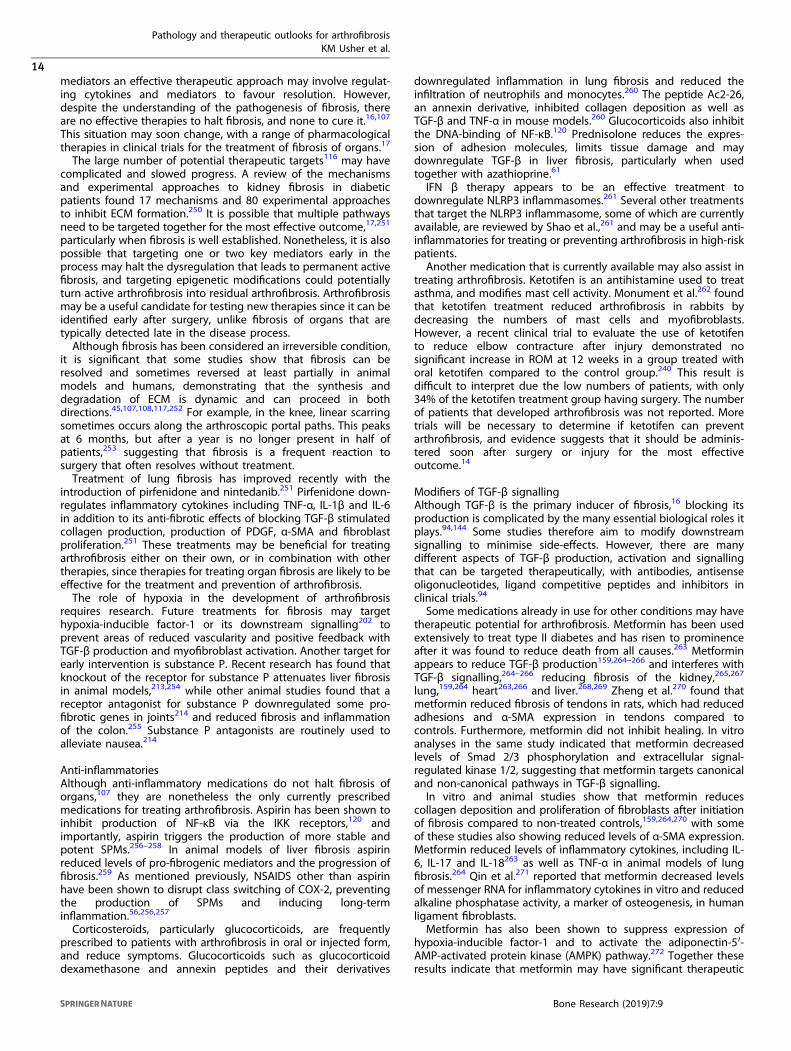
mediators an effective therapeutic approach may involve regulat-ing cytokines and mediators to favour resolution. However,despite the understanding of the pathogenesis of fibrosis, thereare no effective therapies to halt fibrosis, and none to cure it.16,107
This situation may soon change, with a range of pharmacologicaltherapies in clinical trials for the treatment of fibrosis of organs.17
The large number of potential therapeutic targets116 may havecomplicated and slowed progress. A review of the mechanismsand experimental approaches to kidney fibrosis in diabeticpatients found 17 mechanisms and 80 experimental approachesto inhibit ECM formation.250 It is possible that multiple pathwaysneed to be targeted together for the most effective outcome,17,251
particularly when fibrosis is well established. Nonetheless, it is alsopossible that targeting one or two key mediators early in theprocess may halt the dysregulation that leads to permanent activefibrosis, and targeting epigenetic modifications could potentiallyturn active arthrofibrosis into residual arthrofibrosis. Arthrofibrosismay be a useful candidate for testing new therapies since it can beidentified early after surgery, unlike fibrosis of organs that aretypically detected late in the disease process.Although fibrosis has been considered an irreversible condition,
it is significant that some studies show that fibrosis can beresolved and sometimes reversed at least partially in animalmodels and humans, demonstrating that the synthesis anddegradation of ECM is dynamic and can proceed in bothdirections.45,107,108,117,252 For example, in the knee, linear scarringsometimes occurs along the arthroscopic portal paths. This peaksat 6 months, but after a year is no longer present in half ofpatients,253 suggesting that fibrosis is a frequent reaction tosurgery that often resolves without treatment.Treatment of lung fibrosis has improved recently with the
introduction of pirfenidone and nintedanib.251 Pirfenidone down-regulates inflammatory cytokines including TNF-α, IL-1β and IL-6in addition to its anti-fibrotic effects of blocking TGF-β stimulatedcollagen production, production of PDGF, α-SMA and fibroblastproliferation.251 These treatments may be beneficial for treatingarthrofibrosis either on their own, or in combination with othertherapies, since therapies for treating organ fibrosis are likely to beeffective for the treatment and prevention of arthrofibrosis.The role of hypoxia in the development of arthrofibrosis
requires research. Future treatments for fibrosis may targethypoxia-inducible factor-1 or its downstream signalling202 toprevent areas of reduced vascularity and positive feedback withTGF-β production and myofibroblast activation. Another target forearly intervention is substance P. Recent research has found thatknockout of the receptor for substance P attenuates liver fibrosisin animal models,213,254 while other animal studies found that areceptor antagonist for substance P downregulated some pro-fibrotic genes in joints214 and reduced fibrosis and inflammationof the colon.255 Substance P antagonists are routinely used toalleviate nausea.214
Anti-inflammatoriesAlthough anti-inflammatory medications do not halt fibrosis oforgans,107 they are nonetheless the only currently prescribedmedications for treating arthrofibrosis. Aspirin has been shown toinhibit production of NF-κB via the IKK receptors,120 andimportantly, aspirin triggers the production of more stable andpotent SPMs.256–258 In animal models of liver fibrosis aspirinreduced levels of pro-fibrogenic mediators and the progression offibrosis.259 As mentioned previously, NSAIDS other than aspirinhave been shown to disrupt class switching of COX-2, preventingthe production of SPMs and inducing long-terminflammation.56,256,257
Corticosteroids, particularly glucocorticoids, are frequentlyprescribed to patients with arthrofibrosis in oral or injected form,and reduce symptoms. Glucocorticoids such as glucocorticoiddexamethasone and annexin peptides and their derivatives
downregulated inflammation in lung fibrosis and reduced theinfiltration of neutrophils and monocytes.260 The peptide Ac2-26,an annexin derivative, inhibited collagen deposition as well asTGF-β and TNF-α in mouse models.260 Glucocorticoids also inhibitthe DNA-binding of NF-κB.120 Prednisolone reduces the expres-sion of adhesion molecules, limits tissue damage and maydownregulate TGF-β in liver fibrosis, particularly when usedtogether with azathioprine.61
IFN β therapy appears to be an effective treatment todownregulate NLRP3 inflammasomes.261 Several other treatmentsthat target the NLRP3 inflammasome, some of which are currentlyavailable, are reviewed by Shao et al.,261 and may be a useful anti-inflammatories for treating or preventing arthrofibrosis in high-riskpatients.Another medication that is currently available may also assist in
treating arthrofibrosis. Ketotifen is an antihistamine used to treatasthma, and modifies mast cell activity. Monument et al.262 foundthat ketotifen treatment reduced arthrofibrosis in rabbits bydecreasing the numbers of mast cells and myofibroblasts.However, a recent clinical trial to evaluate the use of ketotifento reduce elbow contracture after injury demonstrated nosignificant increase in ROM at 12 weeks in a group treated withoral ketotifen compared to the control group.240 This result isdifficult to interpret due the low numbers of patients, with only34% of the ketotifen treatment group having surgery. The numberof patients that developed arthrofibrosis was not reported. Moretrials will be necessary to determine if ketotifen can preventarthrofibrosis, and evidence suggests that it should be adminis-tered soon after surgery or injury for the most effectiveoutcome.14
Modifiers of TGF-β signallingAlthough TGF-β is the primary inducer of fibrosis,16 blocking itsproduction is complicated by the many essential biological roles itplays.94,144 Some studies therefore aim to modify downstreamsignalling to minimise side-effects. However, there are manydifferent aspects of TGF-β production, activation and signallingthat can be targeted therapeutically, with antibodies, antisenseoligonucleotides, ligand competitive peptides and inhibitors inclinical trials.94
Some medications already in use for other conditions may havetherapeutic potential for arthrofibrosis. Metformin has been usedextensively to treat type II diabetes and has risen to prominenceafter it was found to reduce death from all causes.263 Metforminappears to reduce TGF-β production159,264–266 and interferes withTGF-β signalling,264–266 reducing fibrosis of the kidney,265,267
lung,159,264 heart263,266 and liver.268,269 Zheng et al.270 found thatmetformin reduced fibrosis of tendons in rats, which had reducedadhesions and α-SMA expression in tendons compared tocontrols. Furthermore, metformin did not inhibit healing. In vitroanalyses in the same study indicated that metformin decreasedlevels of Smad 2/3 phosphorylation and extracellular signal-regulated kinase 1/2, suggesting that metformin targets canonicaland non-canonical pathways in TGF-β signalling.In vitro and animal studies show that metformin reduces
collagen deposition and proliferation of fibroblasts after initiationof fibrosis compared to non-treated controls,159,264,270 with someof these studies also showing reduced levels of α-SMA expression.Metformin reduced levels of inflammatory cytokines, including IL-6, IL-17 and IL-18263 as well as TNF-α in animal models of lungfibrosis.264 Qin et al.271 reported that metformin decreased levelsof messenger RNA for inflammatory cytokines in vitro and reducedalkaline phosphatase activity, a marker of osteogenesis, in humanligament fibroblasts.Metformin has also been shown to suppress expression of
hypoxia-inducible factor-1 and to activate the adiponectin-5′-AMP-activated protein kinase (AMPK) pathway.272 Together theseresults indicate that metformin may have significant therapeutic
Pathology and therapeutic outlooks for arthrofibrosisKM Usher et al.
14
Bone Research (2019) 7:9
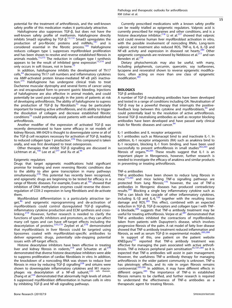
potential for the treatment of arthrofibrosis, and the well-knownsafety profile of this medication makes it particularly attractive.Halofuginone also suppresses TGF-β, but does not have the
well-known safety profile of metformin. Halofuginone directlyinhibits Smad3 signalling by TGF-β.15,114 Smad3 upregulates theproduction of pro-fibrotic proteins and miRNAs,147 and isconsidered essential in the fibrotic process.144 Halofuginonereduces collagen type I, suppresses myofibroblast proliferationand has been shown to resolve and reverse established fibrosis inanimals models.15,273 The reduction in collagen type I synthesisappears to be the result of inhibited gene expression15,274 andonly occurs in soft tissues, not in bone.15
In addition, halofuginone inhibits the development of Th17cells,44 decreasing Th17 cell numbers and inflammatory cytokinesvia AMP-activated protein kinase-mediated NF-κB p65 inactiva-tion.275 Halofuginone has undergone clinical trials to treatDuchenne muscular dystrophy and several forms of cancer usingan oral encapsulated form to prevent gastric bleeding. Injectionsof halofuginone are also effective in animal models, and couldpotentially be used post-surgically in the joints of patients at riskof developing arthrofibrosis. The ability of halofuginone to supressthe production of TGF-β by fibroblasts15 may be particularlyimportant for treating active arthrofibrosis, and its ability to triggerthe dissolution of collagen and decrease established fibroticconditions15 could potentially assist patients with well-establishedarthrofibrosis.Another modifier of the expression of activated TGF-β was
recently demonstrated to have some efficacy in rat models ofkidney fibrosis. MK-0429 is thought to downregulate some or all ofthe TGF-β cell receptors necessary for activation of TGF-β, leadingto reduced collagen type 1 production.276 This compound is takenorally, and was first developed to treat osteoporosis.Other therapies that inhibit TGF-β signalling are discussed in
Lichtman et al.,148 Lee et al.277 and Xu et al.94
Epigenetic regulatorsDrugs that target epigenetic modifications hold significantpromise for treating and even reversing fibrotic conditions dueto the ability to alter gene transcription in many pathwayssimultaneously.278 This potential has recently been recognised,and epigenetic drugs are beginning to be tested for efficacy in arange of fibrotic diseases. For example, Evans et al.195 showed thatinhibition of DNA methylation enzymes could reverse the down-regulation of COX-2 expression in lung fibroblasts and de-activatethem.Myofibroblast differentiation is a particularly attractive tar-
get,46 and epigenetic reprogramming and de-activation ofmyofibroblasts could control dysregulated TGF-β signalling,inflammatory cytokine production and ECM synthesis and cross-linking.219 However, further research is needed to clarify thefunctions of specific inhibitors and promoters, as they can affectmany cell types and can have off-target effects including thedeacetylation of proteins.220,279 Zeybel et al.278 demonstratedthat myofibroblasts in liver fibrosis could be targeted usingliposomes coated with myofibroblast-specific antibodies todeliver epigenetic drugs, potentially side-stepping potentialissues with off-target effects.Histone deacetylase inhibitors have been effective in treating
liver and kidney fibrosis in rodents,218 and Schuetze et al.279
demonstrated that diverse histone deacetylase inhibitors were ableto suppress proliferation of cardiac fibroblasts in vitro. In addition,the knockdown of a noncoding RNA was shown to reduce liverfibrosis in mice by reducing TGF-β signalling228 and sirtuins wereshown to downregulate inflammatory cytokines and M1 macro-phages via deacetylation of a NF-κB subunit,220 refs therein.Zhang et al.224 demonstrated that abnormally high levels of sirtuin6 suppressed myofibroblast differentiation in human cells in vitroby inhibiting TGF-β and NF-κB signalling pathways.
Currently prescribed medications with a known safety profilecan be readily trialled as epigenetic regulators. Valproic acid iscurrently prescribed for migraines and other conditions, and is ahistone deacetylase inhibitor.218 Li et al.227 showed that valproicacid could reverse human liver myofibroblast activation in vitro,with the possible involvement of noncoding RNAs. Long-termvalproic acid treatment also reduced ROS, TNF-α, IL-6, IL-1β andNF-κB activity and expression in diseased rat hearts.280 Otherepigenetic compounds are reviewed by Nebbioso et al.217 and vanBeneden et al.218
Dietary phytochemicals may also be useful, with many,including polyphenols, curcumin, quercetin, soy isoflavones,lycopene and resveratrol shown to reverse epigenetic modifica-tions, often acting on more than one class of epigeneticmodification.281
BIOLOGICSTGF-β antibodiesA number of TGF-β neutralising antibodies have been developedand tested in a range of conditions including OA. Neutralisation ofTGF-β may be a powerful therapy that interrupts the positive-feedback loop between this cytokine and myofibroblasts,94 andcould potentially lead to the resolution of active arthrofibrosis.Several TGF-β neutralising antibodies as well as receptor blockingantibodies have been developed and have passed early clinicaltrials for fibrotic diseases and cancer.94
IL-1 antibodies and IL receptor antagonistsIL-1 antibodies such as Rilonacept bind to and inactivate IL-1. Inaddition, IL-1 receptor antagonists (RA) such as anakinra bind toIL-1 receptors, blocking IL-1 from binding, and have been usedsuccessfully to prevent arthrofibrosis in small studies282,283 andfibrosis of organs.98,284 These results suggest that IL-1 is animportant player in fibrogenesis, however, further research isneeded to investigate the efficacy of anakinra and similar productsin preventing or treating arthrofibrosis.
TNF-α antibodiesTNF-α antibodies have been shown to reduce lung fibrosis inmice173,285 and mice lacking TNF-α signalling pathways areprotected from lung fibrosis;170 however, the use of TNF-αantibodies in fibrogenic diseases has produced contradictoryresults.285 Blocking a single key inflammatory cytokine such asTNF-α can block the cascade of other inflammatory cytokines,including IL-1β and IL-6,158 together with the resulting tissuedamage and ROS.285 This effect, combined with an expectedreduction in TGF-β, TGF-β receptors and collagen type I with TNF-α blockade,286 suggests that TNF-α antibody treatment may beuseful for treating arthrofibrosis. Verjee et al.287 demonstrated thatTNF-α antibodies inhibited the contractions of myofibroblaststaken from patients with Dupuytren’s disease, which involvesprogressive fibrosis of the palm. A rat model of fatty liver diseaseshowed that TNF-α antibody treatment reduced inflammation andfibrosis, as well as serum TGF-β in experimental models.285,288
In support of this, one patient on the patient websiteKNEEguru241 reported that TNF-α antibody treatment waseffective for managing the pain associated with active arthrofi-brosis. TNF-α induces peripheral pain sensitisation56,187,289 so it isexpected that TNF-α antibodies will assist in pain management.However, the usefulness TNF-α antibody therapy for managingarthrofibrosis in the wider patient community is unknown. TNF-αhas pleiotropic effects, and its role in organ fibrosis remainscontroversial.285,286 In addition, it may have different effects indifferent organs.285 The importance of TNF-α in establishedfibrosis remains to be clarified, and further research is requiredto understand the effectiveness of TNF-α antibodies as atherapeutic agent for treating fibrosis.
Pathology and therapeutic outlooks for arthrofibrosisKM Usher et al.
15
Bone Research (2019) 7:9

For a review of potential pharmacological therapies to treatfibrosis see Nanthakumar et al.17
MESENCHYMAL STEM CELLSMesenchymal stem cells (MSCs) are able to home in on injuredtissue and differentiate into different tissue types.290 Theymodulate the immune system by altering the activation andproliferation of immune cells, and are being tested in clinical trialsfor the treatment of lung fibrosis.174,291,292
Some studies have reported positive results using MSCs to treatfibrosis of organs in mice, however, MSC treatment remainscontroversial. Bone marrow-derived MSCs transform into myofi-broblasts in rats293 and organ-resident MSC-like cells have beenshown by genetic lineage tracing to transform into myofibroblastsand contribute to fibrosis progression in mice.107 Mice with liverfibrosis treated with bone marrow-derived human MSCs showedthat some of the donated cells appeared to differentiate intomyofibroblasts in the liver.294,295 It is not known how closely thesecells resemble the MSC lineages used in in vitro studies.Nonetheless, the few studies of MSCs in humans to date havenot demonstrated worsening fibrosis.296,297
It is known that MSCs can contribute to the growth oftumours,290 but a recent small phase I clinical trial of MSCs inthe treatment of pulmonary fibrosis reported no adverse safetyoutcomes.292 Different preparations of MSCs differ in their efficacy,perhaps because of variation in their expression of anti-inflammatory genes.298 The age and origin of MSCs may affectthe outcome of fibrosis therapy, as MSCs from different lineagesexpress different proteins that affect their therapeutic potential.299
MSCs from the IFP of patients with OA appear to inhibit theproduction of inflammatory cytokines in vitro.300
However, MSCs from the IFP are also capable of differentiatinginto fibroblasts25 in a similar wasy to MSCs from bone marrow.113
Furthermore, MSCs from the synovial membrane are positive forthe TGF-β receptor CD 105,299 raising the possibility that thesecells may induce fibrosis under inflammatory conditions. Indeed,TGF-β is understood to be a key factor that recruits MSCs todamaged tissue, and the demonstrated differentiation of MSCsinto myofibroblasts94 suggests that MSCs are often involved in thepathology of fibrosis.
DIETA number of dietary additions may assist those about to undergosurgery to avoid arthrofibrosis, or reduce symptoms in those withan existing condition, although data is lacking. A diet rich inomega 3 fatty acids is recommended for inflammatory conditions(see above in Resolvins). Capsaicin (found in chilli and peppers)and sulphoraphane (found in cruciferous vegetables) have beendemonstrated to reverse differentiation of myofibroblasts in vivo.Sulphoraphane has an anti-fibrosis effect via the activation ofnuclear factor erythroid 2-related 2, which may be important inpreventing fibroblast differentiation.301 A diet high in resistantfibre is also likely to be beneficial for preventing inflammation andfibrosis. Gut bacteria produce short-chain fatty acids from non-digestible dietary fibre, which counter inflammation and suppressthe cleavage of protease caspase-1 and secretion of IL-18.302
In addition, consumption of soy products may help reduce thelevels of inflammatory cytokines. The breakdown products of soyisoflavones, daidzein and genistein, are known to be antioxidantand anti-inflammatory compounds, but their usefulness in treatingarthrofibrosis has had only limited testing in non-human animals.Liu et al.303 found that in rabbits the topical application ofdaidzein to exposed tissue during surgery reduced fibroblastdensity, collagen formation and adhesions. Daidzein reduced ROSand levels of TGF-β and when given subcutaneously to rats,reducing lung fibrosis.150
Some of the dietary compounds mentioned above also changeepigenetic modifications, and were recently reviewed by Khanet al.281
Vitamin D is required for immune system homoeostasis,reducing TGF-β, suppressing the Th17 profile, and supportingregulatory T cells that suppress autoreactive T cells.304 Vitamin Ddeficiency is correlated with fibrosis of the liver and vitamin D3inhibits the production of collagen type 1 in the liver305 and in thelung.306 This suggests that this important vitamin may be a usefulanti-fibrotic agent.103
Fibrosis may be promoted by a reduced dietary intake ofpotassium (K+) and low serum K+ levels were associated with liverfibrosis.307 K+ efflux from cells can result from cellular damage andthe release of adenosine triphosphate.308 Upregulation of theintermediate/small-conductance Ca2+-activated K+ channel altersthe membrane potential of cells and promotes fibrogenesis, witheffects including higher levels of Ca2+ entry into cells andexpression of Ca2+-dependant growth factor genes, cyclins andkinases involved in cell division.309 High-intracellular levels of Ca2+
are associated with cardiac fibrosis202and KCa3.1 silencing inanimal models of renal fibrosis decreased the numbers ofmyofibroblasts and attenuated the development of fibrosis.310
This result appears to be mediated via the Smad2/3 pathway,since KCa3.1 blockade reduced levels of TGF-β1, and TGF-β1receptor II.311
The loss of intracellular K+ also activates NLRP3 inflammasomes,which are known to promote fibrosis. Sun et al. found that lowlevels of K+ promotes vascular calcification and osteogenicdifferentiation.312 This may have implications for the calcificationthat sometimes occurs within ECM in arthrofibrosis.Intermittent fasting has profound positive effects on many
health measures and can improve functional outcomes for manydiseases.313 Animal research shows that intermittent fasting isprotective against fibrosis of organs,314 and it is also knownsuppress inflammation313,315 and downregulate the inflammatorycytokines IL-1, IL-6 and TNF-α in humans.316 Intermittent fastingsuppresses the activity of NLRP3 inflammasomes,313 providingwhat appears to be a cumulative anti-fibrotic effect. Intermittentfasting can be approached in a number of ways, includingrestricting calorie intake 2 days of the week, restricting food intaketo 8 h or less per day, and periods of 1 or 2 days in which there islittle or no calorie intake on a recurring basis.313 Nutrient depletionprior to surgery can protect against damage from ischaemicconditions.317 Intermittent fasting is also known to reduce levelsof insulin-like growth factor-1, which promotes the survival ofmyofibroblasts in liver fibrosis.252
Intermittent fasting may also trigger the amino acid limitationresponse, which alters immune function by regulating T- and B-cell proliferation, activation and differentiation.318 The amino acidL-proline is required for fibrosis, as it is a necessary pre-curser ofcollagen.319 L-proline production can be upregulated by argininemetabolism, resulting in macrophage switching and promotingTh2 cells and fibrosis.319
OTHER TREATMENTSRecent research suggests that injections of collagenase, aproteolytic bacterial enzyme that specifically breaks downcollagen, can increase ROM in arthrofibrosis of the shoulder320,321
and in animal models of arthrofibrosis of the knee, however,further research is needed to address concerns relating todegradation of articular cartilage, ligaments and tendons.322 Inaddition, the signalling effects of collagen fragments created bycleavage also needs to be considered, as some of these fragmenthave biological activity.104 However, collagenase has beenapproved for Dupuytren disease, a fibroproliferative disease ofthe palm.323 Collagenase in slow release nanocapsules have beendeveloped and tested in an animal model of skin fibrosis, showing
Pathology and therapeutic outlooks for arthrofibrosisKM Usher et al.
16
Bone Research (2019) 7:9

sustained release over 10 days.324 This could lower the number ofdoses required.Soft tissue mobilisation techniques using tools designed to
exert shear force, break down ECM and stimulate blood flow havebeen developed, and have several trademark names includingASTYM and the Graston Technique. These tools can be applied tothe joint and may assist recovery from arthrofibrosis32,325,326 andtendon damage,327 particularly before the ECM matures. Somepatients on the website KNEEguru report good results,241
however, there are few large well-controlled trials, and moreresearch is needed to determine how effective these methodsare.326
Interventions that increase tissue vascularisation may slow theprogression of fibrotic processes.13 A small study involving threepatients undergoing revision TKR for arthrofibrosis suggested thatlow dose irradiation prior to surgery may result in improvedROM.30
PATIENT PERSPECTIVESThe experiences of arthrofibrosis patients are seldom heard in thescientific literature, and can provide useful insights into thecondition. Arthrofibrosis patients on the website KNEEguru241
show that there are a group of patients with active arthrofibrosis,for whom surgical intervention to lyse ECM has proven to bedetrimental. Another group may have had initially detrimentalresults from surgery, however, subsequent surgery led toimportant improvements in symptoms. The reasons for thesesignificantly different outcomes are not clear, but may include thelevel of inflammation, the time between surgeries, surgicaltechnique, rehabilitation protocols and individual predispositionthrough genetic or other factors. Unfortunately, as with researchpublications, these forums are largely silent on the long-termoutcomes for patients with permanent active arthrofibrosis.Many patients believe that overly aggressive exercise rehabilita-
tion soon after surgery was detrimental to their recovery, andstrongly advocate that arthrofibrosis sufferers “listen to theirknee”, and do not push too hard to fit in with exercise regimesand expected recovery timetables. As mentioned previously, thisfits with views expressed recently that rehabilitation should beprogressed conservatively, and based on the inflammatoryresponse it provokes.12,328
One patient provides a useful case study into the causes andtypes of arthrofibrosis. Following bilateral TKR a 45-year-oldfemale immediately began intensive post-operative use of CPMin addition to exercise rehabilitation. Initially the right knee hadrestricted ROM of around 70°. After several months of intensivedaily CPM use the knee regained a functional amount of flexionand CPM use was discontinued. The ROM on the left knee wasinitially 105°, but at 2 weeks post-surgery a minor forced bendingon a CPM lacking digital control likely caused internal bleeding,and the knee immediately lost ROM, with maximum flexion fallingto 70°. Intensive CPM use gradually increased flexion, however, theknee remained painful and intensive CPM use was required tomaintain functional ROM.After 5 and half months the arthrofibrosis resolved, and the
knee became fully functional without pain and with stable activeflexion of about 110°. However, 4 weeks after full resolution ofarthrofibrosis the patient became ill with suspected influenza.Active arthrofibrosis spontaneously and permanently returnedin the left knee, with pain and difficulty maintaining active flexionof 80°.Several factors are significant in this history. Firstly, the patient
has one knee with residual arthrofibrosis (resolved but stiff), whilethe other knee has active arthrofibrosis, triggered initially byinternal bleeding shortly after surgery. Vascular damage is knownto promote fibroblast activation.329 Secondly, after the resolutionof early symptoms influenza appears to have triggered the
permanent return of active arthrofibrosis. Many of the inflamma-tory cytokines that are produced in response to influenza,particularly IL-1, IL-6 and TNF-α330,331 are known to cause thedifferentiation of fibroblasts into myofibroblasts103 and areimportant cytokines in arthrofibrosis (see above). In addition,influenza directly activates NLRP3 inflammasomes,332,333 whichare implicated in fibrosis. Influenza also activates biologicallyinactive TGF-β,146,331 the primary driver of fibrosis. This suggeststhat this virus is a significant risk factor for arthrofibrosis, with therisk potentially higher for post-operative patients and those withresidual fibrosis.
CONCLUSION AND FUTURE DIRECTIONSArthrofibrosis is a fibrotic disease caused by excessive myofibro-blast proliferation with defective apoptosis, primarily induced bydysregulated TGF-β signalling. Once established these factors andothers, including extensive collagen cross-linking, create acomplex web of positive feedback processes that establish anew pathological homoeostasis that maintains excessive ECMaccumulation and low-grade inflammation. These processes areinitiated by chronic low-grade or acute inflammatory conditions orevents.Surgical lysis and removal of ECM, and MUA, remain the primary
treatments for arthrofibrosis. The surgical removal of ECM can bebeneficial, not only because it removes the physical stress andrestriction to ROM but also because it removes pro-fibroticmediators bound to the ECM that can fuel the cycle of ECMformation. However, the potential benefits of surgery must beweighed against the risks, which include increased dysregulationof fibrogenesis in response to the surgical injury. This can result inthe rapid return of arthrofibrosis with increased severity ofsymptoms. MUA also carries significant risks such as fracturedbones, torn tendons and damaged prostheses and can increasesymptom severity.The development of a minimally invasive diagnostic tool kit that
assesses the levels of cytokines, collagen fragments and othermediators of fibrosis in serum and synovial fluid may help topredict which patients are at greatest risk of post-operativearthrofibrosis. This research could provide relatively fast resultsthat may help prevent permanent disability for thousands.Patients found to be at risk could receive anti-fibrotic therapiesand intensive monitoring by a rheumatologist to controlinflammation.Early intervention to prevent fibrosis is likely to be important,14
halting the process before extensive epigenetic modificationsoccur and a significant amount of ECM has formed and becomestrongly cross-linked. This could potentially stop the pathologicalcascade of dysregulation and positive feedback that results inpermanent active fibrosis,214 while also preventing damage tojoint structures such as cartilage and ligaments that occurs withtissue contraction and altered biomechanics.Future research should investigate the mechanism of potentia-
tion (memory) from previous surgeries and injuries. Elucidation ofthis mechanism may indicate the best timing and mostappropriate treatment targets to prevent post-operative arthrofi-brosis. Anti-fibrotic coatings on surgical implants may prove usefulin preventing arthrofibrosis.248 In addition, the lack of an effectivetherapeutic agent to halt or reverse fibrosis after it developsremains an issue of enormous importance for arthrofibrosis andfibrosis of organs. Recent research shows that it is possible toreverse fibrosis but it is still not understood how this occurs.Until these research priorities are addressed risk factors such as
many previous surgeries, pre-existing stiffness or inflammation,early onset OA, childhood adversity and female gender should beconsidered prior to surgery. In addition, a conservative approachto rehabilitation exercises is recommended with adjustmentsmade according to how the joint responds. Aspirin provides a
Pathology and therapeutic outlooks for arthrofibrosisKM Usher et al.
17
Bone Research (2019) 7:9
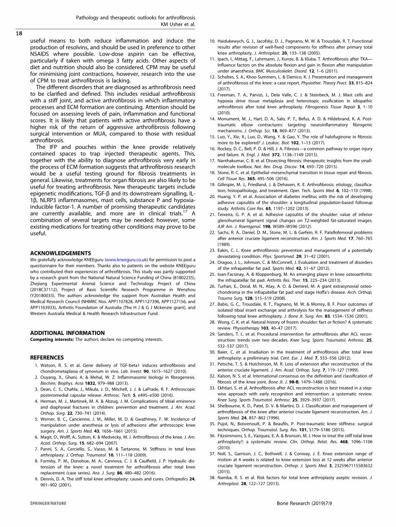
useful means to both reduce inflammation and induce theproduction of resolvins, and should be used in preference to otherNSAIDS where possible. Low-dose aspirin can be effective,particularly if taken with omega 3 fatty acids. Other aspects ofdiet and nutrition should also be considered. CPM may be usefulfor minimising joint contractions, however, research into the useof CPM to treat arthrofibrosis is lacking.The different disorders that are diagnosed as arthrofibrosis need
to be clarified and defined. This includes residual arthrofibrosiswith a stiff joint, and active arthrofibrosis in which inflammatoryprocesses and ECM formation are continuing. Attention should befocused on assessing levels of pain, inflammation and functionalscores. It is likely that patients with active arthrofibrosis have ahigher risk of the return of aggressive arthrofibrosis followingsurgical intervention or MUA, compared to those with residualarthrofibrosis.The IFP and pouches within the knee provide relatively
contained spaces to trap injected therapeutic agents. This,together with the ability to diagnose arthrofibrosis very early inthe process of ECM formation suggests that arthrofibrosis researchwould be a useful testing ground for fibrosis treatments ingeneral. Likewise, treatments for organ fibrosis are also likely to beuseful for treating arthrofibrosis. New therapeutic targets includeepigenetic modifications, TGF-β and its downstream signalling, IL-1β, NLRP3 inflammasomes, mast cells, substance P and hypoxia-inducible factor-1. A number of promising therapeutic candidatesare currently available, and more are in clinical trials.17 Acombination of several targets may be needed; however, someexisting medications for treating other conditions may prove to beuseful.
ACKNOWLEDGEMENTSWe gratefully acknowledge KNEEguru (www.kneeguru.co.uk) for permission to post aquestionnaire for their members. Thanks also to patients on the website KNEEguruwho contributed their experiences of arthrofibrosis. This study was partly supportedby a research grant from the National Natural Science Funding of China (81802235),Zhejiang Experimental Animal Science and Technology Project of China(2018C37112), Project of Basic Scientific Research Programme in Wenzhou(Y20180033). The authors acknowledge the support from Australian Health andMedical Research Council (NHMRC Nos. APP1107828, APP1127396, APP1127156, andAPP1163933), Arthritis Foundation of Australia (The H J & G J Mckenzie grant), andWestern Australia Medical & Health Research Infrastructure Fund.
ADDITIONAL INFORMATIONCompeting interests: The authors declare no competing interests.
REFERENCES1. Watson, R. S. et al. Gene delivery of TGF-beta1 induces arthrofibrosis and
chondrometaplasia of synovium in vivo. Lab. Invest. 90, 1615–1627 (2010).2. Ouyang, X., Ghani, A. & Mehal, W. Z. Inflammasome biology in fibrogenesis.
Biochim. Biophys. Acta 1832, 979–988 (2013).3. Dean, C. S., Chahla, J., Mikula, J. D., Mitchell, J. J. & LaPrade, R. F. Arthroscopic
posteromedial capsular release. Arthrosc. Tech. 5, e495–e500 (2016).4. Herman, M. J., Martinek, M. A. & Abzug, J. M. Complications of tibial eminence
and diaphyseal fractures in children: prevention and treatment. J. Am. Acad.Orthop. Surg. 22, 730–741 (2014).
5. Werner, B. C., Cancienne, J. M., Miller, M. D. & Gwathmey, F. W. Incidence ofmanipulation under anesthesia or lysis of adhesions after arthroscopic kneesurgery. Am. J. Sports Med. 43, 1656–1661 (2015).
6. Magit, D., Wolff, A., Sutton, K. & Medvecky, M. J. Arthrofibrosis of the knee. J. Am.Acad. Orthop. Surg. 15, 682–694 (2007).
7. Panni, S. A., Cerciello, S., Vasso, M. & Tartarone, M. Stiffness in total kneearthroplasty. J. Orthop. Traumatol. 10, 111–118 (2009).
8. Formby, P. M., Donohue, M. A., Cannova, C. J. & Caulfield, J. P. Hydraulic dis-tension of the knee: a novel treatment for arthrofibrosis after total kneereplacement (case series). Anz. J. Surg. 86, 480–482 (2016).
9. Dennis, D. A. The stiff total knee arthroplasty: causes and cures. Orthopedics 24,901–902 (2001).
10. Haidukewych, G. J., Jacofsky, D. J., Pagnano, M. W. & Trousdale, R. T. Functionalresults after revision of well-fixed components for stiffness after primary totalknee arthroplasty. J. Arthroplast. 20, 133–138 (2005).
11. Ipach, I., Mittag, F., Lahrmann, J., Kunze, B. & Kluba, T. Arthrofibrosis after TKA—Influence factors on the absolute flexion and gain in flexion after manipulationunder anaesthesia. BMC Musculoskelet. Disord. 12, 1–6 (2011).
12. Scholtes, S. A., Khoo-Summers, L. & Damico, K. J. Presentation and managementof arthrofibrosis of the knee: a case report. Physiother. Theory Pract. 33, 815–824(2017).
13. Freeman, T. A., Parvizi, J., Dela Valle, C. J. & Steinbeck, M. J. Mast cells andhypoxia drive tissue metaplasia and heterotopic ossification in idiopathicarthrofibrosis after total knee arthroplasty. Fibrogenesis Tissue Repair 3, 1–10(2010).
14. Monument, M. J., Hart, D. A., Salo, P. T., Befus, A. D. & Hildebrand, K. A. Post-traumatic elbow contractures: targeting neuroinflammatory fibrogenicmechanisms. J. Orthop. Sci. 18, 869–877 (2013).
15. Luo, Y., Xie, X., Luo, D., Wang, Y. & Gao, Y. The role of halofuginone in fibrosis:more to be explored? J. Leukoc. Biol. 102, 1–13 (2017).
16. Rockey, D. C., Bell, P. D. & Hill, J. A. Fibrosis—a common pathway to organ injuryand failure. N. Engl. J. Med. 372, 1138–1149 (2015).
17. Nanthakumar, C. B. et al. Dissecting fibrosis: therapeutic insights from the small-molecule toolbox. Nat. Rev. Drug. Discov. 14, 693–720 (2015).
18. Stone, R. C. et al. Epithelial–mesenchymal transition in tissue repair and fibrosis.Cell Tissue Res. 365, 495–506 (2016).
19. Gillespie, M. J., Friedland, J. & Dehaven, K. E. Arthrofibrosis: etiology, classifica-tion, histopathology, and treatment. Oper. Tech. Sports Med. 6, 102–110 (1998).
20. Huang, Y. P. et al. Association of diabetes mellitus with the risk of developingadhesive capsulitis of the shoulder: a longitudinal population-based followupstudy. Arthritis Care Res. 65, 1197–1202 (2013).
21. Teixeira, G. P. A. et al. Adhesive capsulitis of the shoulder: value of inferiorglenohumeral ligament signal changes on T2-weighted fat-saturated images.AJR Am. J. Roentgenol. 198, W589–W596 (2012).
22. Sachs, R. A., Daniel, D. M., Stone, M. L. & Garfein, R. F. Patellofemoral problemsafter anterior cruciate ligament reconstruction. Am. J. Sports Med. 17, 760–765(1989).
23. Eakin, C. L. Knee arthrofibrosis: prevention and management of a potentiallydevastating condition. Phys. Sportsmed. 29, 31–42 (2001).
24. Dragoo, J. L., Johnson, C. & McConnell, J. Evaluation and treatment of disordersof the infrapatellar fat pad. Sports Med. 42, 51–67 (2012).
25. Ioan-Facsinay, A. & Kloppenburg, M. An emerging player in knee osteoarthritis:the infrapatellar fat pad. Arthritis Res. Ther. 15, 225–234 (2013).
26. Turhan, E., Doral, M. N., Atay, A. O. & Demirel, M. A giant extrasynovial osteo-chondroma in the infrapatellar fat pad: end stage Hoffa’s disease. Arch. Orthop.Trauma Surg. 128, 515–519 (2008).
27. Babis, G. C., Trousdale, R. T., Pagnano, M. W. & Morrey, B. F. Poor outcomes ofisolated tibial insert exchange and arthrolysis for the management of stiffnessfollowing total knee arthroplasty. J. Bone Jt. Surg. Am. 83, 1534–1536 (2001).
28. Wong, C. K. et al. Natural history of frozen shoulder: fact or fiction? A systematicreview. Physiotherapy 103, 40–47 (2017).
29. Sanders, T. L. et al. Procedural intervention for arthrofibrosis after ACL recon-struction: trends over two decades. Knee Surg. Sports Traumatol. Arthrosc. 25,532–537 (2017).
30. Baier, C. et al. Irradiation in the treatment of arthrofibrosis after total kneearthroplasty: a preliminary trial. Cent. Eur. J. Med. 7, 553–556 (2012).
31. Petsche, T. S. & Hutchinson, M. R. Loss of extension after reconstruction of theanterior cruciate ligament. J. Am. Acad. Orthop. Surg. 7, 119–127 (1999).
32. Kalson, N. S. et al. International consensus on the definition and classification offibrosis of the knee joint. Bone Jt. J. 98-B, 1479–1488 (2016).
33. Ekhtiari, S. et al. Arthrofibrosis after ACL reconstruction is best treated in a step-wise approach with early recognition and intervention: a systematic review.Knee Surg. Sports Traumatol. Arthrosc. 25, 3929–3937 (2017).
34. Shelbourne, K. D., Patel, D. V. & Martini, D. J. Classification and management ofarthrofibrosis of the knee after anterior cruciate ligament reconstruction. Am. J.Sports Med. 24, 857–862 (1996).
35. Pujol, N., Boisrenoult, P. & Beaufils, P. Post-traumatic knee stiffness: surgicaltechniques. Orthop. Traumatol. Surg. Res. 101, S179–S186 (2015).
36. Fitzsimmons, S. E., Vazquez, E. A. & Bronson, M. J. How to treat the stiff total kneearthroplasty?: a systematic review. Clin. Orthop. Relat. Res. 468, 1096–1106(2010).
37. Noll, S., Garrison, J. C., Bothwell, J. & Conway, J. E. Knee extension range ofmotion at 4 weeks is related to knee extension loss at 12 weeks after anteriorcruciate ligament reconstruction. Orthop. J. Sports Med. 3, 2325967115583632(2015).
38. Namba, R. S. et al. Risk factors for total knee arthroplasty aseptic revision. J.Arthroplast. 28, 122–127 (2013).
Pathology and therapeutic outlooks for arthrofibrosisKM Usher et al.
18
Bone Research (2019) 7:9

39. Ranawat, C. S., Ranawat, A. S. & Mehta, A. Total knee arthroplasty rehabilitationprotocol: whatmakes the difference? J. Arthroplast. 18, 27–30 (2003).
40. Abdul, N. et al. Fibrosis is a common outcome following total knee arthroplasty.Sci. Rep. 5, 16469 (2015).
41. Tjoumakaris, F. P. et al. Arthroscopic lysis of adhesions for the stiff total knee:results after failed manipulation. Orthopedics 37, e482–e487 (2014).
42. Daluga, D., Lombardi, A. V., Mallory, T. H. & Vaughn, B. K. Knee manipulationfollowing total knee arthroplasty. J. Arthroplast. 6, 119–128 (1991).
43. Remst, D. F., Blaney Davidson, E. N. & van der Kraan, P. M. Unravellingosteoarthritis-related synovial fibrosis: a step closer to solving joint stiffness.Rheumatology 54, 1954–1963 (2015).
44. Pines, M. Halofuginone for fibrosis, regeneration and cancer in the gastro-intestinal tract. World J. Gastroenterol. 20, 14778–14786 (2014).
45. Iredale, J. P., Thompson, A. & Henderson, N. C. Extracellular matrix degradationin liver fibrosis: biochemistry and regulation. Biochim. Biophys. Acta 1832,876–883 (2013).
46. Wynn, T. A. & Ramalingam, T. R. Mechanisms of fibrosis: therapeutic translationfor fibrotic disease. Nat. Med. 18, 1028–1040 (2012).
47. Kurosaka, M., Yoshiya, S., Mizuno, K. & Yamamoto, T. Maximizing flexion aftertotal knee arthroplasty: the need and the pitfalls. J. Arthroplast. 17, 59–62 (2002).
48. Cheuy, V. A. et al. Arthrofibrosis associated with total knee arthroplasty. J.Arthroplast. 32, 2604–2611 (2017).
49. Millet, P. J., Johnson, B., Carlson, J., Krishnan, S. & Steadman, J. R. Rehabilitationof the arthrofibrotic knee. Am. J. Orthop. 32, 531–538 (2003).
50. Heinemeier, K., Langberg, H., Olesen, J. L. & Kjaer, M. Role of TGF-β1 in relation toexercise-induced type I collagen synthesis in human tendinous tissue. J. Appl.Physiol. 95, 2390–2397 (2003).
51. Mackey, A. L. et al. Sequenced response of extracellular matrix deadhesion andfibrotic regulators after muscle damage is involved in protection against futureinjury in human skeletal muscle. FASEB J. 25, 1943–1959 (2011).
52. Kim, D. H., Gill, T. J. & Millett, P. J. Arthroscopic treatment of the arthrofibroticknee. Arthroscopy 20, 187–194 (2004).
53. Pan, L.-H., Ohtani, H., Yamauchi, K. & Nagura, H. Co-expression of TNFα and IL-1βin human acute pulmonary fibrotic diseases: an immunohistochemical analysis.Pathol. Int. 46, 91–99 (1996).
54. Rho, R. H., Brewer, R. P., Lamer, T. J. & Wilson, P. R. Complex regional painsyndrome. Mayo Clin. Proc. 77, 174–180 (2002).
55. Borchers, A. T. & Gershwin, M. E. Complex regional pain syndrome: a compre-hensive and critical review. Autoimmun. Rev. 13, 242–265 (2014).
56. Ji, R.-R., Xu, Z.-Z., Strichartz, G. & Serhan, C. N. Emerging roles of resolvins in theresolution of inflammation and pain. Trends Neurosci. 34, 599–609 (2011).
57. Koeck, F. X. et al. Predominance of synovial sensory nerve fibers in arthrofibrosisfollowing total knee arthroplasty compared to osteoarthritis of the knee. J.Orthop. Surg. Res. 11, 1–8 (2016).
58. Solbak, N. M. et al. Alterations in Hoffa’s fat pad induced by an inflammatoryresponse following idealized anterior cruciate ligament surgery. Inflamm. Res.64, 615–626 (2015).
59. Lee, J. S., Shin, J. H. & Choi, B. S. Serum levels of IL-8 and ICAM-1 as biomarkersfor progressive massive fibrosis in coal workers’ pneumoconiosis. J. Korean Med.Sci. 30, 140–144 (2015).
60. Kisseleva, T. et al. Myofibroblasts revert to an inactive phenotype duringregression of liver fibrosis. Proc. Natl Acad. Sci. USA 109, 9448–9453 (2012).
61. Czaja, A. J. Hepatic inflammation and progressive liver fibrosis in chronic liverdisease. World J. Gastroenterol. 20, 2515–2532 (2014).
62. Wynn, T. A. Fibrotic disease and the T(H)1/T(H)2 paradigm. Nat. Rev. Immunol. 4,583–594 (2004).
63. White, D., Choi, H., Peloquin, C., Zhu, Y. & Zhang, Y. Secular trend of adhesivecapsulitis. Arthritis Care Res. 63, 1571–1575 (2011).
64. Nwachukwu, B. U. et al. Arthrofibrosis after anterior cruciate ligament recon-struction in children and adolescents. J. Pediatr. Orthop. 31, 811–817 (2011).
65. Hemsley, K. Neuromuscular and Psychological Influences on Range of MotionRecovery in Anterior Cruciate Ligament Reconstruction Rehabilitation Patients.3293223 thesis (Temple University, Ann Arbor, USA, 2007).
66. Klein, S. L. & Flanagan, K. L. Sex differences in immune responses. Nat. Rev.Immunol. 16, 626–638 (2016).
67. Snelling, S. J. et al. Presence of IL-17 in synovial fluid identifies a potentialinflammatory osteoarthritic phenotype. PLoS ONE 12, 1–13 (2017).
68. Bourne, R. B., Maloney, W. J. & Wright, J. G. An AOA critical issue the outcome ofthe outcomes movement. J. Bone Jt. Surg. Am. 86, 633–640 (2004).
69. Yeranosian, M. G., Petrigliano, F. A., Terrell, R. D., Wang, J. C. & McAllister, D. R.Incidence of postoperative infections requiring reoperation after arthroscopicknee surgery. Arthroscopy 29, 1355–1361 (2013).
70. Roved, J., Westerdahl, H. & Hasselquist, D. Sex differences in immune responses:hormonal effects, antagonistic selection, and evolutionary consequences. Horm.Behav. 88, 95–105 (2017).
71. Maurya, V. K. et al. Transforming growth factor-beta 1 (TGF-B1) liberation fromits latent complex during embryo implantation and its regulation by estradiol inmouse. Biol. Reprod. 89, 1–17 (2013).
72. Sanjabi, S., Oh, S. A. & Li, M. O. Regulation of the immune response by TGF-beta:from conception to autoimmunity and infection. Cold Spring Harb. Perspect. Biol.9, 1–33 (2017).
73. Zhang, W., Ouyang, H., Dass, C. R. & Xu, J. Current research onpharmacologic and regenerative therapies for osteoarthritis. Bone Res. 4, 1–14(2016).
74. Scanzello, C. R. et al. Local cytokine profiles in knee osteoarthritis: elevatedsynovial fluid interleukin-15 differentiates early from end-stage disease.Osteoarthr. Cartil. 17, 1040–1048 (2009).
75. Oehler, S., Neureiter, D., Meyer-Scholten, C. & Aigner, T. Subtyping of osteoar-thritic synoviopathy. Clin. Exp. Rheumatol. 20, 633–640 (2002).
76. Scanzello, C. R. & Goldring, S. R. The role of synovitis in osteoarthritis patho-genesis. Bone 51, 249–257 (2012).
77. Shen, J., Li, S. & Chen, D. TGF-beta signaling and the development of osteoar-thritis. Bone Res. 2, 1–7 (2014).
78. Zhen, G. et al. Inhibition of TGF-beta signaling in mesenchymal stemcells of subchondral bone attenuates osteoarthritis. Nat. Med. 19, 704–712(2013).
79. Finnson, K. W. et al. Endoglin differentially regulates TGF-B-induced Smad2/3and Smad1/5 signalling and its expression correlates with extracellular matrixproduction and cellular differentiation state in human chondrocytes. Osteoarthr.Cartil. 18, 1518–1527.
80. Klein-Wieringa, I. R. et al. The infrapatellar fat pad of patients with osteoarthritishas an inflammatory phenotype. Ann. Rheum. Dis. 70, 851–857 (2011).
81. Wynn, T. A. & Barron, L. Macrophages: master regulators of inflammation andfibrosis. Semin. Liver Dis. 30, 245–257 (2010).
82. Marks, P. H. & Donaldson, M. L. Inflammatory cytokine profiles associated withchondral damage in the anterior cruciate ligament-deficient knee. Arthroscopy21, 1342–1347 (2005).
83. Chen, C. Z. & Raghunath, M. Focus on collagen: in vitro systems to studyfibrogenesis and antifibrosis state of the art. Fibrogenesis Tissue Repair 2, 1–10(2009).
84. Goodman, S. B., Yao, Z., Keeney, M. & Yang, F. The future of biologic coatings fororthopaedic implants. Biomaterials 34, 3174–3183 (2013).
85. Felitti, V. J. M. D. et al. Relationship of childhood abuse and household dys-function to many of the leading causes of death in adults. Am. J. Prev. Med. 14,245–258.
86. Elwenspoek, M. M. C. et al. Proinflammatory T cell status associated with earlylife adversity. J. Immunol. 13, ji1701082 (2017).
87. Bufalino, C., Hepgul, N., Aguglia, E. & Pariante, C. M. The role of immune genes inthe association between depression and inflammation: a review of recentclinical studies. Brain Behav. Immun. 31, 31–47 (2013).
88. Raison, C. L., Capuron, L. & Miller, A. H. Cytokines sing the blues: inflammationand the pathogenesis of depression. Trends Immunol. 27, 24–31 (2006).
89. Sivakumar, P., Kitson, C. & Jarai, G. Modeling and measuring extracellular matrixalterations in fibrosis: challenges and perspectives for antifibrotic drug dis-covery. Connect. Tissue Res. 60, 62–70 (2018).
90. Trautwein, C., Friedman, S. L., Schuppan, D. & Pinzani, M. Hepatic fibrosis: con-cept to treatment. J. Hepatol. 62, S15–S24 (2015).
91. Su, E. P. & Su, S. L. The stiff total knee replacement: evaluation and treatment.Semin. Arthroplast. 24, 142–148 (2013).
92. Hakim, A. J., Cherkas, L. F., Spector, T. D. & MacGregor, A. J. Genetic associationsbetween frozen shoulder and tennis elbow: a female twin study. Rheumatology42, 739–742 (2003).
93. Skutek, M. et al. Screening for arthrofibrosis after anterior cruciate ligamentreconstruction: analysis of association with human leukocyte antigen. Arthro-scopy 20, 469–473 (2004).
94. Xu, X. et al. Transforming growth factor-beta in stem cells and tissue home-ostasis. Bone Res. 6, 1–31 (2018).
95. van de Laar, I. M. et al. Mutations in SMAD3 cause a syndromic form of aorticaneurysms and dissections with early-onset osteoarthritis. Nat. Genet. 43,121–126 (2011).
96. van der Linde, D. et al. Aneurysm-osteoarthritis syndrome with visceral and iliacartery aneurysms. J. Vasc. Surg. 57, 96–102 (2013).
97. Aref-Eshghi, E. et al. SMAD3 is associated with the total burden of radiographicosteoarthritis: the Chingford study. PLoS ONE 9, e97786 (2014).
98. Gasse, P. et al. IL-1R1/MyD88 signaling and the inflammasome are essential inpulmonary inflammation and fibrosis in mice. J. Clin. Invest. 117, 3786–3799(2007).
99. Di Vita, G. et al. Cytokines and growth factors in wound drainage fluid frompatients undergoing incisional hernia repair. Wound Repair Regen. 14, 259–264(2006).
Pathology and therapeutic outlooks for arthrofibrosisKM Usher et al.
19
Bone Research (2019) 7:9

100. Grellner, W., Georg, T. & Wilske, J. Quantitative analysis of proinflammatorycytokines (IL-1β, IL-6, TNF-α) in human skin wounds. Forensic Sci. Int. 113,251–264 (2000).
101. Liu, R. M. & Gaston Pravia, K. A. Oxidative stress and glutathione in TGF-beta-mediated fibrogenesis. Free Radic. Biol. Med. 48, 1–15 (2010).
102. Chaudhary, K. & Madaio, M. P. Amino acid limitation stress response ininflammation. Transl. Cancer Res. 5, 220–222 (2016).
103. Kendall, R. T. & Feghali-Bostwick, C. A. Fibroblasts in fibrosis: novel roles andmediators. Front. Pharmacol. 5, 1–13 (2014).
104. Ricard-Blum, S., Baffet, G. & Theret, N. Molecular and tissue alterations of col-lagens in fibrosis. Matrix Biol. 68-69, 122–149 (2018).
105. van der Slot, A. J. et al. Increased formation of pyridinoline cross-links due tohigher telopeptide lysyl hydroxylase levels is a general fibrotic phenomenon.Matrix Biol. 23, 251–257 (2004).
106. Wu, J., Chitapanarux, T., Chen, Y., Soon, R. K. Jr. & Yee, H. F. Jr. Intestinal myo-fibroblasts produce nitric oxide in response to combinatorial cytokine stimula-tion. J. Cell. Physiol. 228, 572–580 (2013).
107. El Agha, E. et al. Mesenchymal stem cells in fibrotic disease. Cell Stem Cell 21,166–177 (2017).
108. Hinz, B. et al. Recent developments in myofibroblast biology: paradigms forconnective tissue remodeling. Am. J. Pathol. 180, 1340–1355 (2012).
109. Fullerton, J. N., O’Brien, A. J. & Gilroy, D. W. Lipid mediators in immune dys-function after severe inflammation. Trends Immunol. 35, 12–21 (2014).
110. Sziksz, E. et al. Fibrosis related inflammatory mediators: role of the IL-10 cytokinefamily. Mediat. Inflamm. 2015, 1–15 (2015).
111. Monument, M. J., Hart, D. A., Salo, P. T., Befus, A. D. & Hildebrand, K. A. Neu-roinflammatory mechanisms of connective tissue fibrosis: targeting neurogenicand mast cell contributions. Adv. Wound Care 4, 137–151 (2015).
112. Pines, M. & Spector, I. Halofuginone—the multifaceted molecule. Molecules 20,573–594 (2015).
113. Hinz, B. et al. The myofibroblast: one function, multiple origins. Am. J. Pathol.170, 1807–1816 (2007).
114. Ao, M. et al. Stretching fibroblasts remodels fibronectin and alters cancer cellmigration. Sci. Rep. 5, 1–9 (2015).
115. Tomasek, J. J., Gabbiani, G., Hinz, B., Chaponnier, C. & Brown, R. A. Myofibroblastsand mechano-regulation of connective tissue remodelling. Nat. Rev. Mol. CellBiol. 3, 349–363 (2002).
116. Kolahian, S., Fernandez, I. E., Eickelberg, O. & Hartl, D. Immune mechanisms inpulmonary fibrosis. Am. J. Respir. Cell Mol. Biol. 55, 309–322 (2016).
117. Issa, R. et al. Spontaneous recovery from micronodular cirrhosis: evidence forincomplete resolution associated with matrix cross-linking. Gastroenterology126, 1795–1808 (2004).
118. Li, M. O. et al. Emergence of fibroblasts with a proinflammatory epigeneticallyaltered phenotype in severe hypoxic pulmonary hypertension. J. Immunol. 187,2711–2722 (2011).
119. Duffield, J. S. et al. Selective depletion of macrophages reveals distinct,opposing roles during liver injury and repair. J. Clin. Invest. 115, 56–65 (2005).
120. Liu, T., Zhang, L., Joo, D. & Sun, S.-C. NF-κB signaling in inflammation. SignalTransduct. Target Ther. 2, e17023 (2017).
121. Jester, J. V., Huang, J., Matthew Petroll, W. & Dwight Cavanagh, H. TGFβ inducedmyofibroblast differentiation of rabbit keratocytes requires synergistic TGFβ,PDGF and integrin signaling. Exp. Eye Res. 75, 645–657 (2002).
122. Boström, H. et al. PDGF-A signaling is a critical event in lung alveolar myofi-broblast development and alveogenesis. Cell 85, 863–873 (1996).
123. Zhao, T. et al. Platelet-derived growth factor-D promotes fibrogenesis of cardiacfibroblasts. Am. J. Physiol. Heart Circ. Physiol. 304, H1719–H1726 (2013).
124. Li, Y. et al. Severe lung fibrosis requires an invasive fibroblast phenotyperegulated by hyaluronan and CD44. J. Exp. Med. 208, 1459–1471 (2011).
125. Bradding, P. & Pejler, G. The controversial role of mast cells in fibrosis. Immunol.Rev. 282, 198–231 (2018).
126. Hugle, T. Beyond allergy: the role of mast cells in fibrosis. Swiss Med. Wkly 144,w13999 (2014).
127. Pesci, A., Bertorelli, G., Gabrielli, M. & Olivieri, D. Mast cells in fibrotic lungdisorders. Chest 103, 989–996 (1993).
128. Trautmann, A., Krohne, G., Bröcker, E.-B. & Klein, C. E. Human mast cells augmentfibroblast proliferation by heterotypic cell-cell adhesion and action of IL-4. J.Immunol. 160, 5053–5057 (1998).
129. Monteiro, M., Almeida, C. F., Agua-Doce, A. & Graca, L. Induced IL-17–producinginvariant NKT cells require activation in presence of TGF-β and IL-1β. J. Immunol.190, 805–811 (2013).
130. Ludwig-Portugall, I. et al. An NLRP3-specific inflammasome inhibitor attenuatescrystal-induced kidney fibrosis in mice. Kidney Int. 90, 525–539 (2016).
131. Inzaugarat, M. E. et al. Direct activation of Nlrp3 inflammasome inhepatic stellate cells leads to upregulation of fibrotic markers. J. Hepatol. 66, S39(2017).
132. Wree, A. et al. NLRP3 inflammasome activation is required for fibrosis devel-opment in NAFLD. J. Mol. Med. 92, 1069–1082 (2014).
133. Stout-Delgado, H. W. et al. Age-dependent susceptibility to pulmonary fibrosis Isassociated with NLRP3 inflammasome activation. Am. J. Respir. Cell Mol. Biol. 55,252–263 (2016).
134. Zhong, Z. et al. NF-kappaB restricts inflammasome activation via elimination ofdamaged mitochondria. Cell 164, 896–910 (2016).
135. Johnson, Z. I., Schoepflin, Z. R., Choi, H., Shapiro, I. M. & Risbud, M. V. Disc inflames: roles of TNF-α and IL-1β in intervertebral disc degeneration. Eur. CellMater. 30, 104–117 (2015).
136. Afonina, I. S., Zhong, Z., Karin, M. & Beyaert, R. Limiting inflammation-thenegative regulation of NF-kappaB and the NLRP3 inflammasome. Nat. Immunol.18, 861–869 (2017).
137. Naik, E. & Dixit, V. M. Mitochondrial reactive oxygen species drive proin-flammatory cytokine production. J. Exp. Med. 208, 417–420 (2011).
138. Bracey, N. A. et al. Mitochondrial NLRP3 protein induces reactive oxygen speciesto promote Smad protein signaling and fibrosis independent from the inflam-masome. J. Biol. Chem. 289, 19571–19584 (2014).
139. Deng, Y. Q. et al. Selected cytokines serve as potential biomarkers for predictingliver inflammation and fibrosis in chronic hepatitis B patients with normal tomildly elevated aminotransferases. Medicines 94, e2003 (2015).
140. Liu, X. Inflammatory cytokines augments TGF-beta1-induced epithelial–mesenchymal transition in A549 cells by up-regulating TbetaR-I. Cell Motil. Cytos-kelet. 65, 935–944 (2008).
141. Yang, X., Chen, B., Liu, T. & Chen, X. Reversal of myofibroblast differentiation: areview. Eur. J. Pharmacol. 734, 83–90 (2014).
142. Border, W. A. & Noble, N. A. Transforming growth factor β in tissue fibrosis. N.Engl. J. Med. 331, 1286–1292 (1994).
143. Schafer, S. et al. IL-11 is a crucial determinant of cardiovascular fibrosis. Nature552, 110–115 (2017).
144. Flanders, K. C. Smad3 as a mediator of the fibrotic response. Int. J. Exp. Pathol.85, 47–64 (2004).
145. Bonniaud, P. et al. TGF-B and Smad3 signaling link inflammation to chronicfibrogenesis. J. Immunol. 175, 5390–5395 (2005).
146. Carlson, C. M. et al. Transforming growth factor-beta: activation by neur-aminidase and role in highly pathogenic H5N1 influenza pathogenesis. PLoSPathog. 6, e1001136 (2010).
147. Meng, X. M., Nikolic-Paterson, D. J. & Lan, H. Y. TGF-beta: the master regulator offibrosis. Nat. Rev. Nephrol. 12, 325–338 (2016).
148. Lichtman, M. K., Otero-Vinas, M. & Falanga, V. Transforming growth factor beta(TGF-beta) isoforms in wound healing and fibrosis. Wound Repair Regen. 24,215–222 (2016).
149. Robertson, I. B. et al. Latent TGF-beta-binding proteins. Matrix Biol. 47, 44–53(2015).
150. Soumyakrishnan, S., Divya, T., Kalayarasan, S., Sriram, N. & Sudhandiran, G.Daidzein exhibits anti-fibrotic effect by reducing the expressions of proteinaseactivated receptor 2 and TGFbeta1/smad mediated inflammation and apoptosisin Bleomycin-induced experimental pulmonary fibrosis. Biochimie 103, 23–36(2014).
151. Koli, K., Myllarniemi, M., Keski-Oja, J. & Kinnula, V. L. Transforming growth factor-beta activation in the lung: focus on fibrosis and reactive oxygen species.Antioxid. Redox Signal. 10, 333–342 (2008).
152. Robert, S., Gicquel, T., Bodin, A., Lagente, V. & Boichot, E. Characterization of theMMP/TIMP imbalance and collagen production induced by IL-1beta or TNF-alpha release from human hepatic stellate cells. PLoS ONE 11, e0153118 (2016).
153. Pardo, A., Cabrera, S., Maldonado, M. & Selman, M. Role of matrix metallopro-teinases in the pathogenesis of idiopathic pulmonary fibrosis. Respir. Res. 17,1–10 (2016).
154. Giannandrea, M. & Parks, W. C. Diverse functions of matrix metalloproteinasesduring fibrosis. Dis. Model Mech. 7, 193–203 (2014).
155. Pardo, A. & Selman, M. Matrix metalloproteases in aberrant fibrotic tissueremodeling. Proc. Am. Thorac. Soc. 3, 383–388 (2006).
156. Hengartner, N. E., Fiedler, J., Schrezenmeier, H., Huber-Lang, M. & Brenner, R. E.Crucial role of IL1beta and C3a in the in vitro-response of multipotentmesenchymal stromal cells to inflammatory mediators of polytrauma. PLoS ONE10, e0116772 (2015).
157. Seki, E. & Schwabe, R. F. Hepatic inflammation and fibrosis: functional links andkey pathways. Hepatology 61, 1066–1079 (2015).
158. Dinarello, C. A. Anti-inflammatory agents: present and future. Cell 140, 935–950(2010).
159. Li, L. et al. Metformin attenuates gefitinib-induced exacerbation of pulmonaryfibrosis by inhibition of TGF-β signaling pathway. Oncotarget 6, 43605–43619(2015).
160. Saito, F. et al. Role of interleukin-6 in bleomycin-induced lung inflammatorychanges in mice. Am. J. Respir. Cell Mol. Biol. 38, 566–571 (2008).
Pathology and therapeutic outlooks for arthrofibrosisKM Usher et al.
20
Bone Research (2019) 7:9

161. Fielding, C. A. et al. Interleukin-6 signaling drives fibrosis in unresolvedinflammation. Immunity 40, 40–50 (2014).
162. Luckett-Chastain, L. R., Cottrell, M. L., Kawar, B. M., Ihnat, M. A. & Gallucci, R. M.Interleukin (IL)-6 modulates transforming growth factor-beta receptor I and II(TGF-betaRI and II) function in epidermal keratinocytes. Exp. Dermatol. 26,697–704 (2017).
163. Wang, Y., van Boxel-Dezaire, A. H. H., Cheon, H., Yang, J. & Stark, G. R. STAT3activation in response to IL-6 is prolonged by the binding of IL-6 receptor to EGFreceptor. Proc. Natl Acad. Sci. USA 110, 16975–16980 (2013).
164. Akdis, M. et al. Interleukins (from IL-1 to IL-38), interferons, transforming growthfactor beta, and TNF-alpha: Receptors, functions, and roles in diseases. J. AllergyClin. Immunol. 138, 984–1010 (2016).
165. Putoczki, T. L. et al. Interleukin-11 is the dominant IL-6 family cytokine duringgastrointestinal tumorigenesis and can be targeted therapeutically. Cancer Cell.24, 257–271 (2013).
166. Connolly, M. K. et al. In liver fibrosis, dendritic cells govern hepatic inflammationin mice via TNF-alpha. J. Clin. Invest. 119, 3213–3225 (2009).
167. Saperstein, S., Chen, L., Oakes, D., Pryhuber, G. & Finkelstein, J. IL-1beta aug-ments TNF-alpha-mediated inflammatory responses from lung epithelial cells. J.Interferon Cytokine Res. 29, 273–284 (2009).
168. Sullivan, D. E., Ferris, M., Pociask, D. & Brody, A. R. Tumor necrosis factor-alphainduces transforming growth factor-beta expression in lung fibroblasts throughthe extracellular signal-regulated kinase pathway. Am. J. Respir. Cell Mol. Biol. 32,342–349 (2005).
169. MacEwan, D. J. TNF receptor subtype signalling: differences and cellular con-sequences. Cell Signal. 14, 477–492 (2002).
170. Oikonomou, N. et al. Soluble TNF mediates the transition from pulmonaryinflammation to fibrosis. PLoS ONE 1, e108 (2006).
171. Zhang, Y., Lee, T. C., Guillemin, B., Yu, M. C. & Rom, W. N. Enhanced IL-1 beta andtumor necrosis factor-alpha release and messenger RNA expression in macro-phages from idiopathic pulmonary fibrosis or after asbestos exposure. J.Immunol. 150, 4188–4196 (1993).
172. Levy, B. D., Clish, C. B., Schmidt, B., Gronert, K. & Serhan, C. N. Lipid mediatorclass switching during acute inflammation: signals in resolution. Nat. Immunol.2, 612–619 (2001).
173. Piguet, P. F., Collart, M. A., Grau, G. E., Sappino, A.-P. & Vassalli, P. Requirement oftumour necrosis factor for development of silica-induced pulmonary fibrosis.Nature 344, 245–247 (1990).
174. Milosavljevic, N. et al. Mesenchymal stem cells attenuate liver fibrosis by sup-pressing Th17 cells—an experimental study. Transpl. Int. 31, 102–115 (2018).
175. Shahrara, S. et al. IL-17–mediated monocyte migration occurs partially throughCC chemokine ligand 2/monocyte chemoattractant protein-1 induction. J.Immunol. 184, 4479–4487 (2010).
176. Wilke, C. M., Bishop, K., Fox, D. & Zou, W. Deciphering the role of Th17 cells inhuman disease. Trends Immunol. 32, 603–611 (2011).
177. Koshy, P. J. et al. Interleukin 17 induces cartilage collagen breakdown: novelsynergistic effects in combination with proinflammatory cytokines. Ann. Rheum.Dis. 61, 704–713 (2002).
178. Jovanovic, D. V. et al. IL-17 stimulates the production and expression ofproinflammatory cytokines, IL-β and TNF-α, by human macrophages. J. Immu-nol. 160, 3513–3521 (1998).
179. Mikhed, Y., Gorlach, A., Knaus, U. G. & Daiber, A. Redox regulation of genomestability by effects on gene expression, epigenetic pathways and DNA damage/repair. Redox Biol. 5, 275–289 (2015).
180. Jiang, S. et al. AMPK orchestrates an elaborate cascade protecting tissue fromfibrosis and aging. Ageing Res. Rev. 38, 18–27 (2017).
181. Sharma, K. Obesity oxidative stress, and fibrosis in chronic kidney disease. Kid-ney Int. Suppl. (2011) 4, 113–117 (2014).
182. Daskalopoulos, E. P., Dufeys, C., Bertrand, L., Beauloye, C. & Horman, S. AMPK incardiac fibrosis and repair: Actions beyond metabolic regulation. J. Mol. Cell.Cardiol. 91, 188–200 (2016).
183. Serhan, C. N., Dalli, J., Colas, R. A., Winkler, J. W. & Chiang, N. Protectins andmaresins: New pro-resolving families of mediators in acute inflammation andresolution bioactive metabolome. Biochim. Biophys. Acta 1851, 397–413 (2015).
184. Dalli, J. Does promoting resolution instead of inhibiting inflammation representthe new paradigm in treating infections? Mol. Asp. Med. 58, 12–20 (2017).
185. Molfino, A., Amabile, M. I., Monti, M. & Muscaritoli, M. Omega-3 polyunsaturatedfatty acids in critical illness: anti-inflammatory, proresolving, or both? Oxid. Med.Cell. Longev. 2017, 1–6 (2017).
186. Furman, D. et al. Expression of specific inflammasome gene modules stratifiesolder individuals into two extreme clinical and immunological states. Nat. Med.23, 174–184 (2017).
187. Yoo, S., Lim, J. Y. & Hwang, S. W. Resolvins: endogenously-generated potentpainkilling substances and their therapeutic perspectives. Curr. Neuropharmacol.11, 664–676 (2013).
188. Mas, E., Croft, K. D., Zahra, P., Barden, A. & Mori, T. A. Resolvins D1, D2, and othermediators of self-limited resolution of inflammation in human blood followingn-3 fatty acid supplementation. Clin. Chem. 58, 1476–1484 (2012).
189. Barden, A. E. et al. Specialised pro-resolving mediators of inflammation ininflammatory arthritis. Prostaglandins Leukot. Essent. Fatty Acids 107, 24–29(2016).
190. Qu, X. et al. Resolvins E1 and D1 inhibit interstitial fibrosis in the obstructedkidney via inhibition of local fibroblast proliferation. J. Pathol. 228, 506–519(2012).
191. Qiu, W. et al. Resolvin E1 reduces hepatic fibrosis in mice with Schistosomajaponicum infection. Exp. Ther. Med. 7, 1481–1485 (2014).
192. Kieran, N. E., Maderna, P. & Godson, C. Lipoxins: Potential anti-inflammatory,proresolution, and antifibrotic mediators in renal disease. Kidney Int. 65,1145–1154.
193. Gilroy, D. W. et al. Inducible cyclooxygenase may have anti-inflammatoryproperties. Nat. Med. 5, 698–701 (1999).
194. Schwab, J. M., Chiang, N., Arita, M. & Serhan, C. N. Resolvin E1 and protectin D1activate inflammation-resolution programmes. Nature 447, 869–874 (2007).
195. Evans, I. C. et al. Epigenetic regulation of cyclooxygenase-2 by methylation ofc8orf4 in pulmonary fibrosis. Clin. Sci. 130, 575–586 (2016).
196. Horl, W. H. Nonsteroidal anti-inflammatory drugs and the kidney. Pharmaceu-ticals 3, 2291–2321 (2010).
197. Favejee, M. M., Huisstede, B. M. A. & Koes, B. W. Frozen shoulder: the effec-tiveness of conservative and surgical interventions—systematic review. Br. J.Sports Med. 45, 49–56 (2011).
198. Ruthenborg, R. J., Ban, J. J., Wazir, A., Takeda, N. & Kim, J. W. Regulation of woundhealing and fibrosis by hypoxia and hypoxia-inducible factor-1. Mol. Cells 37,637–643 (2014).
199. Chu, S. J., Zhang, Z. H., Wang, M. & Xu, H. F. Effect of bevacizumab on theexpression of fibrosis-related inflammatory mediators in ARPE-19 cells. Int. J.Ophthalmol. 10, 366–371 (2017).
200. Shi, Y.-F. et al. Hypoxia induces the activation of human hepatic stellate cells LX-2 through TGF-β signaling pathway. FEBS Lett. 581, 203–210 (2007).
201. Kottmann, R. M. et al. Lactic acid is elevated in idiopathic pulmonary fibrosisand induces myofibroblast differentiation via pH-dependent activation oftransforming growth factor-beta. Am. J. Respir. Crit. Care Med. 186, 740–751(2012).
202. Kumar, S. et al. Hypoxia-induced mitogenic factor promotes cardiac hyper-trophy via calcium-dependent and hypoxia-inducible factor-1alpha mechan-isms. Hypertension 72, 1–12 (2018).
203. Basu, R. K. et al. Interdependence of HIF-1α and TGF-β/Smad3 signaling innormoxic and hypoxic renal epithelial cell collagen expression. Am. J. Physiol.Ren. Physiol. 300, F898–F905 (2011).
204. Zhang, H. et al. Cellular response to hypoxia involves signaling via Smad pro-teins. Blood 101, 2253–2260 (2003).
205. Copple, B. L., Bai, S., Burgoon, L. D. & Moon, J. O. Hypoxia-inducible factor-1alpharegulates the expression of genes in hypoxic hepatic stellate cells important forcollagen deposition and angiogenesis. Liver. Int. 31, 230–244 (2011).
206. Wang, Z. et al. Hypoxia-inducible factor-1alpha contributes to the profibroticaction of angiotensin II in renal medullary interstitial cells. Kidney Int. 79,300–310 (2011).
207. Mesarwi, O. A. et al. Hepatocyte hypoxia inducible factor-1 mediates thedevelopment of liver fibrosis in a mouse model of nonalcoholic fatty liver dis-ease. PLoS ONE 11, e0168572 (2016).
208. Thannickal, V. J. & Fanburg, B. L. Reactive oxygen species in cell signaling. Am. J.Physiol. Lung Cell. Mol. Physiol. 279, L1005–L1028 (2000).
209. Boudreau, H. E., Emerson, S. U., Korzeniowska, A., Jendrysik, M. A. & Leto, T. L.Hepatitis C virus (HCV) proteins induce NADPH oxidase 4 expression in atransforming growth factor beta-dependent manner: a new contributor to HCV-induced oxidative stress. J. Virol. 83, 12934–12946 (2009).
210. Proell, V. et al. TGF-beta dependent regulation of oxygen radicals duringtransdifferentiation of activated hepatic stellate cells to myofibroblastoid cells.Comp. Hepatol. 6, 1–12 (2007).
211. Brito, H. O. et al. Evidence of substance P autocrine circuitry that involves TNF-alpha, IL-6, and PGE2 in endogenous pyrogen-induced fever. J. Neuroimmunol.293, 1–7 (2016).
212. Ansel, J. C., Brown, J. R., Payan, D. G. & Brown, M. A. Substance P selectivelyactivates TNF-alpha gene expression in murine mast cells. J. Immunol. 150,4478–4485 (1993).
213. Wan, Y. et al. Substance P increases liver fibrosis by differential changes insenescence of cholangiocytes and hepatic stellate cells. Hepatology 66, 528–541(2017).
214. Morrey, M. E. et al. Intra-articular injection of a substance P inhibitor affectsgene expression in a joint contracture model. J. Cell. Biochem. 119, 1326–1336(2018).
Pathology and therapeutic outlooks for arthrofibrosisKM Usher et al.
21
Bone Research (2019) 7:9

215. Glaser, S. et al. Knockout of the neurokinin-1 receptor reduces cholangiocyteproliferation in bile duct-ligated mice. Am. J. Physiol. Gastrointest. Liver Physiol.301, G297–G305 (2011).
216. Rodriguez, P. L., Jiang, S., Fu, Y., Avraham, S. & Avraham, H. K. The proin-flammatory peptide substance P promotes blood–brain barrier breaching bybreast cancer cells through changes in microvascular endothelial cell tightjunctions. Int. J. Cancer 134, 1034–1044 (2014).
217. Nebbioso, A., Carafa, V., Benedetti, R. & Altucci, L. Trials with ‘epigenetic’ drugs:an update. Mol. Oncol. 6, 657–682 (2012).
218. Van Beneden, K., Mannaerts, I., Pauwels, M., Van den Branden, C. & vanGrunsven, L. A. HDAC inhibitors in experimental liver and kidney fibrosis.Fibrogenes. Tissue Repair 6, 1–14 (2013).
219. Wilson, C. L., Mann, D. A. & Borthwick, L. A. Epigenetic reprogramming in liverfibrosis and cancer. Adv. Drug Deliv. Rev. 121, 124–132 (2017).
220. Felisbino, M. B. & McKinsey, T. A. Epigenetics in cardiac fibrosis: emphasis oninflammation and fibroblast activation. JACC Basic Transl. Sci. 3, 704–715 (2018).
221. Agrawal, K., Das, V., Vyas, P., Hajduch, M. & Nucleosidic, D. N. A. demethylatingepigenetic drugs—a comprehensive review from discovery to clinic. Pharmacol.Ther. 188, 45–79 (2018).
222. Zhang, X. et al. DNA methylation regulated gene expression in organ fibrosis.Biochim Biophys. Acta Mol. Basis Dis. 1863, 2389–2397 (2017).
223. Bian, E. B. et al. New advances of DNA methylation in liver fibrosis, with specialemphasis on the crosstalk between microRNAs and DNA methylation machin-ery. Cell. Signal. 25, 1837–1844 (2013).
224. Zhang, Q. et al. Sirtuin 6 inhibits myofibroblast differentiation via inactivatingtransforming growth factor-beta1/Smad2 and nuclear factor-kappaB signalingpathways in human fetal lung fibroblasts. J. Cell. Biochem. 120, 93–104 (2019).
225. Li, J., Qu, X., Ricardo, S. D., Bertram, J. F. & Nikolic-Paterson, D. J. Resveratrolinhibits renal fibrosis in the obstructed kidney: potential role in deacetylation ofSmad3. Am. J. Pathol. 177, 1065–1071 (2010).
226. Elkouris, M. et al. SET9-mediated regulation of TGF-beta signaling links proteinmethylation to pulmonary fibrosis. Cell Rep. 15, 2733–2744 (2016).
227. Li, X. Q. et al. Key anti-fibrosis associated long noncoding RNAs identified inhuman hepatic stellate cell via transcriptome sequencing analysis. Int. J. Mol. Sci.19, 1–17 (2018).
228. Zhang, K. et al. The liver-enriched lnc-LFAR1 promotes liver fibrosis by activatingTGFbeta and Notch pathways. Nat. Commun. 8, 1–16 (2017).
229. Nanduri, J. et al. Epigenetic regulation of redox state mediates persistent car-diorespiratory abnormalities after long-term intermittent hypoxia. J. Physiol.595, 63–77 (2017).
230. Murakami, S., Muneta, T., Ezura, Y., Furuya, K. & Yamamoto, H. Quantitativeanalysis of synovial fibrosis in the infrapatellar fat pad before and after anteriorcruciate ligament reconstruction. Am. J. Sports Med. 25, 29–34 (1997).
231. Noyes, F., Wojtys, E. M. & Marshall, M. T. The early diagnosis and treatment ofdevelopmental patella infera syndrome. Clin. Orthop. Relat. Res. 265, 241–252(1991).
232. Faust, I. et al. Human xylosyltransferases—mediators of arthrofibrosis? Newpathomechanistic insights into arthrofibrotic remodeling after knee replace-ment therapy. Sci. Rep. 5, 1–11 (2015).
233. Unterhauser, F. N., Bosch, U., Zeichen, J. & Weiler, A. α-Smooth muscle actincontaining contractile fibroblastic cells in human knee arthrofibrosis tissue. Arch.Orthop. Trauma Surg. 124, 585–591 (2004).
234. Ruppert, M. et al. Histopathological, immunohistochemical criteria and confocallaser-scanning data of arthrofibrosis. Pathol. Res. Pract. 209, 681–688 (2013).
235. Morrey, B. F. The posttraumatic stiff elbow. Clin. Orthop. Relat. Res. 431, 26–35(2005).
236. Harvey, L. A., Brosseau, L. & Herbert, R. D. Continuous passive motion followingtotal knee arthroplasty in people with arthritis. Cochrane Database Syst. Rev.CD004260, https://doi.org/10.1002/14651858.CD004260.pub3 (2014).
237. Chaudhry, H. & Bhandari, M. Cochrane in CORR ((R)): continuous passive motionfollowing total knee arthroplasty in people with arthritis (review). Clin. Orthop.Relat. Res. 473, 3348–3354 (2015).
238. Ferretti, M. et al. Anti-inflammatory effects of continuous passive motion onmeniscal fibrocartilage. J. Orthop. Res. 23, 1165–1171 (2005).
239. Mack, M. Inflammation and fibrosis. Matrix Biol. 68-69, 106–121 (2018).240. Schneider, P. S. et al. Randomized, placebo-controlled clinical trial evaluating
ketotifen fumarate in reduction of post-traumatic elbow joint contracture. J.Hand Surg. 42, S50–S51 (2017).
241. The KNEEguru, http://www.kneeguru.co.uk/KNEEtalk/ (1997).242. Schroer, W. C. et al. Why are total knees failing today? Etiology of total knee
revision in 2010 and 2011. J. Arthroplast. 28, 116–119 (2013).243. Evidence, D. & Standards Branch, H. Q. O. Arthroscopic debridement of the knee:
an evidence update. Ont. Health Technol. Assess. Ser. 14, 1–43 (2014).244. Bisson, L. J. et al. Patient outcomes after observation versus debridement of
unstable chondral lesions during partial meniscectomy: the chondral lesions
and meniscus procedures (ChAMP) randomized controlled trial. J. Bone Jt. Surg.Am. 99, 1078–1085 (2017).
245. Moseley, J. B. et al. A controlled trial of arthroscopic surgery for osteoarthritis ofthe knee. N. Engl. J. Med. 347, 81–88 (2002).
246. Kirkley, A. M. D. et al. A randomized trial of arthroscopic surgery for osteoar-thritis of the knee. N. Engl. J. Med. 359, 1097–1107 (2008).
247. Salzler, M. J. et al. Complications after arthroscopic knee surgery. Am. J. SportsMed. 42, 292–296 (2014).
248. Zeplin, P., Larena-Avellaneda, A. & Schmidt, K. Surface modification of siliconebreast implants by binding the antifibrotic drug halofuginone reduces capsularfibrosis. Plast. Reconstr. Surg. 26, 266–274 (2010).
249. Arsoy, D. et al. Joint contracture is reduced by intra-articular implantation ofrosiglitazone-loaded hydrogels in a rabbit model of arthrofibrosis. J. Orthop. Res.36, 2949–2955 (2018).
250. Boor, P., Šebeková, K., Ostendorf, T. & Floege, J. Treatment targets in renalfibrosis. Nephrol. Dial. Transplant. 22, 3391–3407 (2007).
251. Margaritopoulos, G. A., Vasarmidi, E. & Antoniou, K. M. Pirfenidone in thetreatment of idiopathic pulmonary fibrosis: an evidence-based review of itsplace in therapy. Core Evid. 11, 11–22 (2016).
252. Issa, R. et al. Apoptosis of hepatic stellate cells: involvement in resolution ofbiliary fibrosis and regulation by soluble growth factors. Gut 48, 548–557 (2001).
253. Tang, G., Niitsu, M., Ikeda, K., Endo, H. & Itai, Y. Fibrous scar in the infrapatellar fatpad after arthroscopy: MR imaging. Radiat. Med. 18, 1–5 (2000).
254. Giang, T. et al. 510—Knockout of the substance P/neurokinin-1 receptor (SP/NK-1R) axis reduces liver fibrosis and biliary damage in the murine model of pri-mary sclerosing cholangitis (PSC). Gastroenterology 154, S-1095 (2018).
255. Koon, H. W. et al. Substance P modulates colitis-associated fibrosis. Am. J. Pathol.177, 2300–2309 (2010).
256. Serhan, C. N., Chiang, N. & Dalli, J. The resolution code of acute inflammation:novel pro-resolving lipid mediators in resolution. Semin. Immunol. 27, 200–215(2015).
257. Serhan, C. N. et al. Novel functional sets of lipid-derived mediators with anti-inflammatory actions generated from omega-3 fatty acids via cyclooxygenase 2-nonsteroidal antiinflammatory drugs and transcellular processing. J. Exp. Med.192, 1197–1204 (2000).
258. Serhan, C. N. et al. Resolvins: a family of bioactive products of omega-3 fatty acidtransformation circuits initiated by aspirin treatment that counter proin-flammation signals. J. Exp. Med. 196, 1025–1037 (2002).
259. Yoshida, S. et al. Extrahepatic platelet-derived growth factor-beta, delivered byplatelets, promotes activation of hepatic stellate cells and biliary fibrosis in mice.Gastroenterology 147, 1378–1392 (2014).
260. Trentin, P. G. et al. Annexin A1 mimetic peptide controls the inflammatory andfibrotic effects of silica particles in mice. Br. J. Pharmacol. 172, 3058–3071 (2015).
261. Shao, B. Z., Xu, Z. Q., Han, B. Z., Su, D. F. & Liu, C. NLRP3 inflammasome and itsinhibitors: a review. Front. Pharmacol. 6, 1–9 (2015).
262. Monument, M. J. et al. The mast cell stabilizer ketotifen fumarate lessens con-tracture severity and myofibroblast hyperplasia: a study of a rabbit model ofposttraumatic joint contractures. J. Bone Jt. Surg. Am. 92, 1468–1477 (2010).
263. Mummidi, S. et al. Metformin inhibits aldosterone-induced cardiac fibroblastactivation, migration and proliferation in vitro, and reverses aldosterone+salt-induced cardiac fibrosis in vivo. J. Mol. Cell. Cardiol. 98, 95–102 (2016).
264. Gamad, N. et al. Metformin alleviates bleomycin-induced pulmonary fibrosis inrats: pharmacological effects and molecular mechanisms. Biomed. Pharmac-other. 97, 1544–1553 (2018).
265. Feng, Y., Wang, S., Zhang, Y. & Xiao, H. Metformin attenuates renal fibrosis inboth AMPKalpha2-dependent and independent manners. Clin. Exp. Pharmacol.Physiol. 44, 648–655 (2017).
266. Xiao, H. et al. Metformin attenuates cardiac fibrosis by inhibiting the TGFbeta1-Smad3 signalling pathway. Cardiovasc. Res. 87, 504–513 (2010).
267. Kim, H. et al. Activation of AMP-activated protein kinase inhibits ER stress andrenal fibrosis. Am. J. Physiol. Ren. Physiol. 308, F226–F236 (2015).
268. Ko, M. T. et al. Metformin reduces intrahepatic fibrosis and intrapulmonaryshunts in biliary cirrhotic rats. J. Chin. Med. Assoc. 80, 467–475 (2017).
269. Kita, Y. et al. Metformin prevents and reverses inflammation in a non-diabeticmouse model of nonalcoholic steatohepatitis. PLoS ONE 7, e43056 (2012).
270. Zheng, W. et al. Metformin prevents peritendinous fibrosis by inhibiting trans-forming growth factor-β signaling. Oncotarget 8, 101784–101794 (2017).
271. Qin, X. et al. Effect of metformin on ossification and inflammation of fibroblastsin ankylosing spondylitis: An in vitro study. J. Cell. Biochem. 119, 1074–1082(2018).
272. Mu, Q. et al. Metformin inhibits proliferation and cytotoxicity and inducesapoptosis via AMPK pathway in CD19-chimeric antigen receptor-modifiedT cells. Onco Targets Ther. 11, 1767–1776 (2018).
273. Spira, G. et al. Halofuginone, a collagen type I inhibitor improves liver regen-eration in cirrhotic rats. J. Hepatol. 37, 331–339 (2002).
Pathology and therapeutic outlooks for arthrofibrosisKM Usher et al.
22
Bone Research (2019) 7:9

274. Levi-Schaffer, F., Nagler, A., Slavin, S., Knopov, V. & Pines, M. Inhibition of col-lagen synthesis and changes in skin morphology in murine graft-versus-hostdisease and tight skin mice: effect of halofuginone. J. Invest. Dermatol. 106,84–88 (1996).
275. Zhan, W. et al. Halofuginone ameliorates inflammation in severe acute hepatitisB virus (HBV)-infected SD rats through AMPK activation. Drug Des. Devel Ther. 11,2947–2955 (2017).
276. Zhou, X. et al. An integrin antagonist (MK‐0429) decreases proteinuria and renalfibrosis in the ZSF1 rat diabetic nephropathy model. Pharmacol. Res. Perspect. 5,e00354 (2017).
277. Lee, S. Y., Kim, S. I. & Choi, M. E. Therapeutic targets for treating fibrotic kidneydiseases. Transl. Res. 165, 512–530 (2015).
278. Zeybel, M. et al. A proof-of-concept for epigenetic therapy of tissue fibrosis:Inhibition of liver fibrosis progression by 3-deazaneplanocin A. Mol. Ther. 25,218–231 (2017).
279. Schuetze, K. B. et al. Overlapping and divergent actions of structurally distincthistone deacetylase inhibitors in cardiac fibroblasts. J. Pharmacol. Exp. Ther. 361,140–150 (2017).
280. Cardinale, J. P. et al. HDAC inhibition attenuates inflammatory, hypertrophic,and hypertensive responses in spontaneously hypertensive rats. Hypertension56, 437–444 (2010).
281. Khan, M. I., Rath, S., Adhami, V. M. & Mukhtar, H. Targeting epigenome withdietary nutrients in cancer: Current advances and future challenges. Pharmacol.Res. 129, 375–387 (2018).
282. Brown, C. A., Toth, A. P. & Magnussen, B. Clinical benefits of intra-articularanakinra for arthrofibrosis. Orthopedics 33, 1–5 (2010).
283. Magnussen, R. A. et al. Intra-articular anakinra for the treatment of persistentinflammation and arthrofibrosis following anterior cruciate ligament recon-struction. Duke Orthop. J. 1, 47–52 (2011).
284. Lan, H. Y., Nikolic-Paterson, D. J., Mu, W., Vannice, J. L. & Atkins, R. C. Interleukin-1receptor antagonist halts the progression of established crescentic glomer-ulonephritis in the rat. Kidney Int. 47, 1303–1309 (1995).
285. Altintas, N. et al. Protective effect of Infliximab, a tumor necrosis factor-alfainhibitor, on bleomycin-induced lung fibrosis in rats. Inflammation 39, 65–78(2016).
286. Yang, Y. M. & Seki, E. TNFalpha in liver fibrosis. Curr. Pathobiol. Rep. 3, 253–261(2015).
287. Verjee, L. S. et al. Unraveling the signaling pathways promoting fibrosis inDupuytren’s disease reveals TNF as a therapeutic target. Proc. Natl Acad. Sci. USA110, E928–E937 (2013).
288. Koca, S. S. et al. The treatment with antibody of TNF-alpha reducesthe inflammation, necrosis and fibrosis in the non-alcoholic steatohepatitisinduced by methionine- and choline-deficient diet. Inflammation 31, 91–98(2008).
289. Lim, J. Y., Park, C. K. & Hwang, S. W. Biological roles of resolvins and relatedsubstances in the resolution of pain. Biomed. Res. Int. 2015, 1–14 (2015).
290. Yagi, H. et al. Mesenchymal stem cells: mechanisms of immunomodulation andhoming. Cell Transplant. 19, 667–679 (2010).
291. Zhang, C. et al. Clinical observation of umbilical cord mesenchymal stem celltreatment of severe idiopathic pulmonary fibrosis: a case report. Exp. Ther. Med.13, 1922–1926 (2017).
292. Glassberg, M. K. et al. Allogeneic human mesenchymal stem cells in patientswith idiopathic pulmonary fibrosis via intravenous delivery (AETHER): a phase Isafety clinical trial. Chest 151, 971–981 (2017).
293. Broekema, M. et al. Bone marrow-derived myofibroblasts contribute to the renalinterstitial myofibroblast population and produce procollagen I after ischemia/reperfusion in rats. J. Am. Soc. Nephrol. 18, 165–175 (2007).
294. di Bonzo, L. V. et al. Human mesenchymal stem cells as a two-edged sword inhepatic regenerative medicine: engraftment and hepatocyte differentiationversus profibrogenic potential. Gut 57, 223–231 (2008).
295. Baertschiger, R. M. et al. Fibrogenic potential of human multipotent mesench-ymal stromal cells in injured liver. PLoS ONE 4, e6657 (2009).
296. Limper, A. H. Safety of IV human mesenchymal stem sells in patients withidiopathic pulmonary fibrosis. Chest 151, 951–952 (2017).
297. Hostettler, K. E. et al. Multipotent mesenchymal stem cells in lung fibrosis. PLoSOne 12, e0181946 (2017).
298. Prockop, D. J. Inflammation, fibrosis, and modulation of the process bymesenchymal stem/stromal cells. Matrix Biol. 51, 7–13 (2016).
299. Arufe, M. C., la Fuente, De, Fuentes-Boquete, A., De Toro, I. & Blanco, F. J.Differentiation of synovial CD-105(+) human mesenchymal stem cells intochondrocyte-like cells through spheroid formation. J. Cell. Biochem. 108,145–155 (2009).
300. Manferdini, C. et al. Adipose-derived mesenchymal stem cells exert antiin-flammatory effects on chondrocytes and synoviocytes from osteoarthritispatients through prostaglandin E2. Arthritis Rheum. 65, 1271–1281 (2013).
301. Artaud-Macari, E. et al. Nuclear factor erythroid 2-related factor 2 nucleartranslocation induces myofibroblastic dedifferentiation in idiopathic pulmonaryfibrosis. Antioxid. Redox Signal. 18, 66–79 (2013).
302. Blander, J. M., Longman, R. S., Iliev, I. D., Sonnenberg, G. F. & Artis, D. Regulationof inflammation by microbiota interactions with the host. Nat. Immunol. 18,851–860 (2017).
303. Liu, X., Jia, H. & Xia, H. Reduction of intra-articular adhesion by topical appli-cation of Daidzein following knee surgery in rabbits. Afr. J. Tradit. Complement.Altern. Med. 14, 265–271 (2017).
304. Szodoray, P. et al. The complex role of vitamin D in autoimmune diseases.Scand. J. Immunol. 68, 261–269 (2008).
305. Potter, J. J., Liu, X., Koteish, A. & Mezey, E. 1,25‐dihydroxyvitamin D3 and itsnuclear receptor repress human α1(I) collagen expression and type I collagenformation. Liver. Int. 33, 677–686 (2013).
306. Ramirez, A. M. et al. Vitamin D inhibition of pro-fibrotic effects of transforminggrowth factor β1 in lung fibroblasts and epithelial cells. J. Steroid Biochem. Mol.Biol. 118, 142–150 (2010).
307. Tabbaa, A. et al. Low serum potassium levels associated with disease severity inchildren with nonalcoholic fatty liver disease. Pediatr. Gastroenterol. Hepatol.Nutr. 18, 168–174 (2015).
308. Duong, B. H. et al. A20 restricts ubiquitination of pro-interleukin-1beta proteincomplexes and suppresses NLRP3 inflammasome activity. Immunity 42, 55–67(2015).
309. Mene, P. & Pirozzi, N. Potassium channels, renal fibrosis, and diabetes. Diabetes62, 2648–2650 (2013).
310. Grgic, I. et al. Renal fibrosis is attenuated by targeted disruption of KCa3.1potassium channels. Proc. Natl Acad. Sci. USA 106, 14518–14523 (2009).
311. Huang, C. et al. Blockade of KCa3.1 ameliorates renal fibrosis throughthe TGF-beta1/Smad pathway in diabetic mice. Diabetes 62, 2923–2934(2013).
312. Sun, Y. et al. Dietary potassium regulates vascular calcification and arterialstiffness. JCI Insight 2, https://doi.org/10.1172/jci.insight.94920 (2017).
313. Fann, D. Y. et al. Intermittent fasting attenuates inflammasome activity inischemic stroke. Exp. Neurol. 257, 114–119 (2014).
314. Castello, L. et al. Alternate-day fasting protects the rat heart against age-inducedinflammation and fibrosis by inhibiting oxidative damage and NF-kB activation.Free Radic. Biol. Med. 48, 47–54 (2010).
315. Mattson, M. P., Longo, V. D. & Harvie, M. Impact of intermittent fasting on healthand disease processes. Ageing Res. Rev. 39, 46–58 (2017).
316. Faris, Me. A.-I. E. et al. Intermittent fasting during Ramadan attenuates proin-flammatory cytokines and immune cells in healthy subjects. Nutr. Res. 32,947–955 (2012).
317. Young, S. K., Baird, T. D. & Wek, R. C. Translation regulation of the glutamyl-prolyl-tRNA synthetase gene EPRS through bypass of upstream open readingframes with noncanonical initiation codons. J. Biol. Chem. 291, 10824–10835(2016).
318. Bronte, V. & Zanovello, P. Regulation of immune responses by L-argininemetabolism. Nat. Rev. Immunol. 5, 641–654 (2005).
319. Gordon, S. Alternative activation of macrophages. Nat. Rev. Immunol. 3, 23–35(2003).
320. Wang, E. D. et al. Phase 2a study of safety/efficacy of collagenase (CCH) inpatients with adhesive capsulitis. J. Hand Surg. 40, e50 (2015).
321. Badalamente, M. A. & Wang, E. D. CORR((R)) ORS Richard A. Brand Award: clinicaltrials of a new treatment method for adhesive capsulitis. Clin. Orthop. Relat. Res.474, 2327–2336 (2016).
322. Wong, K., Trudel, G. & Laneuville, O. Intra-articular collagenase injectionincreases range of motion in a rat knee flexion contracture model. Drug Des.Devel Ther. 12, 15–24 (2018).
323. Thomas, A. & Bayat, A. The emerging role of Clostridium histolyticumcollagenase in the treatment of Dupuytren disease. Ther. Clin. Risk Manag. 6,557–572 (2010).
324. Villegas, M. R. et al. Collagenase nanocapsules: an approach to fibrosis treat-ment. Acta Biomater. 74, 430–438 (2018).
325. Chughtai, M. et al. A novel, nonoperative treatment demonstrates success forstiff total knee arthroplasty after failure of conventional therapy. J. Knee Surg. 29,188–193 (2016).
326. Seffrin, C. B. Comparison of Various Outcomes Following Instrument-AssistedSoft Tissue Mobilization Treatment: A Systematic Review and Meta-Analysis.10648054 thesis (West Chester University of Pennsylvania, Ann Arbor, USA,2017).
327. Sevier, T. L. & Stegink-Jansen, C. W. Astym treatment vs. eccentric exercise forlateral elbow tendinopathy: a randomized controlled clinical trial. PeerJ 3, e967(2015).
328. Itoi, E. et al. Shoulder stiffness: current concepts and concerns. Arthroscopy 32,1402–1414 (2016).
Pathology and therapeutic outlooks for arthrofibrosisKM Usher et al.
23
Bone Research (2019) 7:9

329. Kajihara, I. et al. Scleroderma dermal fibroblasts overexpress vascular endo-thelial growth factor due to autocrine transforming growth factor β signaling.Mod. Rheumatol. 23, 516–524 (2013).
330. Reeth, K. V. Cytokines in the pathogenesis of influenza. Vet. Microbiol. 74,109–116 (2000).
331. Schultz-Cherry, S. & Hinshaw, V. S. Influenza virus neuraminidaseactivates latent transforming growth factor beta. J. Virol. 70, 8624–8629(1996).
332. Tate, M. D. et al. Reassessing the role of the NLRP3 inflammasome duringpathogenic influenza A virus infection via temporal inhibition. Sci. Rep. 6, 1–8(2016).
333. Ichinohe, T., Pang, I. K. & Iwasaki, A. Influenza virus activatesinflammasomes via its intracellular M2 ion channel. Nat. Immunol. 11,404–410 (2010).
Open Access This article is licensed under a Creative CommonsAttribution 4.0 International License, which permits use, sharing,
adaptation, distribution and reproduction in anymedium or format, as long as you giveappropriate credit to the original author(s) and the source, provide a link to the CreativeCommons license, and indicate if changes were made. The images or other third partymaterial in this article are included in the article’s Creative Commons license, unlessindicated otherwise in a credit line to the material. If material is not included in thearticle’s Creative Commons license and your intended use is not permitted by statutoryregulation or exceeds the permitted use, you will need to obtain permission directlyfrom the copyright holder. To view a copy of this license, visit http://creativecommons.org/licenses/by/4.0/.
© The Author(s) 2019
Pathology and therapeutic outlooks for arthrofibrosisKM Usher et al.
24
Bone Research (2019) 7:9