NSA08 Poster3.ppt | US EPA ARCHIVE DOCUMENT
2
Transcript of NSA08 Poster3.ppt | US EPA ARCHIVE DOCUMENT
LOC
US
AY50
9253
207
439
bp
DN
A
lin
ear
04-A
PR
-200
DE
FIN
ITIO
N O
streid
her
pesv
irus
1,co
mpl
ete
geno
me.
ACC
ES
SIO
NAY
5092
53re
peat
_reg
ion
com
plem
ent(2
0688
9..2
0714
4
/
rpt_
unit_
rang
e=20
7072
..207
14B
AS
EU
NT
652
05 a
399
05 c
404
43 g
6
1
ccc
cccacc
t ccc
caac
acct
cccc
catc
ctcc
ccac
ctc
ctcc
ccct
cc
6
1 cc
gcgat
ccc
gcca
atac
ccat
aatg
cacc
tggg
cact
ctct
tttt t
cct
121
aagat
gtcc
g cc
catt
gcca
ggt
acag
cct t
ccca
ccgt
gtg
a
18
1ctc
cgag
act t
ccct
gacc
a ga
ttgt
cgta
atcc
aatt
gaca
catt
ctcg
241
cctca
tatt
c tcc
atcg
gcc
aact
gtcg
tctc
tact
catg
gtc
ataa
aca
atc
301
actct
tggc
atc
ccgc
aacc
tttcc
aata
g cc
tccc
gaat
tcgt
ctac
cg c
cgcct
361
gtcgt
ccgt
gct
gcaa
tgtg
gtc
ttacc
gcat
tttta
agc
aatg
cgca
cg cc
actc
tcaa
421
ttcct
gaca
g gt
aatc
tcct
cca
ccgg
ttt cc
tatc
gtgt
aat
agac
tga
ccac
ggcg
gc
481c
atgt
ctct
c ag
ttcct
tgc
tcat
ctca
cc a
ccgcc
aacc
aat tc
agca
gt g
acag
ttac
541
ttcgt
gttg
t tgt
tgag
cca
ttata
ttttg
ttaa
ataa
agtg
tgtg
tgtt
ttttta
acct
601
gttgct
gctt
cttc
tctc
taat
cttt
ataa
aaa
tgaa
aat g
aaac
acat
gtg
tgtt
atat
661a
tttat
ttta
tttttt
tttt
cattc
tctc
gtc
aaat
tccc
cgtc
aaaa
caca
ttgt
ccat
721
cgacc
aaat
c gt
atcc
acag
agc
tcca
att c
cttc
ctct
cca
attc
gtt c
acc
cgct
cgt
781c
t ctc
ttcc
g cc
gcct
ctcg
ccaa
tgcg
tcca
ccaa
cctt
catt g
ccgc
c
84
1t ggc
acta
gg ta
gcgt
gacc
tgtc
ccat
gt a
atcg
tccg
t gtc
ctt g
tcg
901
cgtattt
ctt c
tcgt
aata
c ag
atcc
tcca
ccg
attc
ctc
tgcc
atcg
tt ac
tcgc
tcgc
961
aaaac
tttc
t cat
ggta
tcca
tggt
ctta
t cgt
catt
att c
ttgc
tgct
aat
cacc
atgt
1
021a
tttc
atct
t cct
ccac
aga t
cgac
atcg
t ctt
ccat
caa
agct
ctca
cc a
ccctg
tctt
1
081
ccacc
t gtg
aat
gtgt
ctca
gta
gatc
tgc
aact
gtct
gc a
acga
acctt
tggc
aatg
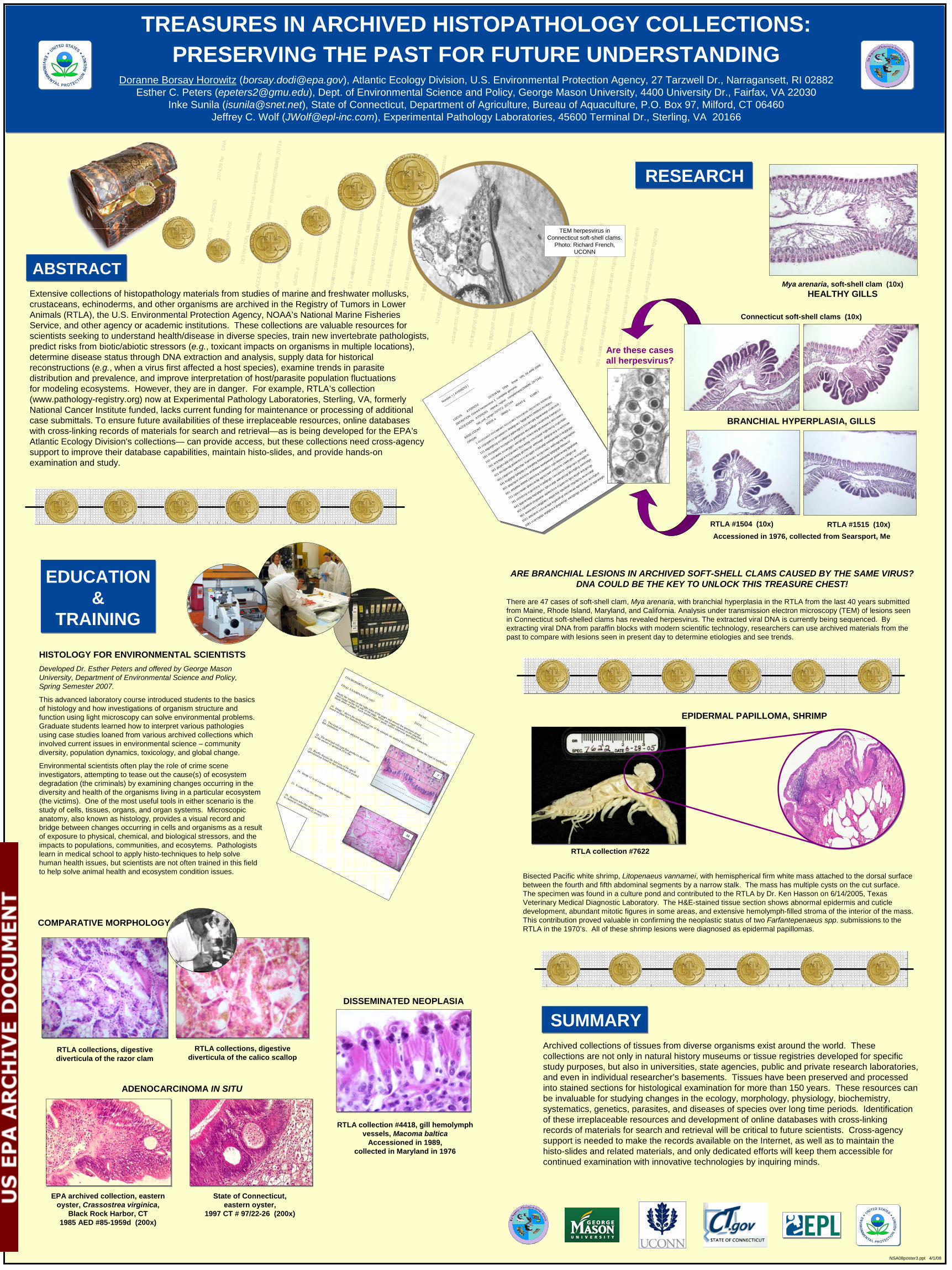
ARE BRANCHIAL LESIONS IN ARCHIVED SOFT-SHELL CLAMS CAUSED BY THE SAME VIRUS?DNA COULD BE THE KEY TO UNLOCK THIS TREASURE CHEST!
There are 47 cases of soft-shell clam, Mya arenaria, with branchial hyperplasia in the RTLA from the last 40 years submitted from Maine, Rhode Island, Maryland, and California. Analysis under transmission electron microscopy (TEM) of lesions seen in Connecticut soft-shelled clams has revealed herpesvirus. The extracted viral DNA is currently being sequenced. By extracting viral DNA from paraffin blocks with modern scientific technology, researchers can use archived materials from the past to compare with lesions seen in present day to determine etiologies and see trends.
RESEARCH
TEM herpesvirus in Connecticut soft-shell clams.
Photo: Richard French, UCONN
Mya arenaria, soft-shell clam (10x)HEALTHY GILLS
Connecticut soft-shell clams (10x)
RTLA #1515 (10x)RTLA #1504 (10x)
BRANCHIAL HYPERPLASIA, GILLS
Are these cases all herpesvirus?
EDUCATION&
TRAINING
HISTOLOGY FOR ENVIRONMENTAL SCIENTISTSDeveloped Dr. Esther Peters and offered by George Mason University, Department of Environmental Science and Policy, Spring Semester 2007.
This advanced laboratory course introduced students to the basics of histology and how investigations of organism structure and function using light microscopy can solve environmental problems. Graduate students learned how to interpret various pathologies using case studies loaned from various archived collections which involved current issues in environmental science – community diversity, population dynamics, toxicology, and global change.
Environmental scientists often play the role of crime scene investigators, attempting to tease out the cause(s) of ecosystem degradation (the criminals) by examining changes occurring in the diversity and health of the organisms living in a particular ecosystem (the victims). One of the most useful tools in either scenario is the study of cells, tissues, organs, and organ systems. Microscopicanatomy, also known as histology, provides a visual record and bridge between changes occurring in cells and organisms as a result of exposure to physical, chemical, and biological stressors, and the impacts to populations, communities, and ecosytems. Pathologists learn in medical school to apply histo-techniques to help solve human health issues, but scientists are not often trained in this field to help solve animal health and ecosystem condition issues.
ABSTRACT
Bisected Pacific white shrimp, Litopenaeus vannamei, with hemispherical firm white mass attached to the dorsal surface between the fourth and fifth abdominal segments by a narrow stalk. The mass has multiple cysts on the cut surface. The specimen was found in a culture pond and contributed to the RTLA by Dr. Ken Hasson on 6/14/2005, Texas Veterinary Medical Diagnostic Laboratory. The H&E-stained tissue section shows abnormal epidermis and cuticle development, abundant mitotic figures in some areas, and extensive hemolymph-filled stroma of the interior of the mass. This contribution proved valuable in confirming the neoplastic status of two Farfantepenaeus spp. submissions to the RTLA in the 1970’s. All of these shrimp lesions were diagnosed as epidermal papillomas.
ENVIRONMENTAL HISTOLOGY NAME ___________________
FINAL EXAMINATION 2007
DATE______________
Study the images on the right sides of the pages and answer the questions about
each image on the left side of the page. Write any longer answers on the attached
blank sheet of paper. Each answer counts 1 point, unless otherwise specified in brackets.
20. Image 7 shows the epidermis of one of the animals we studied this semester. Name the type of epithelium
based on three key features [3 points]:
21. This kind of tissue is adjacent and connecting to
that epithelium:22. The principal cell type that produces the tissue
in the answer to question 29 is the:
23. Briefly discuss the function of the apical
specialization found in this epithelium [4 points]:
24. Image 12 is of a tissue section from this organ:
25. It came from this species:26. Discuss why this organ is susceptible
to chemical contaminants [5 points]:
7
12
RTLA collections, digestive diverticula of the razor clam
RTLA collections, digestive diverticula of the calico scallop
COMPARATIVE MORPHOLOGY
Archived collections of tissues from diverse organisms exist around the world. These collections are not only in natural history museums or tissue registries developed for specific study purposes, but also in universities, state agencies, public and private research laboratories, and even in individual researcher’s basements. Tissues have been preserved and processed into stained sections for histological examination for more than 150 years. These resources can be invaluable for studying changes in the ecology, morphology, physiology, biochemistry, systematics, genetics, parasites, and diseases of species over long time periods. Identification of these irreplaceable resources and development of online databases with cross-linking records of materials for search and retrieval will be critical to future scientists. Cross-agency support is needed to make the records available on the Internet, as well as to maintain the histo-slides and related materials, and only dedicated efforts will keep them accessible for continued examination with innovative technologies by inquiring minds.
SUMMARY
EPIDERMAL PAPILLOMA, SHRIMP
EPA archived collection, eastern oyster, Crassostrea virginica,
Black Rock Harbor, CT 1985 AED #85-1959d (200x)
State of Connecticut,eastern oyster,
1997 CT # 97/22-26 (200x)
ADENOCARCINOMA IN SITU
RTLA collection #4418, gill hemolymphvessels, Macoma baltica
Accessioned in 1989,collected in Maryland in 1976
DISSEMINATED NEOPLASIA
Extensive collections of histopathology materials from studies of marine and freshwater mollusks, crustaceans, echinoderms, and other organisms are archived in the Registry of Tumors in Lower Animals (RTLA), the U.S. Environmental Protection Agency, NOAA’s National Marine Fisheries Service, and other agency or academic institutions. These collections are valuable resources for scientists seeking to understand health/disease in diverse species, train new invertebrate pathologists, predict risks from biotic/abiotic stressors (e.g., toxicant impacts on organisms in multiple locations), determine disease status through DNA extraction and analysis, supply data for historical reconstructions (e.g., when a virus first affected a host species), examine trends in parasite distribution and prevalence, and improve interpretation of host/parasite population fluctuations for modeling ecosystems. However, they are in danger. For example, RTLA’s collection (www.pathology-registry.org) now at Experimental Pathology Laboratories, Sterling, VA, formerly National Cancer Institute funded, lacks current funding for maintenance or processing of additional case submittals. To ensure future availabilities of these irreplaceable resources, online databases with cross-linking records of materials for search and retrieval—as is being developed for the EPA’s Atlantic Ecology Division's collections— can provide access, but these collections need cross-agency support to improve their database capabilities, maintain histo-slides, and provide hands-on examination and study.
TREASURES IN ARCHIVED HISTOPATHOLOGY COLLECTIONS:PRESERVING THE PAST FOR FUTURE UNDERSTANDING
Doranne Borsay Horowitz ([email protected]), Atlantic Ecology Division, U.S. Environmental Protection Agency, 27 Tarzwell Dr., Narragansett, RI 02882Esther C. Peters ([email protected]), Dept. of Environmental Science and Policy, George Mason University, 4400 University Dr., Fairfax, VA 22030
Inke Sunila ([email protected]), State of Connecticut, Department of Agriculture, Bureau of Aquaculture, P.O. Box 97, Milford, CT 06460 Jeffrey C. Wolf ([email protected]), Experimental Pathology Laboratories, 45600 Terminal Dr., Sterling, VA 20166
Accessioned in 1976, collected from Searsport, Me
--------
--------
--------
--------
----
Number = [ A
Y509253 ]
--------
--------
--------
--------
----
LOCUS A
Y509253
207439 bp
DNA linear
VRL 04-APR-2006
DEFINITION Ostreid herpesvirus 1, complete genome.
ACCESSION AY509253 r
epeat_regioncomplement(2
06889..207144)
/rpt_unit_range=207072..207144
BASE COUNT 6
5205 a 3
9905 c 4
0443 g 6
1886 t
ORIGIN
1 ccccccacct ccccaacacc tcccccatcc tccccacctc ctccccctcc tctcttccgc
61 ccgcgatccc gccaataccc ataatgcacc tgggcactct cttttttcct ttccttatcc
121 aagatgtccg cccattgcca ggtacagcct tcccaccgtg tgaacaatgt ccatcctctt
181 ctccgagact tccctgacca gattgtcgta atccaattga cacattctcg tcaatgccct
241 cctcatattctccatcggcc aactgtcgtc tctactcatg gtcataaaca atcccaatcc
301 actcttggca tcccgcaacc tttccaatag cctcccgaat tcgtctaccg ccgccttatc
361 gtcgtccgtg ctgcaatgtg gtcttaccgc atttttaagc aatgcgcacg ccactctcaa
421 ttcctgacag gtaatctcct ccaccggttt cctatcgtgt aatagactga ccacggcggc
481 catgtctctc agttccttgc tcatctcacc accgccaacc aattcagcag tgacagttac
541 ttcgtgttgt tgttgagcca ttatattttg
ttaaataaag tgtgtgtgtt tttttaacct
601 gttgctgctt cttctctcta atctttataa aaatgaaaat gaaacacatg tgtgttatat
661 atttatttta
tttttttttt
cattctctcg tcaaattccc cgtcaaaaca cattgtccat
721 cgaccaaatc gtatccacag agctccaatt ccttcctctc caattcgttcacccgctcgt
781 ctctcttccg ccgcctctcg ccaatgcgtc caccaacctt cattgccgcc gccagttctc
841 tggcactagg tagcgtgacc tgtcccatgt aatcgtccgt gtccttgtcg aatctcttga
901 cgtatttctt ctcgtaatac agatcctcca ccgattcctc tgccatcgtt actcgctcgc
961 aaaactttct catggtatcc atggtcttat cgtcattatt cttgctgcta atcaccatgt
1021 atttcatctt cctccacaga tcgacatcgt cttccatcaa agctctcacc accctgtctt
1081 ccacctgtga atgtgtctca gtagatctgc aactgtctgc aacgaacctt tggcaatgtc
RTLA collection #7622
NSA08poster3.ppt 4/1/08