Novel device for application of continuous mechanical ... · Novel device for application of...
7
RESEARCH ARTICLE Novel device for application of continuous mechanical tensile strain to mammalian cells Satoshi Wada, Hiroyuki Kanzaki*, Tsuyoshi Narimiya and Yoshiki Nakamura ABSTRACT During orthodontic tooth movement, the periodontal ligament (PDL) is exposed to continuous mechanical strain. However, many researchers have applied cyclic tensile strain, not continuous tensile strain, to PDL cells in vitro because there has been no adequate device to apply continuous tensile strain to cultured cells. In this study, we contrived a novel device designed to apply continuous tensile strain to cells in culture. The continuous tensile strain was applied to human immortalized periodontal ligament cell line (HPL cells) and the cytoskeletal structures of HPL cells were examined by immunohistochemistry. The expression of both inflammatory and osteogenic markers was also examined by real-time reverse transcription polymerase chain reaction. The osteogenic protein, Osteopontin (OPN), was also detected by western blot analysis. The actin filaments of HPL cells showed uniform arrangement under continuous tensile strain. The continuous tensile strain increased the expression of inflammatory genes such as IL-1β, IL-6, COX-2 and TNF-α, and osteogenic genes such as RUNX2 and OPN in HPL cells. It also elevated the expression of OPN protein in HPL cells. These results suggest that our new simple device is useful for exploring the responses to continuous tensile strain applied to the cells. KEY WORDS: Simple stretch device, Continuous tensile strain, Periodontal ligament cell, Mechanical stress, Osteoblastic differentiation INTRODUCTION Cells in the body are usually exposed to several types of mechanical stimulation, such as shear stress (Li et al., 2005), compressive stress (MacKelvie et al., 2003; Tschumperlin et al., 2004) and tensile stress (Thomas et al., 2006). For example, vascular endothelial cells are subjected to shear stress from blood flow (Li et al., 2005), vascular smooth muscle cells are exposed to cyclic stretch resulting from pulsatile pressure (Li et al., 2005), cartilage in human joints is subjected to compressive stress during exercise (MacKelvie et al., 2003), and bronchial epithelial cells undergo tensile and compressive stress by breathing (Thomas et al., 2006; Tschumperlin et al., 2004). Thus, many living cells respond to mechanical strain and adapt to their different physical environments. Several studies have reported that mechanical strain regulates various cellular functions such as proliferation, differentiation, apoptosis and migration in mammalian cells (Banes et al., 1995; Lambert et al., 1998; Petrov and Usherwood, 1994). To investigate the cellular response to mechanical stress, numerous researchers have contrived devices to apply mechanical strain, such as shear stress (Jacobs et al., 1998; Yoo et al., 2014), tensile strain (Beckmann et al., 2014; Kanzaki et al., 2006; Shah et al., 2013; Tsuji et al., 2004; Zhu et al., 2008), compression (Kanzaki et al., 2002; Tschumperlin et al., 2004) and hydrostatic pressure (Swartz et al., 2001), to cultured cells in order to mimic the in vivo environment. Much of the mechanical strain in the living body is cyclic in nature, and continuous mechanical strain is only applied in limited situations, such as in orthodontic tooth movement and distraction osteogenesis (Beertsen et al., 1997; Meikle, 2006; Peltomäki, 2009). In orthodontic tooth movement, continuous mechanical strain is systematically applied to the teeth, which are moved by reconstruction of the periodontal ligament (PDL) interposed between the tooth root and alveolar bone (Beertsen et al., 1997; Meikle, 2006). Cells in the PDL are subjected to continuous mechanical strain and are forced to adapt to the new environment by synthesis and secretion of several cytokines and growth factors (Arai et al., 2010; Baba et al., 2011; Saito et al., 1991; Tsuge et al., 2016). As a result, reconstruction of the PDL and alveolar bone occur both in the tension and compression zones of the PDL (Nakamura et al., 2003; Shimpo et al., 2003; Takahashi et al., 2003, 2006). However, the detailed mechanisms of the cellular response in reconstruction of the PDL and alveolar bone have not been clarified. In particular, the relationship between continuous tensile strain and osteogenic markers in the tension zone of the PDL remains unclear during orthodontic tooth movement. In order to clarify the molecular regulatory mechanism of tissue reconstruction in the tension zone of PDL during tooth movement, it is necessary to contrive devices in order to investigate the response of PDL cells to continuous tensile strain in vitro. In this study, we contrived a simple device, ‘Cell Extender’ (ver. 3), designed to apply both continuous and cyclic tensile strain to cultured cells in vitro. Furthermore, this device offers advantageous cost, size, usability and versatility for experiments with a variety of cell types. RESULTS Continuous tensile strain from the device apparently influenced the direction of actin filaments in HPL cells Initially, we observed the tension zone of PDL using perfusion- fixed sections of the PDL of first molar area. The control groups showed that the fibroblasts were scattered in the PDL (Fig. 1A). In contrast, the cellular elements were elongated among the periodontal fibers in the tension zone (Fig. 1B). Next, we applied continuous tensile strain from the device to HPL cells and examined their cytoskeletal structure, because Received 16 December 2016; Accepted 15 March 2017 Department of Orthodontics, School of Dental Medicine, Tsurumi University, 2-1-3 Tsurumi, Tsurumi-ku, Yokohama, Kanagawa 230-8501, Japan. *Author for correspondence ([email protected]) H.K., 0000-0003-3473-1218 This is an Open Access article distributed under the terms of the Creative Commons Attribution License (http://creativecommons.org/licenses/by/3.0), which permits unrestricted use, distribution and reproduction in any medium provided that the original work is properly attributed. 518 © 2017. Published by The Company of Biologists Ltd | Biology Open (2017) 6, 518-524 doi:10.1242/bio.023671 Biology Open by guest on March 13, 2020 http://bio.biologists.org/ Downloaded from
Transcript of Novel device for application of continuous mechanical ... · Novel device for application of...

RESEARCH ARTICLE
Novel device for application of continuous mechanical tensilestrain to mammalian cellsSatoshi Wada, Hiroyuki Kanzaki*, Tsuyoshi Narimiya and Yoshiki Nakamura
ABSTRACTDuring orthodontic tooth movement, the periodontal ligament (PDL)is exposed to continuous mechanical strain. However, manyresearchers have applied cyclic tensile strain, not continuoustensile strain, to PDL cells in vitro because there has been noadequate device to apply continuous tensile strain to cultured cells. Inthis study, we contrived a novel device designed to apply continuoustensile strain to cells in culture. The continuous tensile strain wasapplied to human immortalized periodontal ligament cell line (HPLcells) and the cytoskeletal structures of HPL cells were examined byimmunohistochemistry. The expression of both inflammatory andosteogenic markers was also examined by real-time reversetranscription polymerase chain reaction. The osteogenic protein,Osteopontin (OPN), was also detected by western blot analysis. Theactin filaments of HPL cells showed uniform arrangement undercontinuous tensile strain. The continuous tensile strain increased theexpression of inflammatory genes such as IL-1β, IL-6, COX-2 andTNF-α, and osteogenic genes such asRUNX2 andOPN in HPL cells.It also elevated the expression of OPN protein in HPL cells. Theseresults suggest that our new simple device is useful for exploring theresponses to continuous tensile strain applied to the cells.
KEY WORDS: Simple stretch device, Continuous tensile strain,Periodontal ligament cell, Mechanical stress, Osteoblasticdifferentiation
INTRODUCTIONCells in the body are usually exposed to several types of mechanicalstimulation, such as shear stress (Li et al., 2005), compressive stress(MacKelvie et al., 2003; Tschumperlin et al., 2004) and tensilestress (Thomas et al., 2006). For example, vascular endothelial cellsare subjected to shear stress from blood flow (Li et al., 2005),vascular smooth muscle cells are exposed to cyclic stretch resultingfrom pulsatile pressure (Li et al., 2005), cartilage in human joints issubjected to compressive stress during exercise (MacKelvie et al.,2003), and bronchial epithelial cells undergo tensile andcompressive stress by breathing (Thomas et al., 2006;Tschumperlin et al., 2004). Thus, many living cells respond tomechanical strain and adapt to their different physicalenvironments. Several studies have reported that mechanical strain
regulates various cellular functions such as proliferation,differentiation, apoptosis and migration in mammalian cells(Banes et al., 1995; Lambert et al., 1998; Petrov and Usherwood,1994). To investigate the cellular response to mechanical stress,numerous researchers have contrived devices to apply mechanicalstrain, such as shear stress (Jacobs et al., 1998; Yoo et al., 2014),tensile strain (Beckmann et al., 2014; Kanzaki et al., 2006; Shahet al., 2013; Tsuji et al., 2004; Zhu et al., 2008), compression(Kanzaki et al., 2002; Tschumperlin et al., 2004) and hydrostaticpressure (Swartz et al., 2001), to cultured cells in order to mimic thein vivo environment. Much of the mechanical strain in the livingbody is cyclic in nature, and continuous mechanical strain is onlyapplied in limited situations, such as in orthodontic tooth movementand distraction osteogenesis (Beertsen et al., 1997; Meikle, 2006;Peltomäki, 2009).
In orthodontic tooth movement, continuous mechanical strain issystematically applied to the teeth, which are moved byreconstruction of the periodontal ligament (PDL) interposedbetween the tooth root and alveolar bone (Beertsen et al., 1997;Meikle, 2006). Cells in the PDL are subjected to continuousmechanical strain and are forced to adapt to the new environment bysynthesis and secretion of several cytokines and growth factors(Arai et al., 2010; Baba et al., 2011; Saito et al., 1991; Tsuge et al.,2016). As a result, reconstruction of the PDL and alveolar boneoccur both in the tension and compression zones of the PDL(Nakamura et al., 2003; Shimpo et al., 2003; Takahashi et al., 2003,2006). However, the detailed mechanisms of the cellular response inreconstruction of the PDL and alveolar bone have not been clarified.In particular, the relationship between continuous tensile strain andosteogenic markers in the tension zone of the PDL remains unclearduring orthodontic tooth movement.
In order to clarify the molecular regulatory mechanism of tissuereconstruction in the tension zone of PDL during tooth movement, itis necessary to contrive devices in order to investigate the responseof PDL cells to continuous tensile strain in vitro.
In this study, we contrived a simple device, ‘Cell Extender’ (ver.3), designed to apply both continuous and cyclic tensile strain tocultured cells in vitro. Furthermore, this device offers advantageouscost, size, usability and versatility for experiments with a variety ofcell types.
RESULTSContinuous tensile strain from the device apparentlyinfluenced the direction of actin filaments in HPL cellsInitially, we observed the tension zone of PDL using perfusion-fixed sections of the PDL of first molar area. The control groupsshowed that the fibroblasts were scattered in the PDL (Fig. 1A). Incontrast, the cellular elements were elongated among theperiodontal fibers in the tension zone (Fig. 1B).
Next, we applied continuous tensile strain from the deviceto HPL cells and examined their cytoskeletal structure, becauseReceived 16 December 2016; Accepted 15 March 2017
Department of Orthodontics, School of Dental Medicine, Tsurumi University, 2-1-3Tsurumi, Tsurumi-ku, Yokohama, Kanagawa 230-8501, Japan.
*Author for correspondence ([email protected])
H.K., 0000-0003-3473-1218
This is an Open Access article distributed under the terms of the Creative Commons AttributionLicense (http://creativecommons.org/licenses/by/3.0), which permits unrestricted use,distribution and reproduction in any medium provided that the original work is properly attributed.
518
© 2017. Published by The Company of Biologists Ltd | Biology Open (2017) 6, 518-524 doi:10.1242/bio.023671
BiologyOpen
by guest on March 13, 2020http://bio.biologists.org/Downloaded from

the cytoskeleton reveals changes in cell morphology.Immunohistochemistry demonstrated that actin filaments wereunidirectionally arranged in HPL cells exposed to the continuoustensile strain, though filaments were randomly arranged in HPL
cells in the control group (Fig. 1C,D). To examine the effect ofcontinuous tensile strain from the device on cell viability, therewere no differences in cell viability between both control andexperimental groups (Fig. 1E).
Fig. 1. Effect of continuous tensilestrain on themorphologyof HPL cells.(A-D) Images of the PDL stained withH&E stain. Control group (A and B) and5 days after tooth movement (C and D).B and D are higher magnification imagesof the boxed area in A and C. Arrowindicates the direction of toothmovement. B: bone, D: dentin. Scalebars: 100 μm. The effect of continuoustensile strain on cell morphology andcytoskeletal was assessed by means ofF-actin staining. (E,F) The greenfluorescence indicates the F-actin.Representative photographs of thecontrol (E) and stretched HPL cells (F)are shown. Scale bars: 50 μm. (G) Theeffect of continuous tensile strain fromthe device on cell viability examined byusing cell counting kit-8 (E). Percentageof controls is shown. Mean±s.d.; NS, notsignificant.
519
RESEARCH ARTICLE Biology Open (2017) 6, 518-524 doi:10.1242/bio.023671
BiologyOpen
by guest on March 13, 2020http://bio.biologists.org/Downloaded from

Continuous tensile strain from the device influencedexpression of inflammatory genes in HPL cellsIt has been reported that the expression levels of inflammatory geneswere up-regulated in PDL cells under mechanical strain (Iwasakiet al., 2001; Jacobs et al., 2014; Shimizu et al., 1998; Yamamotoet al., 2006); therefore, we examined the effects of continuous tensilestrain on inflammatory gene expression in HPL cells. Real-time RT-PCR analysis revealed that the expression of IL-1β, IL-6 and COX2mRNAs was significantly upregulated under tensile strain at 24 h incomparison with non-stretched cultures (Fig. 2A-C). TNF-α mRNAwas also significantly upregulated in HPL cells at 12 h and itsupregulation was reduced to control levels at 24 h (Fig. 2D).
Continuous tensile strain from the device also influencedosteogenic gene expression in HPL cellsNumerous studies have shown that mechanical strain inducesosteogenic differentiation of PDL cells by increasing osteogenicgenes such as RUNX2 and OPN (Li et al., 2014; Ren et al., 2015;Tang et al., 2014; Zhang et al., 2015). Therefore, we investigated theeffects of continuous tensile strain from the device on the osteogenicgene expression in HPL cells. Expression of RUNX2 wassignificantly increased by the strain in HPL cells at 24 h whencompared to non-stretched HPL cells (Fig. 3A). Expression of OPNmRNA was also significantly elevated (Fig. 3B).
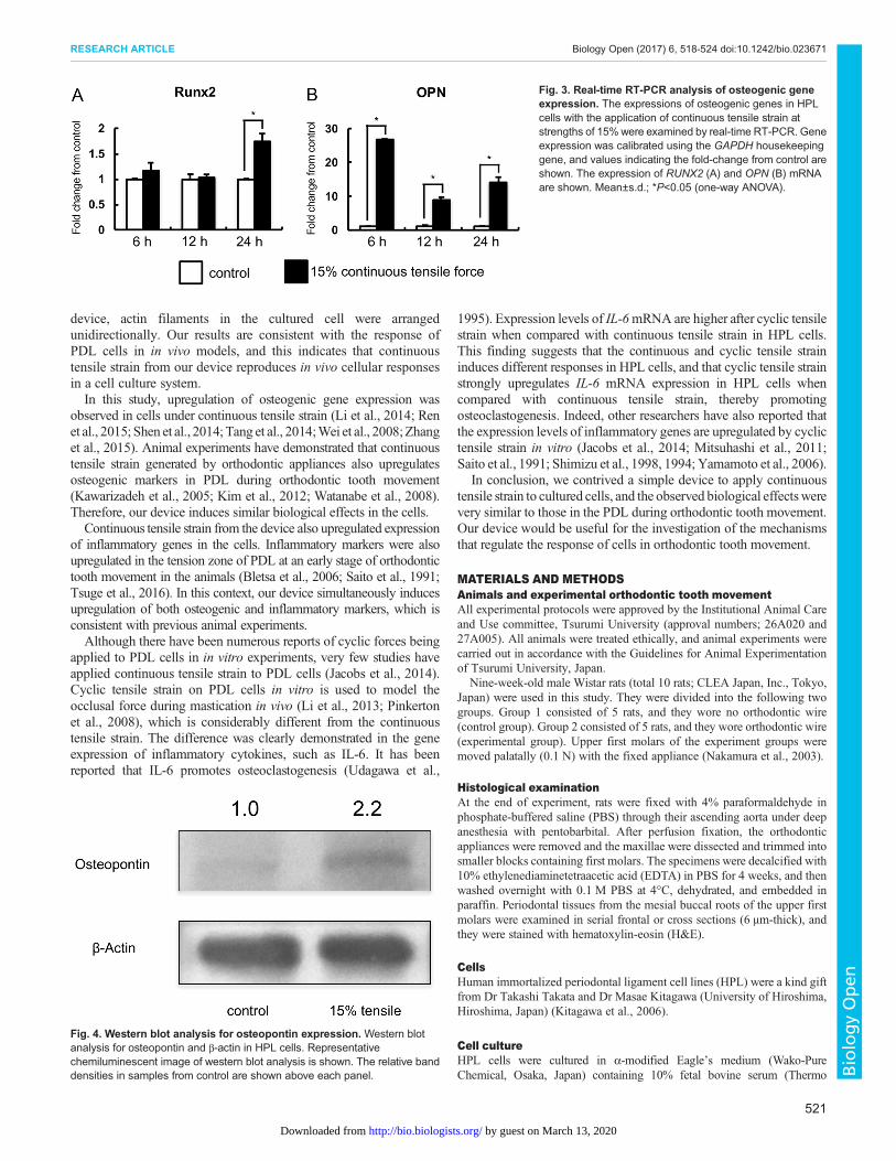
Continuous tensile strain from the device augmented OPN inHPL cellsNext, we examined whether HPL cells induced osteogenic proteinunder continuous tensile strain by western blot analysis. Westernblot analysis for OPN demonstrated that continuous tensile strainaugmented OPN in HPL cells (Fig. 4).
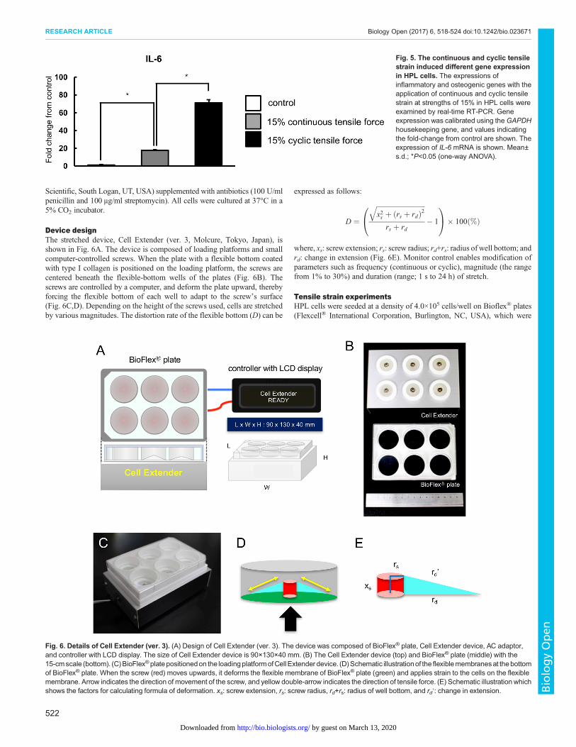
Continuous and cyclic tensile strain from the device inducesdifferent responses in HPL cellsIn order to explore whether there are any differences in cellresponses between continuous and cyclic tensile strains, we
examined the expression levels of IL-6 mRNA in HPL cells, asIL-6 mRNAwas markedly increased by continuous tensile strain inHPL cells. Real-time RT-PCR analysis revealed that the expressionof IL-6 was higher in cyclic tensile strain-applied HPL cells than incontinuous tensile strain-applied HPL cells (Fig. 5).
DISCUSSIONIn this research, we contrived a simple device, whichmade it possibleto apply both continuous and cyclic mechanical strain to culturedcells. The differences between our device and commerciallyavailable devices are size, price, and the mechanism of applyingmechanical strain to cells (Banes et al., 1985; Cheng et al., 2015;Hasegawa et al., 2015; Kanzaki et al., 2006; Long et al., 2002;Suzuki et al., 2014; Tsuji et al., 2004). Though commerciallyavailable devices are useful for exploring cell responses to cyclictensile strain, these devices are cumbersome and expensive. Somedevices for tensile strain, such as the STREX Cell Stretch System(Strex Inc., Osaka, Japan) (Hasegawa et al., 2015; Suzuki et al.,2014) and the ShellPa mechanical cell stretcher (B-BridgeInternational, Inc., Santa Clara, CA, USA) (Cheng et al., 2015),elongate the culture well uniaxially (Naruse et al., 1998; Takedaet al., 2006), but thewidth (orthogonal direction to elongation) of thewell becomes narrow, exerting compressive strain on the cells. Ourdevice deforms the flex bottom vertically and consequently imposesonly uniaxial tensile strain to cells, in a similar manner as FlexcellCulture Systems (Flexcell International Corp., Hillsborough, NC,USA) (Banes et al., 1985; Kanzaki et al., 2006; Long et al., 2002;Tsuji et al., 2004). Our device and Flexcell Culture Systems areexcellent at applying tensile strain to cells. The disadvantage ofFlexcell Culture Systems is the cost; they are approximately tenfoldmore expensive than our device. Taken together, the advantages ofour device are compact size, reasonable price, application of uniaxialtensile force without orthogonal compression, and usability.
We demonstrated that PDL cells were elongated along with theperiodontal fibers in the tension zone during orthodontic toothmovement. After application of continuous tensile strain from our
Fig. 2. Real-time RT-PCR analysis of inflammatorygene expression. The expressions of inflammatorygenes in HPL cells with the application of continuoustensile strain at strengths of 15% were examined by real-time RT-PCR. Gene expression was calibrated using theGAPDH housekeeping gene, and the values indicate thefold-change from control. The expression of IL-1β (A),IL-6 (B), COX-2 (C), and TNF-α (D) mRNA are shown.Mean±s.d.; *P<0.05 (one-way ANOVA).
520
RESEARCH ARTICLE Biology Open (2017) 6, 518-524 doi:10.1242/bio.023671
BiologyOpen
by guest on March 13, 2020http://bio.biologists.org/Downloaded from
device, actin filaments in the cultured cell were arrangedunidirectionally. Our results are consistent with the response ofPDL cells in in vivo models, and this indicates that continuoustensile strain from our device reproduces in vivo cellular responsesin a cell culture system.In this study, upregulation of osteogenic gene expression was
observed in cells under continuous tensile strain (Li et al., 2014; Renet al., 2015; Shen et al., 2014; Tang et al., 2014;Wei et al., 2008;Zhanget al., 2015). Animal experiments have demonstrated that continuoustensile strain generated by orthodontic appliances also upregulatesosteogenic markers in PDL during orthodontic tooth movement(Kawarizadeh et al., 2005; Kim et al., 2012; Watanabe et al., 2008).Therefore, our device induces similar biological effects in the cells.Continuous tensile strain from the device also upregulated expression
of inflammatory genes in the cells. Inflammatory markers were alsoupregulated in the tension zone of PDL at an early stage of orthodontictooth movement in the animals (Bletsa et al., 2006; Saito et al., 1991;Tsuge et al., 2016). In this context, our device simultaneously inducesupregulation of both osteogenic and inflammatory markers, which isconsistent with previous animal experiments.Although there have been numerous reports of cyclic forces being
applied to PDL cells in in vitro experiments, very few studies haveapplied continuous tensile strain to PDL cells (Jacobs et al., 2014).Cyclic tensile strain on PDL cells in vitro is used to model theocclusal force during mastication in vivo (Li et al., 2013; Pinkertonet al., 2008), which is considerably different from the continuoustensile strain. The difference was clearly demonstrated in the geneexpression of inflammatory cytokines, such as IL-6. It has beenreported that IL-6 promotes osteoclastogenesis (Udagawa et al.,
1995). Expression levels of IL-6mRNA are higher after cyclic tensilestrain when compared with continuous tensile strain in HPL cells.This finding suggests that the continuous and cyclic tensile straininduces different responses in HPL cells, and that cyclic tensile strainstrongly upregulates IL-6 mRNA expression in HPL cells whencompared with continuous tensile strain, thereby promotingosteoclastogenesis. Indeed, other researchers have also reported thatthe expression levels of inflammatory genes are upregulated by cyclictensile strain in vitro (Jacobs et al., 2014; Mitsuhashi et al., 2011;Saito et al., 1991; Shimizu et al., 1998, 1994; Yamamoto et al., 2006).
In conclusion, we contrived a simple device to apply continuoustensile strain to cultured cells, and the observed biological effectswerevery similar to those in the PDL during orthodontic tooth movement.Our device would be useful for the investigation of the mechanismsthat regulate the response of cells in orthodontic tooth movement.
MATERIALS AND METHODSAnimals and experimental orthodontic tooth movementAll experimental protocols were approved by the Institutional Animal Careand Use committee, Tsurumi University (approval numbers; 26A020 and27A005). All animals were treated ethically, and animal experiments werecarried out in accordance with the Guidelines for Animal Experimentationof Tsurumi University, Japan.
Nine-week-old male Wistar rats (total 10 rats; CLEA Japan, Inc., Tokyo,Japan) were used in this study. They were divided into the following twogroups. Group 1 consisted of 5 rats, and they wore no orthodontic wire(control group). Group 2 consisted of 5 rats, and they wore orthodontic wire(experimental group). Upper first molars of the experiment groups weremoved palatally (0.1 N) with the fixed appliance (Nakamura et al., 2003).
Histological examinationAt the end of experiment, rats were fixed with 4% paraformaldehyde inphosphate-buffered saline (PBS) through their ascending aorta under deepanesthesia with pentobarbital. After perfusion fixation, the orthodonticappliances were removed and the maxillae were dissected and trimmed intosmaller blocks containing first molars. The specimens were decalcified with10% ethylenediaminetetraacetic acid (EDTA) in PBS for 4 weeks, and thenwashed overnight with 0.1 M PBS at 4°C, dehydrated, and embedded inparaffin. Periodontal tissues from the mesial buccal roots of the upper firstmolars were examined in serial frontal or cross sections (6 μm-thick), andthey were stained with hematoxylin-eosin (H&E).
CellsHuman immortalized periodontal ligament cell lines (HPL) were a kind giftfrom Dr Takashi Takata and Dr Masae Kitagawa (University of Hiroshima,Hiroshima, Japan) (Kitagawa et al., 2006).
Cell cultureHPL cells were cultured in α-modified Eagle’s medium (Wako-PureChemical, Osaka, Japan) containing 10% fetal bovine serum (Thermo
Fig. 3. Real-time RT-PCR analysis of osteogenic geneexpression. The expressions of osteogenic genes in HPLcells with the application of continuous tensile strain atstrengths of 15%were examined by real-time RT-PCR. Geneexpression was calibrated using the GAPDH housekeepinggene, and values indicating the fold-change from control areshown. The expression of RUNX2 (A) and OPN (B) mRNAare shown. Mean±s.d.; *P<0.05 (one-way ANOVA).
Fig. 4. Western blot analysis for osteopontin expression. Western blotanalysis for osteopontin and β-actin in HPL cells. Representativechemiluminescent image of western blot analysis is shown. The relative banddensities in samples from control are shown above each panel.
521
RESEARCH ARTICLE Biology Open (2017) 6, 518-524 doi:10.1242/bio.023671
BiologyOpen
by guest on March 13, 2020http://bio.biologists.org/Downloaded from
Scientific, South Logan, UT, USA) supplemented with antibiotics (100 U/mlpenicillin and 100 μg/ml streptomycin). All cells were cultured at 37°C in a5% CO2 incubator.
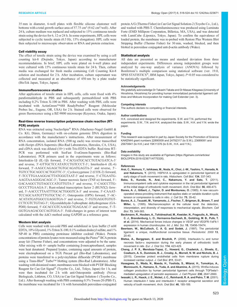
Device designThe stretched device, Cell Extender (ver. 3, Molcure, Tokyo, Japan), isshown in Fig. 6A. The device is composed of loading platforms and smallcomputer-controlled screws. When the plate with a flexible bottom coatedwith type I collagen is positioned on the loading platform, the screws arecentered beneath the flexible-bottom wells of the plates (Fig. 6B). Thescrews are controlled by a computer, and deform the plate upward, therebyforcing the flexible bottom of each well to adapt to the screw’s surface(Fig. 6C,D). Depending on the height of the screws used, cells are stretchedby various magnitudes. The distortion rate of the flexible bottom (D) can be
expressed as follows:
D ¼ffiffiffiffiffiffiffiffiffiffiffiffiffiffiffiffiffiffiffiffiffiffiffiffiffiffiffiffiffix2s þ ðrs þ rdÞ2
q
rs þ rd� 1
0@
1A� 100ð%Þ
where, xs: screw extension; rs: screw radius; rd+rs: radius of well bottom; andrd: change in extension (Fig. 6E). Monitor control enables modification ofparameters such as frequency (continuous or cyclic), magnitude (the rangefrom 1% to 30%) and duration (range; 1 s to 24 h) of stretch.
Tensile strain experimentsHPL cells were seeded at a density of 4.0×10⁵ cells/well on Bioflex® plates(Flexcell® International Corporation, Burlington, NC, USA), which were
Fig. 5. The continuous and cyclic tensilestrain induced different gene expressionin HPL cells. The expressions ofinflammatory and osteogenic genes with theapplication of continuous and cyclic tensilestrain at strengths of 15% in HPL cells wereexamined by real-time RT-PCR. Geneexpression was calibrated using theGAPDHhousekeeping gene, and values indicatingthe fold-change from control are shown. Theexpression of IL-6 mRNA is shown. Mean±s.d.; *P<0.05 (one-way ANOVA).
Fig. 6. Details of Cell Extender (ver. 3). (A) Design of Cell Extender (ver. 3). The device was composed of BioFlex® plate, Cell Extender device, AC adaptor,and controller with LCD display. The size of Cell Extender device is 90×130×40 mm. (B) The Cell Extender device (top) and BioFlex® plate (middle) with the15-cmscale (bottom). (C)BioFlex®platepositionedon the loadingplatformofCell Extender device. (D)Schematic illustrationof the flexiblemembranesat thebottomof BioFlex® plate. When the screw (red) moves upwards, it deforms the flexible membrane of BioFlex® plate (green) and applies strain to the cells on the flexiblemembrane. Arrow indicates the direction of movement of the screw, and yellow double-arrow indicates the direction of tensile force. (E) Schematic illustration whichshows the factors for calculating formula of deformation. xs: screw extension, rs: screw radius, rd+rs: radius of well bottom, and rd′: change in extension.
522
RESEARCH ARTICLE Biology Open (2017) 6, 518-524 doi:10.1242/bio.023671
BiologyOpen
by guest on March 13, 2020http://bio.biologists.org/Downloaded from
35 mm in diameter, 6-well plates with flexible silicone elastomer wellbottoms with a total growth surface area of 57.75 cm2 (9.62 cm2/well). After24 h, culture medium was replaced and subjected to 15% continuous tensilestrain using the device for 6, 12 or 24 h. In some experiments, HPL cells weresubjected to cyclic tensile strain (0.5 Hz, 15% elongation). HPL cells werethen subjected to microscopic observation or RNA and protein extraction.
Cell viability assayThe effect of tensile strain using the device was examined by using a cellcounting kit-8 (Dojindo, Tokyo, Japan) according to manufacturerrecommendations. In brief, HPL cells were plated on 6-well plates andwere cultured with 15% continuous tensile strain for 24 h. Then, culturemedia was exchanged for fresh media containing Cell Counting Kit-8solution and incubated for 2 h. After incubation, culture supernatant wascollected and measured at an absorbance of 450 nm by a plate reader(BioTek Japan, Tokyo, Japan).
Immunofluorescence studiesAfter application of tensile strain in HPL cells, cells were fixed with 4%paraformaldehyde in PBS and subsequently permeabilized with PBSincluding 0.2% Triton X-100 in PBS. After washing with PBS, cells wereincubated with ActinGreen™488 ReadyProbes® Reagent (MolecularProbes Inc., Eugene, OR, USA) for 2 h. Stained cells were observed forgreen fluorescence using a BZ-9000 microscope (Keyence, Osaka, Japan).
Real-time reverse transcription polymerase chain reaction (RT-PCR) analysisRNA was extracted using NucleoSpin® RNA (Macherey-Nagel GmbH &Co. KG, Düren, Germany) with on-column genomic DNA digestion inaccordance with the manufacturer’s instructions. After measurement ofRNA concentration, isolated RNA (500 ng each) was reverse transcribedwith iScript cDNA-Supermix (Bio-Rad Laboratories, Hercules, CA, USA),and cDNA stock was diluted (10×) with Tris-EDTA buffer. Real-time RT-PCR was performed with SsoFast EvaGreen-Supermix (Bio-RadLaboratories). PCR primers used in the experiments were as follows:Interleukin-1β (IL-1β) forward, 5′-CACGATGCACCTGTACGATCA-3′and reverse, 5′-GTTGCTCCATATCCTGTCCCT-3′; Interleulin-6 (IL-6)forward, 5′-AAGCCAGAGCTGTGCAGATGAGTA-3′ and reverse, 5′-TGTCCTGCAGCCACTGGTTC-3′; Cyclooxygenase 2 (COX-2) forward,5′-TCCTTGAAAGGACTTATGGGTAAT-3′ and reverse, 5′-CTGAATG-AAGTAAAGGGACAGC-3′; Tumor necrosis factor (TNF-α) forward, 5′-GACAAGCCTGTAGCCCATGTTGTA-3′ and reverse, 5′-CAGCCTTG-GCCCTTGAAGA-3′; Runt-related transcription factor 2 (RUNX2) forw-ard, 5′-AACCCTTAATTTGCACTGGGTCA-3′ and reverse, 5′-CAAAT-TCCAGCAATGTTTGTGCTAC-3′; Osteopontin (OPN) forward, 5′-AC-ACATATGATGGCCGAGGTGA-3′ and reverse, 5′-TGTGAGGTGTGT-CCTCGTCTGTAG-3′; Glyceraldehyde-3-phosphate dehydrogenase (GA-PDH) forward, 5′-GCACCGTCAAGGCTGAGAAC-3′ and reverse, 5′-T-GGTGAAGACGCCAGTGGA-3′. Fold-changes in genes of interest werecalculated with the ΔΔCt method using GAPDH as a reference gene.
Western blot analysisCells were washed with ice-cold PBS and solubilized in lysis buffer (5 mMEDTA,10%Glycerol, 1%TritonX-100, 0.1%sodiumdodecyl sulfate, and1%NP-40 in PBS) containing proteinase inhibitor cocktail (Wako). Proteinconcentrations of protein lysatesweremeasuredusing thePierce®BCAproteinassay kit (Thermo Fisher), and concentrations were adjusted to be the same.After mixing with 4× sample buffer containing β-mercaptoethanol, sampleswere heat denatured. Prepared lysates, containing equal amounts of protein,were electrophoresed on a TGX Precast gel (Bio-Rad Laboratories), andproteins were transferred to a polyvinylidene difluoride (PVDF) membraneusing a Trans-Blot® Turbo™ blotting system (Bio-Rad Laboratories). Afterwashingwith deionizedwater, membraneswere blockedwith PVDFBlockingReagent for Can Get Signal® (Toyobo Co., Ltd., Tokyo, Japan) for 1 h, andwere then incubated for 2 h with anti-Osteopontin antibody (NovusBiologicals, Littleton, CO, USA) in Can Get Signal Solution-1 (Toyobo Co.,Ltd.). After thorough washing with PBS containing 0.5% Tween-20 (PBS-T),the membrane was incubated for 1 h with horseradish peroxidase-conjugated
proteinA/G (ThermoFisher) inCanGet Signal Solution-2 (ToyoboCo., Ltd.),and washed with PBS-T. Chemiluminescence was produced using LuminataForte (EMD Millipore Corporation, Billerica, MA, USA), and was detectedwith LumiCube (Liponics, Tokyo, Japan). To confirm the equivalence ofloaded protein, the membrane was re-probed with Restore Plus Western BlotStripping Buffer (Thermo Fisher) for 30 min, washed, blocked, and thenblotted in peroxidase conjugated anti-β-actin antibody (Wako).
Statistical analysisAll data are presented as means and standard deviation from threeindependent experiments. Differences among independent groups wereanalyzed by one-way analysis of variance (ANOVA) followed byBonferroni’s multiple comparison using statistical software (ver. 19.0,SPSS STATISTICS®, IBM Japan, Tokyo, Japan). P<0.05 was considered tobe statistically significant.
AcknowledgementsWegratefully acknowledge Dr Takashi Takata and Dr Masae Kitagawa (University ofHiroshima, Hiroshima) for providing human immortalized periodontal ligament celllines and Molcure Incorporation for making Cell Extender (ver. 3).
Competing interestsThe authors declare no competing or financial interests.
Author contributionsH.K. conceived and designed the experiments. S.W. and T.N. performed theexperiments. S.W., T.N. and H.K. analyzed the data. S.W., H.K. and Y.N. wrote thepaper.
FundingThis research was supported in part by Japan Society for the Promotion of ScienceKAKENHI grant numbers 22890009 and 24792317 (to S.W.), 23689081 and25670841 (to H.K.) and 15K11376 (to S.W., H.K. and Y.N.).
Data availabilityImages in this study are available at Figshare (https://figshare.com/articles/BIOLOPEN-2016-023671/4775326).
ReferencesArai, C., Nomura, Y., Ishikawa, M., Noda, K., Choi, J.-W., Yashiro, Y., Hanada, N.
and Nakamura, Y. (2010). HSPA1A is upregulated in periodontal ligament atearly stage of tooth movement in rats. Histochem. Cell Biol. 134, 337-343.
Baba, S., Kuroda, N., Arai, C., Nakamura, Y. and Sato, T. (2011).Immunocompetent cells and cytokine expression in the rat periodontal ligamentat the initial stage of orthodontic tooth movement. Arch. Oral Biol. 56, 466-473.
Banes, A. J., Gilbert, J., Taylor, D. and Monbureau, O. (1985). A new vacuum-operated stress-providing instrument that applies static or variable duration cyclictension or compression to cells in vitro. J. Cell Sci. 75, 35-42.
Banes, A. J., Tsuzaki, M., Yamamoto, J., Fischer, T., Brigman, B., Brown, T. andMiller, L. (1995). Mechanoreception at the cellular level: the detection,interpretation, and diversity of responses to mechanical signals. Biochem. CellBiol. 73, 349-365.
Beckmann, R., Houben, A., Tohidnezhad,M., Kweider, N., Fragoulis, A., Wruck,C. J., Brandenburg, L. O., Hermanns-Sachweh, B., Goldring, M. B., Pufe, T.et al. (2014). Mechanical forces induce changes in VEGF and VEGFR-1/sFlt-1expression in human chondrocytes. Int. J. Mol. Sci. 15, 15456-15474.
Beertsen, W., McCulloch, C. A. G. and Sodek, J. (1997). The periodontalligament: a unique, multifunctional connective tissue. Periodontol. 2000 13,20-40.
Bletsa, A., Berggreen, E. and Brudvik, P. (2006). Interleukin-1α and tumornecrosis factor-α expression during the early phases of orthodontic toothmovement in rats. Eur. J. Oral Sci. 114, 423-429.
Cheng, J. P. X., Mendoza-Topaz, C., Howard, G., Chadwick, J., Shvets, E.,Cowburn, A. S., Dunmore, B. J., Crosby, A., Morrell, N. W. and Nichols, B. J.(2015). Caveolae protect endothelial cells from membrane rupture duringincreased cardiac output. J. Cell Biol. 211, 53-61.
Hasegawa, D., Wada, N., Maeda, H., Yoshida, S., Mitarai, H., Tomokiyo, A.,Monnouchi, S., Hamano, S., Yuda, A. and Akamine, A. (2015). Wnt5a inducescollagen production by human periodontal ligament cells through TGFbeta1-mediated upregulation of periostin expression. J. Cell Physiol. 230, 2647-2660.
Iwasaki, L. R., Haack, J. E., Nickel, J. C., Reinhardt, R. A. and Petro, T. M. (2001).Human interleukin-1 beta and interleukin-1 receptor antagonist secretion andvelocity of tooth movement. Arch. Oral Biol. 46, 185-189.
523
RESEARCH ARTICLE Biology Open (2017) 6, 518-524 doi:10.1242/bio.023671
BiologyOpen
by guest on March 13, 2020http://bio.biologists.org/Downloaded from
Jacobs, C. R., Yellowley, C. E., Davis, B. R., Zhou, Z., Cimbala, J. M. andDonahue, H. J. (1998). Differential effect of steady versus oscillating flow on bonecells. J. Biomech. 31, 969-976.
Jacobs, C., Walter, C., Ziebart, T., Grimm, S., Meila, D., Krieger, E. andWehrbein, H. (2014). Induction of IL-6 and MMP-8 in human periodontalfibroblasts by static tensile strain. Clin. Oral Investig. 18, 901-908.
Kanzaki, H., Chiba, M., Shimizu, Y. and Mitani, H. (2002). Periodontal ligamentcells under mechanical stress induce osteoclastogenesis by receptor activator ofnuclear factor kappaB ligand up-regulation via prostaglandin E2 synthesis.J. Bone Miner. Res. 17, 210-220.
Kanzaki, H., Chiba, M., Sato, A., Miyagawa, A., Arai, K., Nukatsuka, S. andMitani, H. (2006). Cyclical tensile force on periodontal ligament cells inhibitsosteoclastogenesis through OPG induction. J. Dent. Res. 85, 457-462.
Kawarizadeh, A., Bourauel, C., Gotz, W. and Jager, A. (2005). Early responses ofperiodontal ligament cells to mechanical stimulus in vivo. J. Dent. Res. 84,902-906.
Kim, J.-Y., Kim, B.-I., Jue, S.-S., Park, J. H. and Shin, J.-W. (2012). Localization ofosteopontin and osterix in periodontal tissue during orthodontic toothmovement inrats. Angle Orthod. 82, 107-114.
Kitagawa, M., Tahara, H., Kitagawa, S., Oka, H., Kudo, Y., Sato, S., Ogawa, I.,Miyaichi, M. and Takata, T. (2006). Characterization of establishedcementoblast-like cell lines from human cementum-lining cells in vitro and invivo. Bone 39, 1035-1042.
Lambert, C. A., Nusgens, B. V. and Lapiere, C. M. (1998). Mechano-sensing andmechano-reaction of soft connective tissue cells.Adv. SpaceRes. 21, 1081-1091.
Li, Y.-S. J., Haga, J. H. and Chien, S. (2005). Molecular basis of the effects of shearstress on vascular endothelial cells. J. Biomech. 38, 1949-1971.
Li, L., Han, M., Li, S., Wang, L. and Xu, Y. (2013). Cyclic tensile stress duringphysiological occlusal force enhances osteogenic differentiation of humanperiodontal ligament cells via ERK1/2-Elk1 MAPK pathway. DNA Cell Biol. 32,488-497.
Li, L., Han, M. X., Li, S., Xu, Y. and Wang, L. (2014). Hypoxia regulates theproliferation and osteogenic differentiation of human periodontal ligament cellsunder cyclic tensile stress via mitogen-activated protein kinase pathways.J. Periodontol. 85, 498-508.
Long, P., Liu, F., Piesco, N. P., Kapur, R. and Agarwal, S. (2002). Signaling bymechanical strain involves transcriptional regulation of proinflammatory genes inhuman periodontal ligament cells in vitro. Bone 30, 547-552.
MacKelvie, K. J., Khan, K. M., Petit, M. A., Janssen, P. A. and McKay, H. A.(2003). A school-based exercise intervention elicits substantial bone healthbenefits: a 2-year randomized controlled trial in girls. Pediatrics 112, e447-e452.
Meikle, M. C. (2006). The tissue, cellular, and molecular regulation of orthodontictooth movement: 100 years after Carl Sandstedt. Eur. J. Orthod. 28, 221-240.
Mitsuhashi, M., Yamaguchi, M., Kojima, T., Nakajima, R. and Kasai, K. (2011).Effects of HSP70 on the compression force-induced TNF-alpha and RANKLexpression in human periodontal ligament cells. Inflamm. Res. 60, 187-194.
Nakamura, Y., Tanaka, T., Noda, K., Shimpo, S., Oikawa, T., Hirashita, A.,Kawamoto, T. and Kawasaki, K. (2003). Calcification of degenerating tissues inthe periodontal ligament during tooth movement. J. Periodontal. Res. 38,343-350.
Naruse, K., Yamada, T. and Sokabe, M. (1998). Involvement of SA channels inorienting response of cultured endothelial cells to cyclic stretch. Am. J. Physiol.274, H1532-H1538.
Peltomaki, T. (2009). Stability, adaptation and growth following distractionosteogenesis in the craniofacial region. Orthod. Craniofac. Res. 12, 187-194.
Petrov, A. G. andUsherwood, P. N. (1994). Mechanosensitivity of cell membranes.Ion channels, lipid matrix and cytoskeleton. Eur. Biophys. J. 23, 1-19.
Pinkerton, M. N., Wescott, D. C., Gaffey, B. J., Beggs, K. T., Milne, T. J. andMeikle, M. C. (2008). Cultured human periodontal ligament cells constitutivelyexpress multiple osteotropic cytokines and growth factors, several of which areresponsive to mechanical deformation. J. Periodontal Res. 43, 343-351.
Ren, D., Wei, F., Hu, L., Yang, S., Wang, C. and Yuan, X. (2015). Phosphorylationof Runx2, induced by cyclic mechanical tension via ERK1/2 pathway, contributesto osteodifferentiation of human periodontal ligament fibroblasts. J. Cell Physiol.230, 2426-2436.
Saito, M., Saito, S., Ngan, P. W., Shanfeld, J. and Davidovitch, Z. (1991).Interleukin 1 beta and prostaglandin E are involved in the response of periodontalcells to mechanical stress in vivo and in vitro. Am. J. Orthod. Dentofacial Orthop.99, 226-240.
Shah, M. R., Wedgwood, S., Czech, L., Kim, G. A., Lakshminrusimha, S.,Schumacker, P. T., Steinhorn, R. H. and Farrow, K. N. (2013). Cyclic stretchinduces inducible nitric oxide synthase and soluble guanylate cyclase inpulmonary artery smooth muscle cells. Int. J. Mol. Sci. 14, 4334-4348.
Shen, T., Qiu, L., Chang, H., Yang, Y., Jian, C., Xiong, J., Zhou, J. and Dong, S.(2014). Cyclic tension promotes osteogenic differentiation in human periodontalligament stem cells. Int. J. Clin. Exp. Pathol. 7, 7872-7880.
Shimizu, N., Yamaguchi, M., Goseki, T., Ozawa, Y., Saito, K., Takiguchi, H.,Iwasawa, T. and Abiko, Y. (1994). Cyclic-tension force stimulates interleukin-1beta production by human periodontal ligament cells. J. Periodontal Res. 29,328-333.
Shimizu, N., Ozawa, Y., Yamaguchi, M., Goseki, T., Ohzeki, K. and Abiko, Y.(1998). Induction of COX-2 expression by mechanical tension force in humanperiodontal ligament cells. J. Periodontol. 69, 670-677.
Shimpo, S., Horiguchi, Y., Nakamura, Y., Lee, M., Oikawa, T., Noda, K.,Kuwahara, Y. and Kawasaki, K. (2003). Compensatory bone formation in youngand old rats during tooth movement. Eur. J. Orthod. 25, 1-7.
Suzuki, R., Nemoto, E. andShimauchi, H. (2014). Cyclic tensile force up-regulatesBMP-2 expression through MAP kinase and COX-2/PGE2 signaling pathways inhuman periodontal ligament cells. Exp. Cell Res. 323, 232-241.
Swartz, M. A., Tschumperlin, D. J., Kamm, R. D. and Drazen, J. M. (2001).Mechanical stress is communicated between different cell types to elicit matrixremodeling. Proc. Natl. Acad. Sci. USA 98, 6180-6185.
Takahashi, I., Nishimura, M., Onodera, K., Bae, J.-W., Mitani, H., Okazaki, M.,Sasano, Y. and Mitani, H. (2003). Expression of MMP-8 and MMP-13 genes inthe periodontal ligament during toothmovement in rats. J. Dent. Res. 82, 646-651.
Takahashi, I., Onodera, K., Nishimura, M., Mitnai, H., Sasano, Y. and Mitani, H.(2006). Expression of genes for gelatinases and tissue inhibitors ofmetalloproteinases in periodontal tissues during orthodontic tooth movement.J. Mol. Histol. 37, 333-342.
Takeda, H., Komori, K., Nishikimi, N., Nimura, Y., Sokabe, M. and Naruse, K.(2006). Bi-phasic activation of eNOS in response to uni-axial cyclic stretch ismediated by differential mechanisms in BAECs. Life Sci. 79, 233-239.
Tang, M., Peng, Z., Mai, Z., Chen, L., Mao, Q., Chen, Z., Chen, Q., Liu, L., Wang,Y. and Ai, H. (2014). Fluid shear stress stimulates osteogenic differentiation ofhuman periodontal ligament cells via the extracellular signal-regulated kinase 1/2and p38 mitogen-activated protein kinase signaling pathways. J. Periodontol. 85,1806-1813.
Thomas, R. A., Norman, J. C., Huynh, T. T., Williams, B., Bolton, S. J. andWardlaw, A. J. (2006). Mechanical stretch has contrasting effects on mediatorrelease from bronchial epithelial cells, with a rho-kinase-dependent component tothe mechanotransduction pathway. Respir. Med. 100, 1588-1597.
Tschumperlin, D. J., Dai, G., Maly, I. V., Kikuchi, T., Laiho, L. H., McVittie, A. K.,Haley, K. J., Lilly, C. M., So, P. T. C., Lauffenburger, D. A. et al. (2004).Mechanotransduction through growth-factor shedding into the extracellular space.Nature 429, 83-86.
Tsuge, A., Noda, K. and Nakamura, Y. (2016). Early tissue reaction in the tensionzone of PDL during orthodontic tooth movement. Arch. Oral Biol. 65, 17-25.
Tsuji, K., Uno, K., Zhang, G. X. and Tamura, M. (2004). Periodontal ligament cellsunder intermittent tensile stress regulate mRNA expression of osteoprotegerinand tissue inhibitor of matrix metalloprotease-1 and -2. J. Bone Miner. Metab. 22,94-103.
Udagawa, N., Takahashi, N., Katagiri, T., Tamura, T., Wada, S., Findlay, D. M.,Martin, T. J., Hirota, H., Taga, T., Kishimoto, T. et al. (1995). Interleukin (IL)-6induction of osteoclast differentiation depends on IL-6 receptors expressed onosteoblastic cells but not on osteoclast progenitors. J. Exp. Med. 182, 1461-1468.
Watanabe, T., Nakano, N., Muraoka, R., Shimizu, T., Okafuji, N., Kurihara, S.,Yamada, K. and Kawakami, T. (2008). Role of Msx2 as a promoting factor forRunx2 at the periodontal tension sides elicited by mechanical stress. Eur. J. Med.Res. 13, 425-431.
Wei, F., Wang, C., Zhou, G., Liu, D., Zhang, X., Zhao, Y., Zhang, Y. and Yang, Q.(2008). The effect of centrifugal force on the mRNA and protein levels of ATF4 incultured human periodontal ligament fibroblasts. Arch. Oral Biol. 53, 35-43.
Yamamoto, T., Kita, M., Kimura, I., Oseko, F., Terauchi, R., Takahashi, K., Kubo,T. and Kanamura, N. (2006). Mechanical stress induces expression of cytokinesin human periodontal ligament cells. Oral Dis. 12, 171-175.
Yoo, Y.-M., Kwag, J. H., Kim, K. H. and Kim, C. H. (2014). Effects of neuropeptidesand mechanical loading on bone cell resorption in vitro. Int. J. Mol. Sci. 15,5874-5883.
Zhang, C., Lu, Y., Zhang, L., Liu, Y., Zhou, Y., Chen, Y. and Yu, H. (2015).Influence of different intensities of vibration on proliferation and differentiation ofhuman periodontal ligament stem cells. Arch. Med. Sci. 11, 638-646.
Zhu, J., Zhang, X., Wang, C., Peng, X. and Zhang, X. (2008). Different magnitudesof tensile strain induce human osteoblasts differentiation associated with theactivation of ERK1/2 phosphorylation. Int. J. Mol. Sci. 9, 2322-2332.
524
RESEARCH ARTICLE Biology Open (2017) 6, 518-524 doi:10.1242/bio.023671
BiologyOpen
by guest on March 13, 2020http://bio.biologists.org/Downloaded from