Subependymal cell astrocytomas are characterized by mTORC1 ...
Michael R. Clair1
Edwin L. Zalneraitis2 Roger S. Baim1
Ken Goodman 1
Edward A. Perkes 1
This article appears in the November/December 1984 issue of AJNR and the February 1985 issue of AJR.
Received November 9, 1983; accepted after revision April 23, 1984.
1 Department of Radiology, University Hospital , State University of New York at Stony Brook, Health Sciences Center L-4, Stony Brook, NY 11794. Address reprint requests to M. R. Clair.
2 Department of Neurology, University Hospital, State University of New York at Stony Brook, Stony Brook, NY 11794.
AJNR 5:761-764, November/December 1984 0195-6108/84/0506-0761 © American Roentgen Ray Society
Neurosonographic Recognition of Subependymal Cysts in High-Risk Neonates
761
Serial neurosonography was performed in 210 high-risk, preterm neonates. Eleven (5.8%) demonstrated small cystic formations in the subependymal lining of the lateral ventricles. These subependymal cysts were unilateral and were detected at the exact site of a previous subependymal hemorrhage in two cases. These 11 infants were not significantly different in maturity, size, or clinical parameters from our main high-risk newborn population. Ten survivors had marked motor retardation at follow-up ages of 9-13 months, and one died from neonatal sepsis.
Neurosonography is a well established diagnostic technique used in the evaluation of intracranial anatomy and pathology in the fetus and neonate [1-5]. Neurosonography has diagnosed intracranial (ICH) and subependymal (SEH) hemorrhage, hydrocephalus [6, 7] , and miscellaneous ventricular anomalies [8, 9], and inflammatory and postinflammatory changes of cerebral and ependymal tissues [9-14] . Because there is generally a poor concordance between clinical status and intracranial pathology in the high-risk premature infant, routine serial neurosonography was performed in our neonatal intensive-care unit. Although ICH and hydrocephalus were the most common abnormalities seen, 11 newborn infants demonstrated subependymal cysts. The sonographic findings and clinical correlates of this group form the basis of this report .
Subjects and Methods
Serial neurosonography was performed after birth in 210 consecutive high-risk newborns in our neonatal intensive-care nursery. The term high risk was applied to newborn infants assessed by a neonatologist to be less than 35 weeks estimated gestational age (EGA) or weighing less than 2000 g. Additional examination were obtained when deemed necessary by a consulting pediatric neurologist. All examinations were performed with a Philips SDR real-time scanner with a 5 MHz 100° mechanical-sector transducer (Philips Ultrasound, Santa Ana, CAl. Using a trans-anterior fontanelle approach , coronal and sagittal scanning was performed with real-time surveying on the entire ventricular system and freeze-frame recording of images at selected levels . Two coronal scanning planes were used: (1) anterior to the third ventricle, through the heads of the caudate nuclei ; (2) at the level of the third ventricle and foramina of Monro; and (3) at the level of the trigones of the lateral ventricles, through the glomera of the choroid plexus. There were two sagittal scanning planes: (1) midline and (2) modified parasagittal of each lateral ventricle, to include the anterior, occipital , and temporal horns. Almost all examinations were performed in the nursery without sedation or removal of the neonate from the isolette.
Results
Of the 210 high-risk newborn infants evaluated , 188 were premature. Eleven of the premature infants demonstrated subependymal cysts during sonography.
All neonates demonstrating the subependymal cysts were less than 34 weeks

762 CLAIR ET AL. AJNR:5, Nov/Dec 1984
EGA as assessed by a neonatologist, with 82% being 28-32 weeks EGA at birth (table 1). Fifty-four percent of the cysts were seen on the initial neurosonogram, which was obtained within the first 3 days after birth (table 1). The cysts were seen with nearly equal frequency at 28-34 weeks, regardless of the EGA at birth. Birth weights were 940-1750 g. A review of the 11 infants with cysts showed no predictive association of anyone major clinical parameter with the development of subependymal cysts (table 2).
Three newborn infants had well documented SEHs, all at 32 weeks EGA. Two of these three with documented SEH were 3 weeks postpartum (both were 28-29 weeks EGA at
TABLE 1: Clinical Summary of Neonates Exhibiting Subependymal Cysts on Sonography
Estimated gestational age at birth : 28-32 weeks ........... .. .. . . .. . . . ~32 weeks .
Weight at birth (g): $1 000 . 1000- 1500 ... .. ... ... . . . .. .. .. .. . .. . . 1500-1 800 .
Twins ............... . Triplets Estimated gestational age at diagnosis of sub
ependymal cyst: 28- 32 weeks ............... . . . . 32-34 weeks ......... .
Age at diagnosis of subependymal cyst: 1-3 days ............... . . 3-7 days ...... . .. . . . . . . .. . 7-28 days
Nonpredictive clinical parameters: Acute asphyxia (Apgar, pH, seizure) ........ . Chronic asphyxia (Le., small for gestational age . Cerebrospinal fluid analysis
Outcome (9-13 month follow-up): Definite motor impairment and delayed develop
ment consistent with clinical diagnosis of cerebral palsy
Neonatal death from infection and sepsis .
No. of Neonates
9 2
1 7 3 1 set 1 set*
5 6
7t o 4:/:
5 3 6
10 1
• Two had subependymal cysts and were assessed as motor impaired with developmental delay at 9 month follow·up.
t One neonate had a small subependymal hemorrhage at 1 day of age that developed into a cyst at 28 days of age.
t One neonate had a single sonogram at 15 days of age, and one neonate had a single sonogram at 28 days of age (transfers): in addition, two neonates had a subependymal hemorrhage at 21 days of age (both were 28 weeks estimated gestational age at birth).
A B
birth) and one was 24 hr postpartum (32 weeks EGA at birth). Two developed cystic formations at the site of previous SEH (fig. 1); one developed a subependymal cyst at the same time the SEH was documented. All hemorrhages were localized to the germinal matrix. The location of the subependymal cysts and the associated sonographic findings are summarized in table 2.
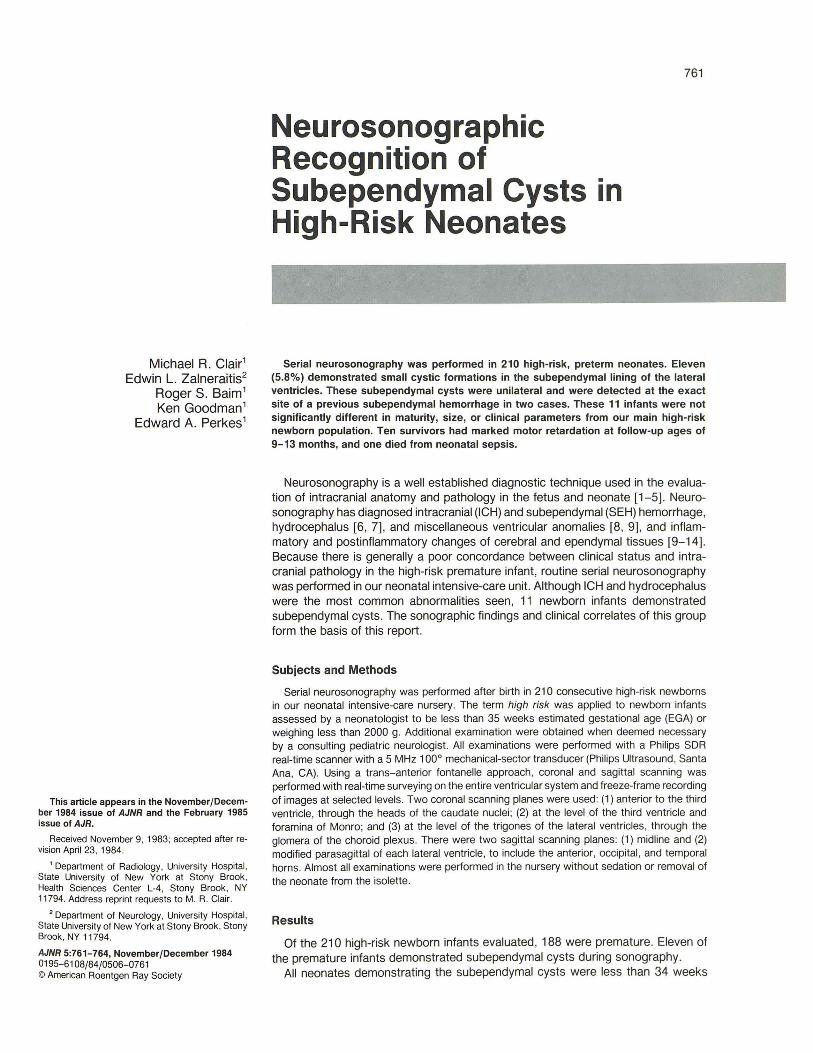
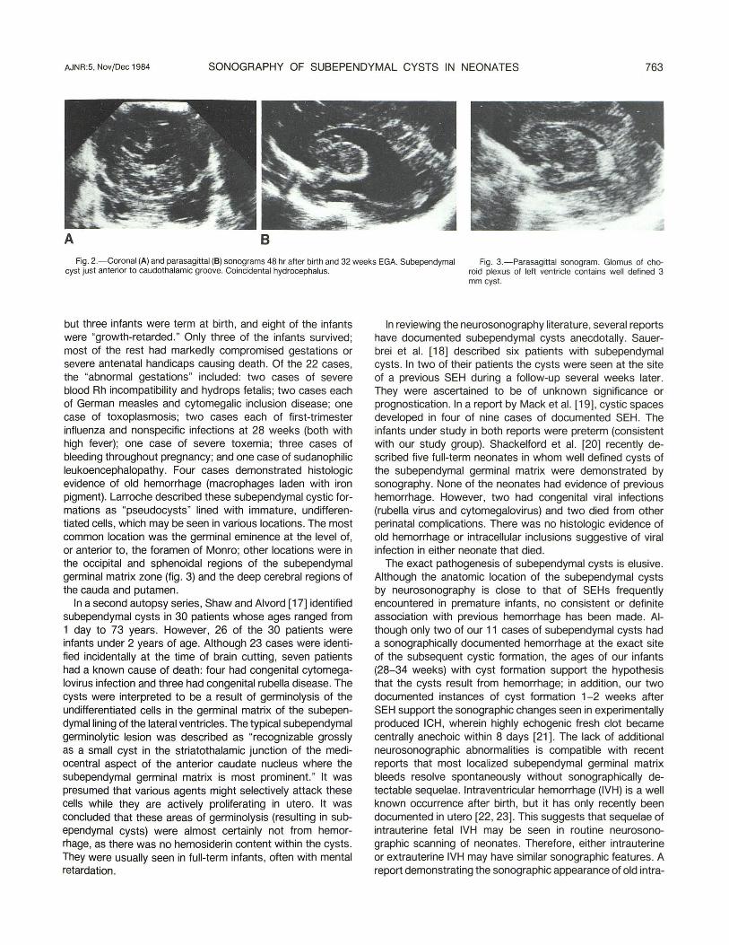
Sonographically, the subependymal cysts were unilocular, unilateral, well defined cystic formations, 10 mm or less in size. The cysts were most often seen adjacent to the foramen of Monro anterior to the caudothalamic grove (fig. 2). In three instances they were located in the glomus of the choroid plexus (fig. 3). Unequivocal hydrocephalus was coincident on initial scanning in one infant (fig . 2). The cystic formation, once detected, did not change in character or size during serial scanning over a 1-2 month period . One infant had a computed tomographic scan of the head that failed to detect the subependymal cyst.
One neonatal death resulted from infection; the 10 other neonates, at follow-up ages of 9-13 months, have motor dysfunction and delayed development.
Discussion
Subependymal cystic formations have been described in neuropathologic studies [15-17]. Larroche [15, 16] reported 22 subependymal cystic formations in the newborn infant. All
TABLE 2: Location, Size, and Associated Findings in Subependymal Cysts as Assessed by Sonography
No. of Neonates
Location: Caudothalamic groove Body or glomus of choroid
Size (mm): 1-3 ....... .. . 3-5 . . . . . . . . . . . . . . . . . . 5-10 ... .................. . . . . . . . . . . . . .
Associated sonographic findings: Uncomplicated ............... . .. . . . . . Subependymal hemorrhage Hydrocephalus
8 3
8 2 1
7 3* 1
• Two neonates had subependymal hemorrhage at site of subsequent subependymal cyst: one neonate had subependymal hemorrhage coincident with documentation of sube· pendymal cyst.
Fig. 1.-Modified parasagittal sonogram of right lateral ventricle within 24 hr after birth shows 14 mm SEH. B, 1 month later. Subependymal cyst in location of prior hemorrhage. Mild ventr:.:ular dilatation.
AJNR :5, Nov/Dec 1984 SONOGRAPHY OF SUBEPENDYMAL CYSTS IN NEONATES 763
A 8 Fig. 2.-Coronal (A) and parasagittal (8) sonograms 48 hr after birth and 32 weeks EGA. Subependymal Fig. 3.-Parasagittal sonogram. Glomus of cho-
cyst just anterior to caudothalamic groove. Coincidental hydrocephalus. roid plexus of left ventricle contains well defined 3 mm cyst.
but three infants were term at birth, and eight of the infants were "growth-retarded. " Only three of the infants survived ; most of the rest had markedly compromised gestations or severe antenatal handicaps causing death. Of the 22 cases, the "abnormal gestations" included: two cases of severe blood Rh incompatibility and hydrops fetalis ; two cases each of German measles and cytomegalic inclusion disease; one case of toxoplasmosis; two cases each of first-trimester influenza and nonspecific infections at 28 weeks (both with high fever); one case of severe toxemia; three cases of bleeding throughout pregnancy; and one case of sudanophilic leukoencephalopathy. Four cases demonstrated histologic evidence of old hemorrhage (macrophages laden with iron pigment). Larroche described these subependymal cystic formations as "pseudocysts" lined with immature, undifferentiated cells, which may be seen in various locations. The most common location was the germinal eminence at the level of, or anterior to, the foramen of Monro; other locations were in the occipital and sphenoidal regions of the subependymal germinal matrix zone (fig . 3) and the deep cerebral regions of the cauda and putamen.
In a second autopsy series, Shaw and Alvord [17] identified subependymal cysts in 30 patients whose ages ranged from 1 day to 73 years. However, 26 of the 30 patients were infants under 2 years of age. Although 23 cases were identified incidentally at the time of brain cutting, seven patients had a known cause of death: four had congenital cytomegalovirus infection and three had congenital rubella disease. The cysts were interpreted to be a result of germinolysis of the undifferentiated cells in the germinal matrix of the subependymallining of the lateral ventricles. The typical subependymal germinolytic lesion was described as "recognizable grossly as a small cyst in the striatothalamic junction of the mediocentral aspect of the anterior caudate nucleus where the subependymal germinal matrix is most prominent." It was presumed that various agents might selectively attack these cells while they are actively proliferating in utero. It was concluded that these areas of germinolysis (resulting in subependymal cysts) were almost certainly not from hemorrhage, as there was no hemosiderin content within the cysts . They were usually seen in full-term infants, often with mental retardation.
In reviewing the neurosonography literature, several reports have documented subependymal cysts anecdotally. Sauerbrei et al. [18] described six patients with subependymal cysts. In two of their patients the cysts were seen at the site of a previous SEH during a follow-up several weeks later. They were ascertained to be of unknown significance or prognostication. In a report by Mack et al. [19], cystic spaces developed in four of nine cases of documented SEH. The infants under study in both reports were preterm (consistent with our study group). Shackelford et al. [20] recently described five full-term neonates in whom well defined cysts of the subependymal germinal matrix were demonstrated by sonography. None of the neonates had evidence of previous hemorrhage. However, two had congenital viral infections (rubella virus and cytomegalovirus) and two died from other perinatal complications. There was no histologiC evidence of old hemorrhage or intracellular inclusions suggestive of viral infection in either neonate that died.
The exact pathogenesis of subependymal cysts is elusive. Although the anatomic location of the subependymal cysts by neurosonography is close to that of SEHs frequently encountered in premature infants, no consistent or definite association with previous hemorrhage has been made. Although only two of our 11 cases of subependymal cysts had a sonographically documented hemorrhage at the exact site of the subsequent cystic formation, the ages of our infants (28-34 weeks) with cyst formation support the hypotheSis that the cysts result from hemorrhage; in addition , our two documented instances of cyst formation 1-2 weeks after SEH support the sonographic changes seen in experimentally produced ICH, wherein highly echogenic fresh clot became centrally anechoic within 8 days [21]. The lack of additional neurosonographic abnormalities is compatible with recent reports that most localized subependymal germinal matrix bleeds resolve spontaneously without sonographically detectable sequelae. Intraventricular hemorrhage (IVH) is a well known occurrence after birth, but it has only recently been documented in utero [22, 23]. This suggests that sequelae of intrauterine fetal IVH may be seen in routine neurosonographic scanning of neonates. Therefore, either intrauterine or extrauterine IVH may have similar sonographic features. A report demonstrating the sonographic appearance of old intra-

764 CLAIR ET AL. AJNR:5, Nov/Dec 1984
ventricular blood present at birth from a fetal IVH, although markedly different in size and bilaterality from our cases, displayed sonographic qualities similar to those of small subependymal cysts [24].
Congenital infection is less easy to hypothesize as the cause of the cysts in our cases. No maternal infections were documented, and only one noncontributory brain sectioning was performed. However, maternal infection may be mild or subclinical and documentable only by serum titers; in only two of our cases were these investigations indicated.
Congenital infection by rubella and cytomegalovirus has been observed in association with subependymal cystic formations, and 23% of the autopsy cases of Shaw and Alvord [17] had proven viral associations. Subependymal cyst formation as part of a generalized disorder of development is supported by their presence in the cerebrohepatorenal syndrome and in the Shaw and Alvord study population, wherein more than one-third displayed a major congenital malformation such as a cardiac anomaly, cleft palate, or congenital cataracts [17, 25].
It becomes evident from our discussion that the formation of subependymal cysts may be caused by a very heterogeneous group of agents with a similar prototypical neuropathologic expression of destruction and lysis of the highly mitotic and vascular ependymal and subependymallayers of the fetal brain. This somewhat subtle and usually incidental lesion may therefore represent focal damage to immature precursor cells of the developing telencephalon. Rarely, it may be part of a more global cerebral or fetal insult. The association of subependymal cysts with neurotropic viruses and SEH, or as part of the pathologic expression of a more generalized syndrome, has not been demonstrated conclusively. However, it may be concluded that patients with subependymal cysts, regardless of the etiology, seem to be at greater risk for motor impairment and delayed development. Further investigation and close follow-up of all newborn infants who demonstrate subependymal cysts on neurosonography is indicated.
ACKNOWLEDGMENTS
We thank Susan Duhaime, Bob Allen, and Kathryn Nugent for technical assistance and Jerry Christiano and Alice Jiminez for help in manuscript preparation.
REFERENCES
1. London DA, Carroll BA, Enzmann DR. Sonography of ventricular size and germinal matrix hemorrhage in premature infants. AJNR 1980;1 :295-300, AJR 1980;135:559-564
2. Grant EG, Schellinger 0, Borts FT, et al. Real-time sonography of the neonatal and infant head. AJNR 1980;1 :487-492 , AJR 1981;136:265-270
3. Edwards MK, Brown DL, Muller J, Grossman CB, Chua GT. Cribside neurosonography: real-time sonography for intracranial investigation of the neonate. AJNR 1980;1 :501-505, AJR 1981 ;136:271-276
4. Babcock OS, Han BK. The accuracy of high resolution, real-time ultrasonography of the head in infancy. Radiology 1981; 139 : 665-676
5. Fleischer AC, Hutchinson AA, Kirchner SG, James AE. Cranial sonography of the preterm neonate. Diagn Imaging 1981 ;3:20-28
6. Garrett WJ, Kossoff G, Warren PS. Cerebral ventricular size in children . Radiology 1980;136:711-715
7. Skolnick ML, Rosenbaum AE, Matzuk T, Guthkelch AN, Heinz ER . Detection of dilated cerebral ventricles in infants: a correlative study between ultrasound and computed tomography. Radiology 1979;131 :447-451
8. Siovis TL, Kuhns LK. Real-time sonography of the brain through the anterior fontanelle. AJR 1981;136:277-286
9. Babcock OS, Han BK, LeQuesne GW. B-mode gray-scale ultrasound of the head in the newborn and young infant. AJNR 1980;1 : 181-192, AJR 1980;134: 457-468
10. Horbar JD, Philip AG, Lucey FJ. Ultrasound scan in neonatal ventriculitis. Lancet 1980;1 :976
11 . Sauerbrei EE, Cooperberg PL. Cystic tumors of the fetal and neonatal cerebrum: ultrasound and computed tomographic evaluation. Radiology 1983;147:689-692
12. Mack LA, Rumack CM, Johnson ML. Ultrasound evaluation of cystic intracranial lesions in the neonate. Radiology 1980; 137 :451-455
13. Sauerbrei EE, Cooperberg PL. Neonatal brain: sonography of congenital abnormalities. AJNR 1981;2 :125-128, AJR 1981 ; 136: 1167-1170
14. Stannard MW, Jimenez JF. Sonographic recognition of multiple cystiC encephalomalacia. AJNR 1983;4: 1111-1114, AJR 1983; 141 :1321-1324
15. Larroche JC. Subependymal pseudo-cysts in the newborn. Bioi Neonate 1972;21: 170-183
16. Larroche JC. Developmental pathology of the neonate. Amsterdam: Elsevier/North Holland, 1977:368-445
17. Shaw CM , Alvord EC. Subependymal germinolysis. Arch Neural 1974;31 :374-381
18. Sauerbrei EE, Digney M. Harrison PB, Cooperberg PL. Ultrasonic evaluation of neonatal intracranial hemorrhage and its complications. Radiology 1981;139:677-685
19. Mack LA, Wright K, Hirsch JH, et al. Intracranial hemorrhage in premature infants: accuracy of sonographic evaluation. AJR 1981;137:245-250
20. Shackelford GO, Fulling KH, Glasier CM. Cysts of the subependymal germinal matrix: sonographic demonstration with pathologic correlation. Radiology 1983;149: 117-121
21 . Enzmann DR, Britt RH, Lyons BE, Buxton JL, Wilson DA. Natural history of experimental intracerebral hemorrhage: sonography, computed tomography and neuropathology. AJNR 1981;2:517-526
22 . Lustig-Gillman I, Young BK, Silverman F, et al. Fetal intraventricular hemorrhage: sonographic diagnosis and clinical implications. JCU 1983;11:277-280
23. Kim MS, Elyarderani MK. Sonographic diagnosis of cerebroventricular hemorrhage in utero. Radiology 1982; 142 : 479-480
24. Donn SM, DiPietro MA, Faix RG, Bowerman RA. The sonographic appearance of old intraventricular hemorrhage present at birth. J Ultrasound Med 1983;2:283-284
25. Sommer A, Bradel EJ, Hamoudi AB. The cerebro-hepato-renal syndrome (Zellweger's syndrome). Bioi Neonate 1974;25 :219-229