Myxomycetes of Mustafa Kemal University campus and...
9
769 Turk J Bot 36 (2012) 769-777 © TÜBİTAK doi:10.3906/bot-1103-10 Myxomycetes of Mustafa Kemal University campus and environs (Turkey) Hayri BABA* Biology Department, Faculty of Science and Arts, Mustafa Kemal University, Alahan-31000, Antakya, Hatay - TURKEY Received: 22.03.2011 ● Accepted: 05.06.2012 Abstract: In this taxonomic study, myxomycetes of Tayfur Sökmen Campus (Hatay) were collected during 2010-2011. As a result of field and laboratory studies we reported 44 species of protosteliomycetes and myxomycetes. ree of these species (Diderma deplanatum Fr., Didymium megalosporum Berk & M.A.Curtis, and Lamproderma atrosporum Meyl.) are recorded for the first time from Turkey. Lamproderma atrosporum was treated with the moist chamber cultures method in the laboratory but Didymium megalosporum and Diderma deplanatum were determined naturally. e distribution, habitat, and collection numbers of the identified species are given. Key words: Hatay, fungal diversity, new records, myxomycetes, Turkey Research Article * E-mail: [email protected] Introduction Myxomycetes (acellular, non-cellular, plasmodial, or true slime moulds) are characterised by an amorphous, multinucleate, protoplasmic mass called the plasmodium as well as fruiting bodies (1-200 mm) with internally borne spores (5-20 µm). ey have been known for more than 350 years based on Pankow’s figure and description of Lycogala epidendrum (L.) Fr. (Martin & Alexopoulos, 1969). Myxomycetes have been classified in the kingdom Plantae (class Myxomycota) and the kingdom Animalia (class Mycetozoa). Because myxomycetes are typically found in the same habitats as fungi, they were treated as taxa within the kingdom Fungi (class Myxomycetes). Unlike fungi, myxomycetes do not excrete extracellular, digestive enzymes, and the role of myxomycetes in the environment is not as decomposers or pathogens (Keller & Braun, 1999). Bauldauf and Doolittle (1997) conducted a phylogenetic analysis of highly conserved, elongation factor 1-alpha (EF-1α) gene sequences and showed that myxomycetes are not fungi. Physiology, morphology, life history, and genetic analysis support the classification of myxomycetes in the kingdom Protoctista along with other eukaryotic micro- organisms (Everhart & Keller, 2008). Myxomycetes are common and relatively cosmopolitan in their distribution. ey have been widely studied in worldwide, but those from Turkey are still poorly known. e number of known myxomycetes species in the world is about 1000 (Lado, 2001). e myxomycetes flora of Turkey has not been fully explored. e first extensive studies in Turkey were carried out by Finnish scientists (Härkönen & Uotila, 1983; Härkönen, 1987; Härkönen & Ukkola, 2000). In recent years some macrofungi and myxomycetes have been added to
-
Upload
vuongtuyen -
Category
Documents
-
view
221 -
download
2
Transcript of Myxomycetes of Mustafa Kemal University campus and...

769
Turk J Bot36 (2012) 769-777© TÜBİTAKdoi:10.3906/bot-1103-10
Myxomycetes of Mustafa Kemal University campus and environs (Turkey)
Hayri BABA*Biology Department, Faculty of Science and Arts, Mustafa Kemal University, Alahan-31000, Antakya, Hatay - TURKEY
Received: 22.03.2011 ● Accepted: 05.06.2012
Abstract: In this taxonomic study, myxomycetes of Tayfur Sökmen Campus (Hatay) were collected during 2010-2011. As a result of field and laboratory studies we reported 44 species of protosteliomycetes and myxomycetes. Three of these species (Diderma deplanatum Fr., Didymium megalosporum Berk & M.A.Curtis, and Lamproderma atrosporum Meyl.) are recorded for the first time from Turkey. Lamproderma atrosporum was treated with the moist chamber cultures method in the laboratory but Didymium megalosporum and Diderma deplanatum were determined naturally. The distribution, habitat, and collection numbers of the identified species are given.
Key words: Hatay, fungal diversity, new records, myxomycetes, Turkey
Research Article
* E-mail: [email protected]
IntroductionMyxomycetes (acellular, non-cellular, plasmodial, or true slime moulds) are characterised by an amorphous, multinucleate, protoplasmic mass called the plasmodium as well as fruiting bodies (1-200 mm) with internally borne spores (5-20 µm). They have been known for more than 350 years based on Pankow’s figure and description of Lycogala epidendrum (L.) Fr. (Martin & Alexopoulos, 1969).
Myxomycetes have been classified in the kingdom Plantae (class Myxomycota) and the kingdom Animalia (class Mycetozoa). Because myxomycetes are typically found in the same habitats as fungi, they were treated as taxa within the kingdom Fungi (class Myxomycetes). Unlike fungi, myxomycetes do not excrete extracellular, digestive enzymes, and the role of myxomycetes in the environment is not as decomposers or pathogens (Keller & Braun,
1999). Bauldauf and Doolittle (1997) conducted a phylogenetic analysis of highly conserved, elongation factor 1-alpha (EF-1α) gene sequences and showed that myxomycetes are not fungi. Physiology, morphology, life history, and genetic analysis support the classification of myxomycetes in the kingdom Protoctista along with other eukaryotic micro-organisms (Everhart & Keller, 2008).
Myxomycetes are common and relatively cosmopolitan in their distribution. They have been widely studied in worldwide, but those from Turkey are still poorly known. The number of known myxomycetes species in the world is about 1000 (Lado, 2001). The myxomycetes flora of Turkey has not been fully explored. The first extensive studies in Turkey were carried out by Finnish scientists (Härkönen & Uotila, 1983; Härkönen, 1987; Härkönen & Ukkola, 2000). In recent years some macrofungi and myxomycetes have been added to

Myxomycetes of Mustafa Kemal University campus and environs (Turkey)
770
the Turkish myxobiota by some researchers (Baba et al., 2008; Doğan & Karadelev, 2009; Alkan et al., 2010; Doğan et al., 2011; Demirel & Kaşık, 2012). So far, 226 species of myxomycetes have been reported from Turkey (Kaşık, 2010). While much research has been done on myxomycetes, the Turkish mycoflora are still incomplete and there have been no previous studies in Hatay. Now 3 taxa have been added to the Turkish myxomycetes flora as new records: Diderma deplanatum Fr., Didymium megalosporum Berk. & M.A.Curtis, and Lamproderma atrosporum Meyl.
Description of the research area
The myxomycete specimens in the current study were collected in and around the Tayfur Sökmen Campus of Mustafa Kemal University during 2010 and 2011. The campus is located outside the city of Hatay, in Alahan village district (Figure 1).
The area is situated in the Mediterranean phytogeographical region and square C6 according to Davis’s grid system (Davis, 1965-1985). The research area is surrounded to the north by the Amanos Mountains and Alahan village, to the south by Amik plain, to the west by Dikmece village, and to the east by the town of Serinyol. The altitude of the land is 100 m in the region of the highway and the village of Alahan is 350 m a.s.l.
The study area is naturally covered with Pinus brutia Ten., scrub vegetation, and furigana formation. In this type of vegetation the woody species Arbutus andrachne L., Quercus coccifera L., and Phillyrea latifolia L. are found; also, in these regions Olea europea L. is grown as a culture plant.
According to meteorological data from the directorate of Hatay, the highest average monthly temperature is in August (27.6 °C), while the lowest
Figure 1. The research area and localities where samples were collected.
1 2 3 4 5 6 7 8 942°
40°
38°
36°26° 28° 30°
0 200 km
32° 34° 36° 38° 40° 42° 44°
A
B
C
N
W E
S
MediterraneanSea

H. BABA
771
average monthly temperature is in December (7.7 °C). The average monthly maximum rainfall of Antakya is in December (192.4 mm), while the lowest average monthly rainfall is in August (3.5 mm) (MGM, 2007).
Materials and methodsThe specimens on natural substrata, bark and debris, the bark of living trees, as well as on decaying bark, wood, leaves, and litter were collected. Natural mature fructifications were gently and directly collected from the substratum and placed in cardboard herbarium boxes. In addition, the fructifications of myxomycetes were as obtained from the moist chamber culture in the laboratory. The cultures were moistened with distilled water. The moist chambers were examined every day under a dissecting microscope. When developing myxomycetes were found, the moist chamber was allowed to dry slowly and the myxomycetes were then dried for 1 week. The same chambers were then rewetted for another 4-week period and examined as before (Martin et al., 1983; Stephenson & Stempen, 2000).
Microscopic and macroscopic features of the samples were determined in the laboratory. The morphological characters examined included fruiting bodies’ shape, size and colour, spore size and ornamentation, capillitium colour and branching, lime crystal size and morphology, and stalk colour and proportion. In addition, some photographs from characteristic qualitative objects were taken. All data were evaluated comparatively for taxonomical aims.
The myxomycete specimens were identified with the aid of Martin and Alexopoulos (1969), Neubert et al. (1993, 1995, 2000), and Sesli and Denchev (2008). The samples were prepared as fungarium material and stored in the laboratory of Department of Biology, Faculty of Science and Arts, Mustafa Kemal University in Hatay, and in the author’s personal collection.
ResultsForty-four species belonging to 11 families of myxomycetes from both the field and moist chamber culture were identified.
Protosteliomycetes
Protosteliales
Ceratiomyxaceae
1. Ceratiomyxa fruticulosa (O.F.Müll.) T.Macbr.
Above Alahan village on wood of conifers, 36°19′55″N, 36°11′05″E, 350 m, 06.11.2010, Baba 18.
Myxomycetes
Echinosteliales
Echinosteliaceae
2. Echinostelium minutum de Bary
Above Alahan village on wood of conifers, 36°19′55″N, 36°11′05″E, 350 m, 06.11.2010, Baba 12,17, 25. In front of Faculty of Ag-riculture on debris of maple, 36°19′30″N, 36°11′45″E, 122 m, 26.01.2010, Baba 11. Across from the Faculty of Medicine on the body of Pinus sp., 36°20′08″N, 36°11′54″E, 113 m, 16.01.2011, Baba 2.
Liceales
Cribrariaceae
3. Cribraria cancellata var. fusca (G.Lister) Nann.-Bremek.
Above Alahan village in coniferous woods, 36°19′55″N, 36°11′05″E, 350 m, 06.11.2010, Baba 9, 30, 41.
4. Cribraria violacea Rex
Over Alahan village in coniferous woods, 36°19′55″N, 36°11′05″E, 350 m, 06.11.2010, Baba 1.
5. Cribraria vulgaris Schrad.
Opposite the Sabancı hostel, on debris, 36°18′27″N, 36°11′58″E, 105 m, 20.10.2010, Baba 24.
Dictydiaethaliaceae
6. Dictydiaethalium plumbeum (Schumach.) Rostaf.
Around the library on debris, 36°19′39″N, 36°11′46″E, 140 m, 24.12.2010, Baba 4.
Myxomycetes of Mustafa Kemal University campus and environs (Turkey)
772
Liceaceae
7. Licea biforis Morgan
Above Alahan village in coniferous woods, 36°19′55″N, 36°11′05″E, 350 m, 06.11.2010, Baba 26.
8. Licea castanea G.Lister
Above Alahan village in coniferous woods, 36°19′55″N, 36°11′05″E, 350 m, 06.11.2010, Baba 30.
9. Licea kleistobolus G.W.Martin
Above Alahan village in coniferous woods, 36°19′55″N, 36°11′05″E, 350 m, 06.11.2010, Baba 26.
10. Licea minima Fr.
Above Alahan village in coniferous woods, 36°19′55″N, 36°11′05″E, 350 m, 06.11.2010, Baba 9, 48.
11. Licea pusilla Schrad.
Across from the Faculty of Medicine on de-bris of Pinus sp., 36°20′08″N, 36°11′54″E, 113 m, 16.01.2011, Baba 2.
12. Licea variabilis Schrad.
Anayazı village on debris, 36°19′00″N, 36°11′30″E, 130 m, 24.11.2010, Baba 11.
Reticulariaceae13. Lycogala epidendrum (J.C.Buxb. ex L.) Fr. Zülüflühan Cemetery on debris of wood,
36°19′00″N, 36°11′30″E, 130 m, 23.12.2010, Baba 10.
PhysaralesDidymiaceae
14. Diderma deplanatum Fr., Syst. Mycol. 3: 110 (1829).
Syn.: Leocarpus deplanatus (Fr.) Fr.; Chondrioderma deplanatum (Fr.) Rostaf.; Chondrioderma niveum var. deplanatum (Fr.) Lister; Diderma niveum var. deplanatum (Fr.) G.Lister; Diderma niveum var. deplanatum (Fr.) Meyl.; Diderma deplanatum f. pulverulentum Meslin.
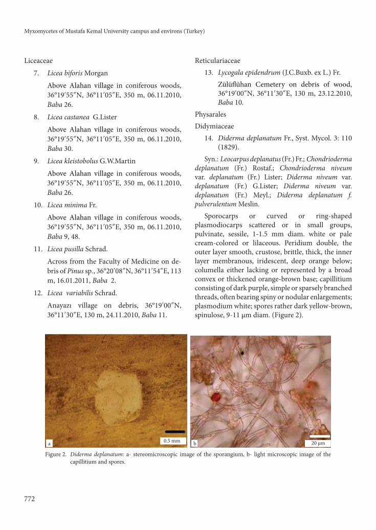
Sporocarps or curved or ring-shaped plasmodiocarps scattered or in small groups, pulvinate, sessile, 1-1.5 mm diam. white or pale cream-colored or lilaceous. Peridium double, the outer layer smooth, crustose, brittle, thick, the inner layer membranous, iridescent, deep orange below; columella either lacking or represented by a broad convex or thickened orange-brown base; capillitium consisting of dark purple, simple or sparsely branched threads, often bearing spiny or nodular enlargements; plasmodium white; spores rather dark yellow-brown, spinulose, 9-11 µm diam. (Figure 2).
0.5 mma b 20 µm
Figure 2. Diderma deplanatum: a- stereomicroscopic image of the sporangium, b- light microscopic image of the capillitium and spores.
H. BABA
773
Locality: Across from the Faculty of Arts and Sciences on debris, 36°19′39″N, 36°11′46″E, 119 m, 23.12.2010, Baba 12.
15. Didymium difforme (Pers.) S.F.Gray Above Alahan village in coniferous woods,
36°19′55″N, 36°11′05″E, 350 m, 06.11.2010, Baba 47. In front of Faculty of Agriculture on debris of maple, 36°19′30″N, 36°11′45″E, 122 m, 22.12.2010, Baba 34, 78. Across from the Faculty of Medicine on debris of Pinus sp., 36°20′08″N, 36°11′54″E, 113 m, 30.12.2010, Baba 22, 58.
16. Didymium megalosporum Berk. & M.A.Curtis, in Berkeley, Grevillea 2:53 (1873).
Syn.: Didymium eximium Peck; Didymium ful-vellum Massee; Didymium nigripes var. eximium (Peck) A.Lister; Didymium discoideum K.S.Thind & H.S.Sehgal.
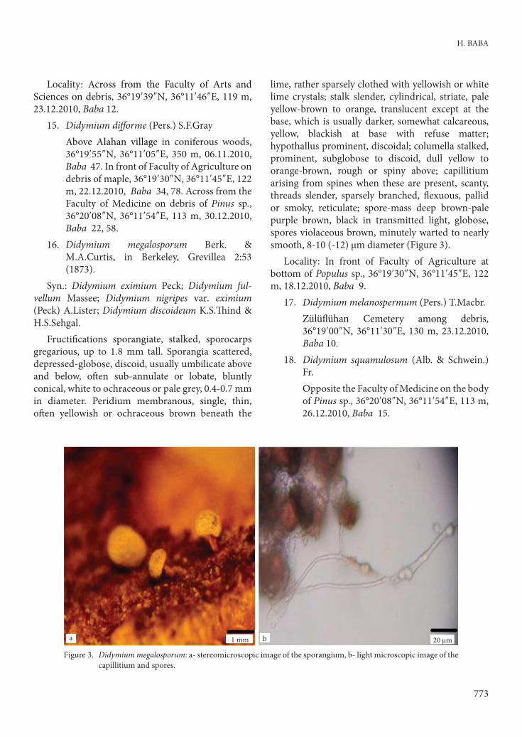
Fructifications sporangiate, stalked, sporocarps gregarious, up to 1.8 mm tall. Sporangia scattered, depressed-globose, discoid, usually umbilicate above and below, often sub-annulate or lobate, bluntly conical, white to ochraceous or pale grey, 0.4-0.7 mm in diameter. Peridium membranous, single, thin, often yellowish or ochraceous brown beneath the
lime, rather sparsely clothed with yellowish or white lime crystals; stalk slender, cylindrical, striate, pale yellow-brown to orange, translucent except at the base, which is usually darker, somewhat calcareous, yellow, blackish at base with refuse matter; hypothallus prominent, discoidal; columella stalked, prominent, subglobose to discoid, dull yellow to orange-brown, rough or spiny above; capillitium arising from spines when these are present, scanty, threads slender, sparsely branched, flexuous, pallid or smoky, reticulate; spore-mass deep brown-pale purple brown, black in transmitted light, globose, spores violaceous brown, minutely warted to nearly smooth, 8-10 (-12) µm diameter (Figure 3).
Locality: In front of Faculty of Agriculture at bottom of Populus sp., 36°19′30″N, 36°11′45″E, 122 m, 18.12.2010, Baba 9.
17. Didymium melanospermum (Pers.) T.Macbr. Zülüflühan Cemetery among debris,
36°19′00″N, 36°11′30″E, 130 m, 23.12.2010, Baba 10.
18. Didymium squamulosum (Alb. & Schwein.) Fr.
Opposite the Faculty of Medicine on the body of Pinus sp., 36°20′08″N, 36°11′54″E, 113 m, 26.12.2010, Baba 15.
1 mma b 20 µm
Figure 3. Didymium megalosporum: a- stereomicroscopic image of the sporangium, b- light microscopic image of the capillitium and spores.

Myxomycetes of Mustafa Kemal University campus and environs (Turkey)
774
Physaraceae19. Badhamia macrocarpa (Ces.) Rostaf. In front of Faculty of Agriculture at bottom
of maple, 36°19′30″N, 36°11′45″E, 122 m, 30.12.2010, Baba 78.
20. Physarum cinereum (Batsch) Pers. In front of Faculty of Agriculture at bottom
of maple, 36°19′30″N, 36°11′45″E, 122 m, 22.12.2010, Baba 34. Across from the Faculty of Arts and Sciences on debris, 36°19′39″N, 36°11′46″E, 119 m, 30.12.2010, Baba 24, 45.
21. Physarum notabile T.Macbr. Anayazı village on debris, 36°19′00″N,
36°11′30″E, 130 m, 24.11.2010, Baba 7.22. Physarum leucopheum Fr. Above Alahan village in coniferous woods,
36°19′55″N, 36°11′05″E, 350 m, 06.11.2010, Baba 42.
StemonitalesStemonitidaceae
23. Collaria elegans (Racib.) Dhillon & Nann.-Bremek.
In front of Faculty of Agriculture at bottom of maple, 36°19′30″N, 36°11′45″E, 122 m, 26.01.2010, Baba 4.
24. Collaria lurida (Lister) Nann.-Bremek. Above Alahan village in coniferous woods,
36°19′55″N, 36°11′05″E, 350 m, 06.11.2010, Baba 8.
25. Comatrichia ellae Härk. Above Alahan village in coniferous woods,
36°19′55″N, 36°11′05″E, 350 m, 06.11.2010, Baba 41.
26. Comatrichia laxa Rostaf. In front of Faculty of Agriculture at bottom
of maple, 36°19′30″N, 36°11′45″E, 122 m, 29.12.2010, Baba 43. Above Alahan village in coniferous woods, 36°19′55″N, 36°11′05″E, 350 m, 06.11.2010, Baba 45.
27. Comatrichia pulchella var. pulchella (C.Bab.) Rostaf.
Across from the Faculty of Arts and Sciences on debris, 36°19′39″N, 36°11′46″E, 119 m, 01.11.2010, Baba 26.
28. Comatrichia tenerrima (Berk. & M.A.Curtis) G.Lister
Anayazı village on debris, 36°19′00″N, 36°11′30″E,130 m, 22.12.2010, Baba 35.
29. Lamproderma atrosporum Meyl. Bull. Soc. Vaud. Sci. Nat. 46: 51; 1910.
Syn.: Lamproderma cribrarioides sensu Kow-alski.
Sporangia dull purplish black, with metallic sheen but not iridescent, sporocarps stipitate, sessile or stalked sporangia are usually ovoid sometimes globose, forming small colonies, plasmodiocarpous. Stalk usually short, black and opaque; peridium is usually evanescent except for a basal cup and small fragments remaining attached to the capillitium, persistent, commonly iridescent and leaving a collar, outermost branchlets of capillitium often attached to peridial fragments by yellowish, funnel-shaped expansions, rest of peridium disappearing except at the base where it forms a cup; columella stout up to about mid-way, usually truncate and with most of the capillitium arising from the top; capillitium often radiating from the centre, frequently branched and sometimes anastomosed, usually with free ends at the periphery. Outermost branchlets of capillitium often attached to peridial fragments by yellowish, funnel-shaped expansions; spore-mass deep brown-pale purple brown, or black in transmitted light, globose, warted or partly to completely spiny-reticulate, 12-18 µm diameter (Figure 4).
Locality: Near the Faculty of Science and Medicine on the twigs and leaves of Eucalyptus sp., 36°20′08″N, 36°11′54″E, 113 m, 16.01.2011, Baba 4.
30. Macbrideola cornea var. cornea (G.Lister & Cran) Alexop.
Above way to Alahan village under Pinus sp., 36°19′30″N, 36°11′45″E, 145 m, 14.10.2010, Baba 10.
31. Stemonitis flavogenita E.Jahn Above Alahan village in coniferous woods,
36°19′55″N, 36°11′05″E, 350 m, 06.11.2010, Baba 1.

H. BABA
775
32. Stemonitis foliicola Ing
In front of Faculty of Agriculture at foot of maple, 36°19′30″N, 36°11′45″E, 122 m, 26.01.2010, Baba 1.
33. Stemonitis fusca Roth
Alahan village in coniferous forest, 36°19′55″N, 36°11′05″E, 222 m, 25.06.2010, Baba 8.
34. Stemonitis herbatica Peck.
Above Alahan village in coniferous woods, 36°19′55″N, 36°11′05″E, 350 m, 06.11.2010, Baba 22.
35. Stemonitopsis amoena (Nann.-Bremek.) Nann.-Bremek.
Opposite the Faculty of Agriculture at bottom of maple, 36°19′30″N, 36°11′45″E, 122 m, 22.12.2010, Baba 14. Above Alahan village in coniferous woods, 36°19′55″N, 36°11′05″E, 350 m, 06.11.2010, Baba 12.
TrichialesArcyriaceae
36. Arcyria cinerea (Bull.) Pers. Opposite the Faculty of Agriculture at bottom
of maple, 36°19′30″N, 36°11′45″E, 122 m, 22.12.2010, Baba 17. Above Alahan village in coniferous woods, 36°19′55″N, 36°11′05″E, 350 m, 06.11.2010, Baba 3, 38.
37. Arcyria denudata (L.) Wettst. Above Alahan village in coniferous woods,
36°19′55″N, 36°11′05″E, 350 m, 06.11.2010, Baba 6.
38. Arcyria incarnata (Pers.) Pers. Above Alahan village in coniferous woods,
36°19′55″N, 36°11′05″E, 350 m, 06.11.2010, Baba 13.
39. Arcyria minuta Buchet Back of Sabancı residence on Quercus sp.
debris, 36°18′27″N, 36°11′58″E, 105 m, 20.10.2010, Baba 5.
0.5 mma b 20 µm
Figure 4. Lamproderma atrosporum: a- stereomicroscopic image of the sporangium, b- light microscopic image of the capillitium and spores.

Myxomycetes of Mustafa Kemal University campus and environs (Turkey)
776
40. Arcyria pomiformis (Leers) Rostaf. Near the Faculty of Arts and Sciences on
debris, 36°19′39″N, 36°11′46″E, 119 m, 01.11.2010, Baba 26.
Trichiaceae41. Perichaena corticalis (Batsch ) Rostaf. Opposite the Faculty of Agriculture at bot-
tom of maple, 36°19′30″N, 36°11′45″E, 122 m, 31.12.2010, Baba 63.
42. Perichaena depressa Lib. Opposite the Faculty of Agriculture un-
der maple, 36°19′30″N, 36°11′45″E, 122 m, 01.11.2010, Baba 42.
43. Perichaena vermicularis (Schwein.) Rostaf. Around the library on debris, 36°19′39″N,
36°11′46″E, 140 m, 24.12.2010, Baba 20.44. Trichia botrytis (J.F.Gmel.) Pers. Above Alahan village in coniferous woods,
36°19′55″N, 36°11′05″E, 350 m, 06.11.2010, Baba 12.
DiscussionForty-four species belonging to 11 families of protosteliomycetes and myxomycetes were identified in this investigation. Three species are new records for Turkey: Diderma deplanatum, Didymium megalosporum, and Lamproderma atrosporum.
In the study area, very rich myxomycete flora was observed. The climatic conditions are suitable for myxomycetes; a very dense flora in both species composition and frequency of myxomycetes was found. Twenty-two myxomycetes were collected in the field. As seen in several reports in the literature, naturally growing myxomycete fructifications are more common in the samples collected in rainy August and September. In fact, as pointed out in many studies, the frequency of myxomycetes from natural habitats is affected by climatic conditions, rainfall, and temperature.
Echinostelium minutum, Arcyria cinerea, and Didymium difforme are the most common species in our investigation but some species are only from certain substrates.
The most important features of Diderma deplanatum are stalk absent, columella absent or represented by thickened base, capillitial thread of one type, dark purple and elastic, often bearing nodular enlargement. Spores rather dark brown, finely spinulose, oblong to ring shaped plasmodiocarps.
The typical features of Didymium megalosporum are fructifications stalked, sporangia subglobose or umbilicate below, spores minutely warted or nearly smooth. Stalk slender, cylindrical, striate, pale yellow-brown to orange, peridium and capillitium has lime crystals. In our study the habitat and distribution of this species are similar to those of species known elsewhere in the world. Habitat is dead leaves and twigs; spore size range is 8-10 µm. According to Martin and Alexopoulos (1969), fructifications vary greatly in shape and the columella is also shaped like the contours of the fruiting bodies. According to Farr (1981), this rather uncommon species occurs on leaf litter and other plant debris mostly in the east. According to Hagelstein (1944), it seems to favour drier locations. The genus Didymium was collected from different substrates, such as Abies Mill., Crataegus L., Juglans L., Juniperus L., Picea Link, Pinus L., Populus L., Quercus L., Sorbus L., and Ulmus L. (Baba et al., 2008; Demirel & Kaşık, 2012); we collected this species from the bark of Populus sp.
Lamproderma atrosporum is stalked, black sporangia are usually ovoid, globose, forming small colonies, peridium is evanescent except for a basal cup and small fragments remaining attached to the capillitium. Capillitium is often attached to peridial fragments by yellowish, funnel-shaped expansions. Spores are warted-reticulate or spiny or partly to completely spiny-reticulate. According to Farr (1981), this is one of the western species, found in melting snowbanks in the mountains. We collected this species on the twigs and leaves of Eucalyptus sp.
With this study 3 new Myxomycetes were added to the myxobiota of Turkey and all of the species are recorded for the first time in Hatay.
AcknowledgementThis study was supported by Mustafa Kemal University Scientific Research Projects (BAP) (Project No: 1001 M 0113).

H. BABA
777
References
Alkan S, Kaşık G & Aktaş S (2010). Macrofungi of Derebucak district (Konya, Turkey). Turkish Journal of Botany 34: 335-350.
Baba H, Tamer AÜ & Kalyoncu F (2008). New myxomycete records for Turkey: one new genus and three new species. Turkish Journal of Botany 32: 329-332
Baldauf SL & Doolittle WF (1997). Origin and evolution of the slime molds. Proceedings of the National Academy of Science 94: 12007-12012.
Davis PH (ed.) (1965-1985). Flora of Turkey and the East Aegean Islands. Vols. 1-9, Edinburgh: Edinburgh Univ. Press.
Demirel G & Kaşık G (2012). Four new records for Physarales from Turkey. Turkish Journal of Botany 36: 95-100.
Doğan HH & Karadelev M (2009). Phellinus sulphurascens (Hymenochaetaceae, Basidiomycota): a very rare wood-decay fungus in Europe collected in Turkey. Turkish Journal of Botany 33: 239-242.
Doğan HH, Karadelev M & Işıloğlu M (2011). Macrofungal diversity associated with the scale-leaf juniper trees, Juniperus excelsa and J. foetidissima, distributed in Turkey. Turkish Journal of Botany 35: 219-237.
Everhart SE & Keller HW (2008). Life history strategies of corticolous Myxomycetes: the life cycle, plasmodial types, fruiting bodies, and taxonomic orders. Fungal Diversity 29: 1-16.
Farr ML (1981). True Slime Molds. p. 132. Dubuque Iowa: Wm. C. Brown Comp. Hagelstein R (1944). The Mycetozoa of North America. Published by the author. Mineola, New York.
Härkönen M & Uotila P (1983). Turkish Myxomycetes developed in moist chamber cultures. Karstenia 23: 1-9.
Härkönen M (1987). Some additions to the knowledge of Turkish Myxomycetes. Karstenia 27: 1-7.
Härkönen M & Ukkola T (2000). Conclusions on Myxomycetes compiled over twenty-five years from 4793 moist chamber cultures. Stapfia 73, zugleich Kataloge des OO. Landesmuseums, Neue Folge Nr. 155: 105-112.
Kaşık G (2010). Mantar Bilimi (1. baskı). Marifet matbaa ve Yay. Selçuk Üniv. Fen Fak. Biyoloji Böl. Konya. (in Turkish).
Keller HW & Braun KL (1999). Myxomycetes of Ohio: Their Systematics, Biology, and Use in Teaching. Ohio Biological Survey Bulletin New Series, Vol. 13, Number 2. Columbus, Ohio.
Lado C (2001). Nomenmyx. A nomenclatural taxabase of Myxomycetes. Madrid.
Martin GW & Alexopoulos CJ (1969). The Myxomycetes. University of Iowa Press, Iowa City.
Martin GW, Alexopoulos CJ & Farr ML (1983). The Genera of Myxomycetes. Univ. Iowa Press, Iowa City.
MGM (2007). Hatay iline ait sıcaklık ve yağış değerleri, Meteoroloji Genel Müdürlüğü, Ankara (in Turkish).
Neubert H, Nowotny W & Baumann K (1993). Die Myxomyceten (Band I). Gomaringen: Karlheinz Baumann Verlag.
Neubert H, Nowotny W & Baumann K (1995). Die Myxomyceten (Band II). Karlheinz Baumann Verlag Gomaringen.
Neubert H, Nowotny W, Baumann K & Marx H (2000). Die Myxo-myceten (Band III). Karlheinz Baumann Verlag Gomaringen.
Sesli E & Denchev CM (2008). Checklists of the Myxomycetes, Larger Ascomycetes and Larger Basidiomycetes in Turkey. Mycotaxon 106: 65-67.
Stephenson SL & Stempen H (2000). A Handbook of Slime Moulds. p. 183. Oregon: Timber Press, Portland.


![Mustafa Kemal Atatürk - resources.saylor.org · Mustafa Kemal Atatürk (pronounced [musˈtafa keˈmaɫ ataˈtyɾk]; 19 May 1881 by a posteriori [1] –10 November 1938) was an Ottoman](https://static.fdocuments.us/doc/165x107/5c7b4a1b09d3f277748b8666/mustafa-kemal-atatuerk-mustafa-kemal-atatuerk-pronounced-mustafa-kema.jpg)
