Multimodal MRI Brain Tumor Image Segmentation Using Sparse ...
Upload
pooja-g-nCategory
view
86download
2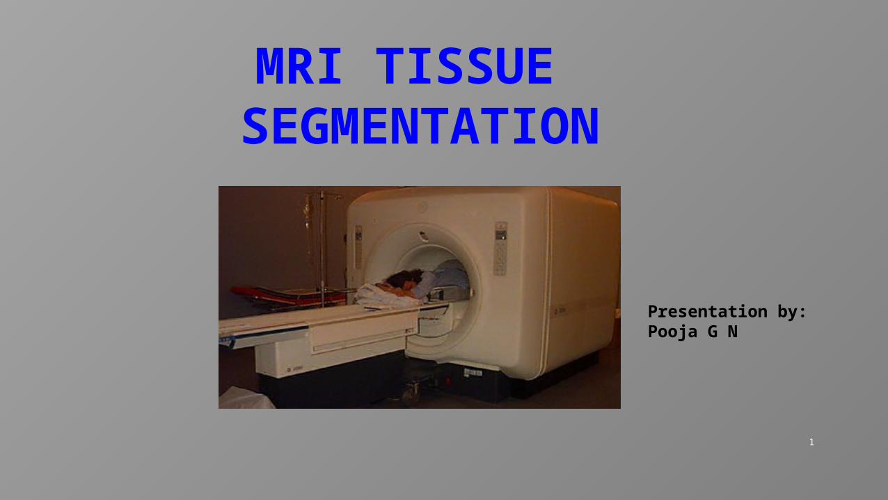
1
MRI TISSUE SEGMENTATION
Presentation by:Pooja G N
2
CONTENTS Image Image processing MRI MRI tissue segmentation Segmentation methods Bias field Bias field correction methods Inhomogeneties Energy minimization K-means algorithm Related work
3
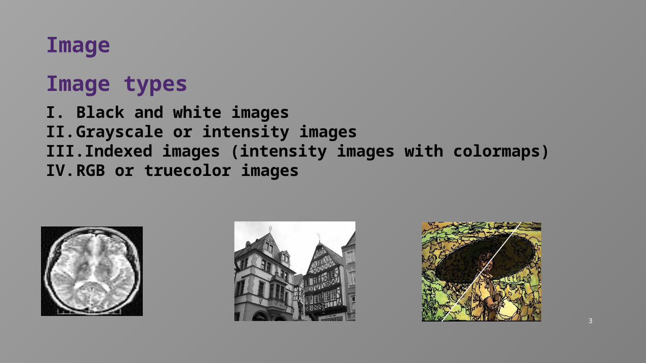
Image
Image typesI. Black and white imagesII. Grayscale or intensity imagesIII. Indexed images (intensity images with colormaps)IV. RGB or truecolor images
4
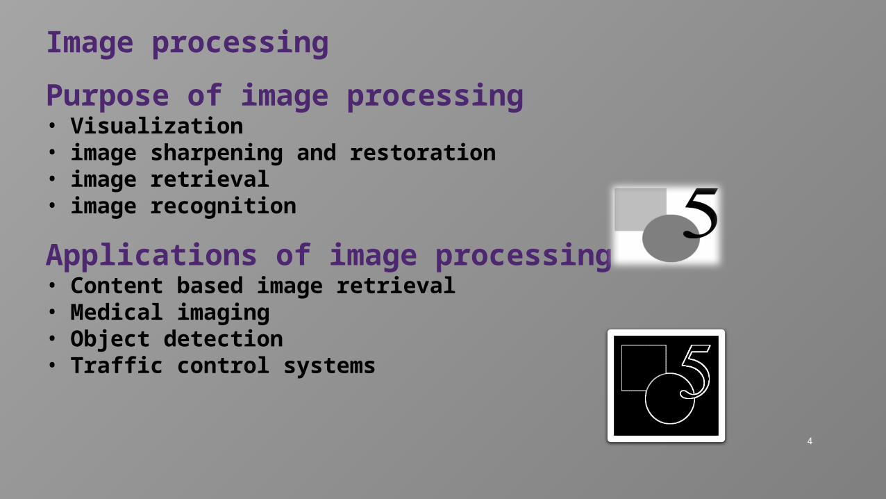
Image processing
Purpose of image processing• Visualization• image sharpening and restoration• image retrieval• image recognition
Applications of image processing• Content based image retrieval• Medical imaging• Object detection• Traffic control systems
5
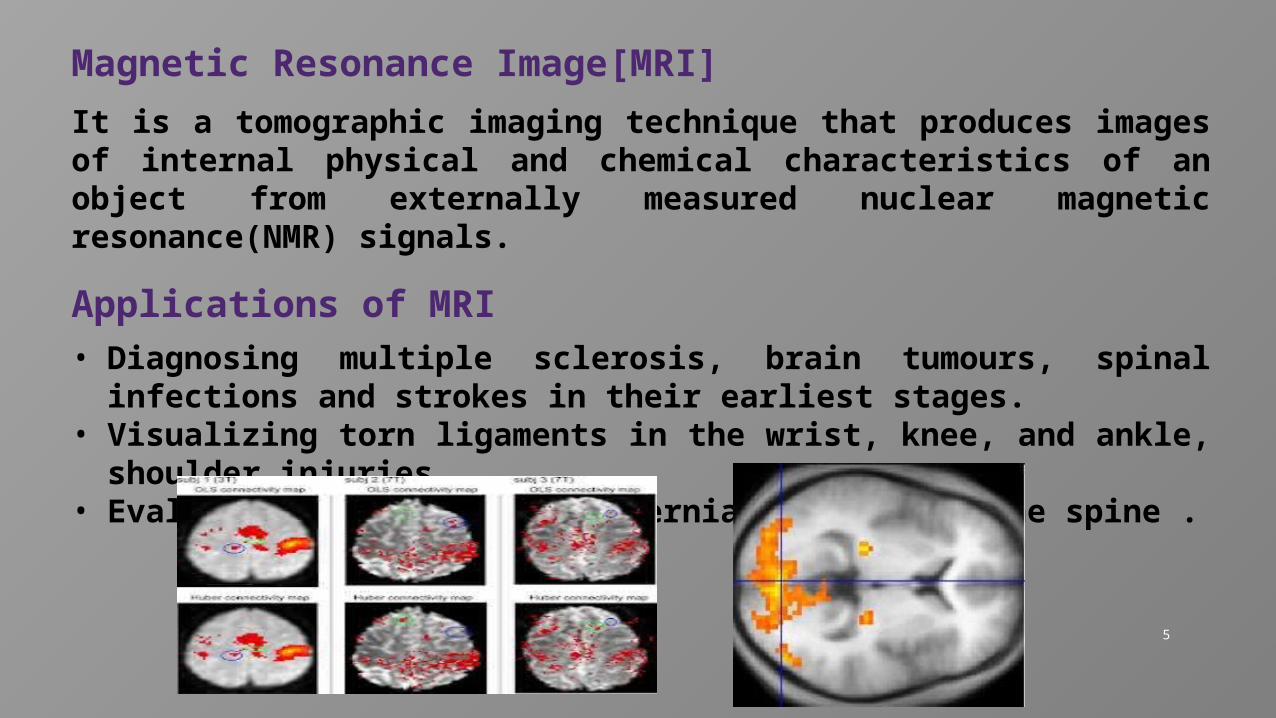
Magnetic Resonance Image[MRI]
It is a tomographic imaging technique that produces images of internal physical and chemical characteristics of an object from externally measured nuclear magnetic resonance(NMR) signals.
Applications of MRI• Diagnosing multiple sclerosis, brain tumours, spinal
infections and strokes in their earliest stages.• Visualizing torn ligaments in the wrist, knee, and ankle,
shoulder injuries.• Evaluating bone tumors and herniated discs in the spine .
6
Limitations of MRI• Machine makes a very loud continuous hammering
noise when operating.
• Extreme precautions must be taken to keep metallic objects out of the room where the machine is operating.
• MRI system are expensive to buy and run.
• People with pacemakers can’t safely be scanned.
• MRI scans require patients to hold very still for long periods of time up to 90 minutes.
7
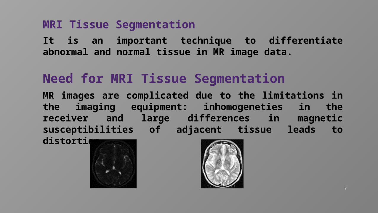
MRI Tissue SegmentationIt is an important technique to differentiate abnormal and normal tissue in MR image data.
Need for MRI Tissue SegmentationMR images are complicated due to the limitations in the imaging equipment: inhomogeneties in the receiver and large differences in magnetic susceptibilities of adjacent tissue leads to distortion.
8
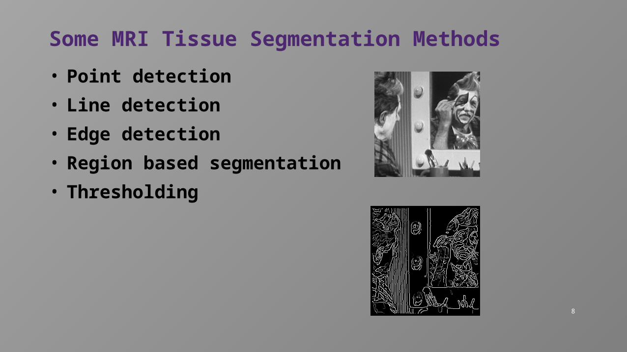
Some MRI Tissue Segmentation Methods
• Point detection
• Line detection
• Edge detection
• Region based segmentation
• Thresholding
9
Bias field estimation for MRI tissue segmentation
A “bias field signal” is a low frequency smooth undesirable signal that corrupts MR images because of the inhomogeneties in the magnetic field of the MRI machine.
Bias field correction methods classified into two classes:
Prospective methodsMain aim is to avoid intensity inhomogeneities in
the image acquisition process.
Retrospective methods Rely only on the information in the acquired images
and thus they can remove intensity inhomogeneties regard less of their sources.
10
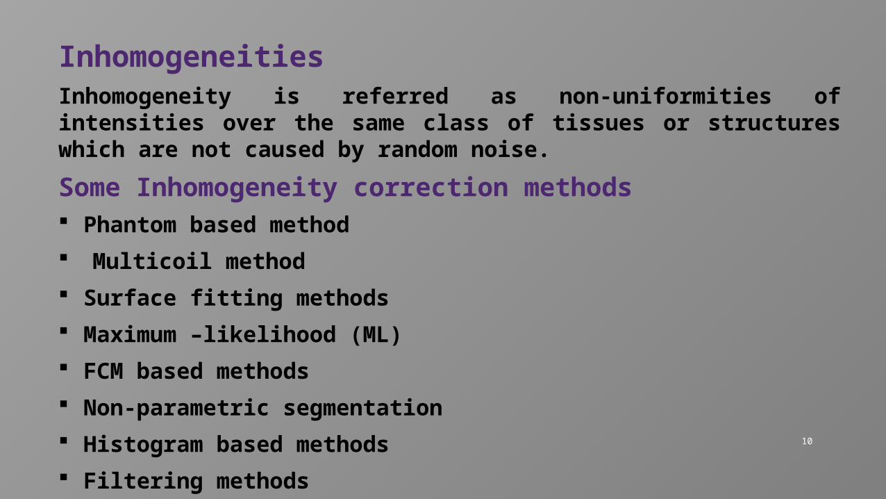
Inhomogeneities Inhomogeneity is referred as non-uniformities of intensities over the same class of tissues or structures which are not caused by random noise.
Some Inhomogeneity correction methods Phantom based method
Multicoil method
Surface fitting methods
Maximum –likelihood (ML)
FCM based methods
Non-parametric segmentation
Histogram based methods
Filtering methods
11
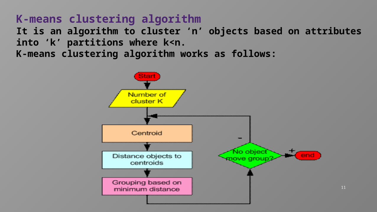
K-means clustering algorithmIt is an algorithm to cluster ‘n’ objects based on attributes into ‘k’ partitions where k<n.K-means clustering algorithm works as follows:
12
Related workSome of the algorithms that were used in the past few years in MRI Tissue Segmentation are:
In 1999, Adaptive fuzzy C-means algorithm In 2001, Expectation maximization algorithm In 2002, Modified fuzzy C-means algorithm
13
Related work(contd.,) In 2008, A Gaussian kernel-based fuzzy C-means algorithm
with spatial bias correction.
In 2009, Coherent Local Intensity Clustering (CLIC) . In 2011, A modified possibilistic fuzzy c-means clustering In 2012, Fuzzy Logic Gaussian Mixture Model(FLGMM)

14
Any queries?

15
Thank you