MRI Tissue Segmentation using MICO
16
MRI TISSUE SEGMENTATION Presentatio n By: POOJA G N 1
-
Upload
pooja-g-n -
Category
Technology
-
view
63 -
download
1
Transcript of MRI Tissue Segmentation using MICO
1
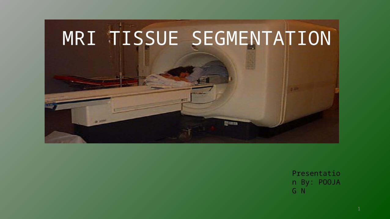
MRI TISSUE SEGMENTATION
Presentation By: POOJA G N
2
Introduction
• MRI : It is a tomographic technique that produces images of internal physical and chemical characteristics of an object from externally measured nuclear magnetic resonance (NMR) signals.
• MRI Tissue Segmentation: It is an important technique to differentiate abnormal and normal tissue in MR image data.
• Need for MRI Tissue Segmentation: MR images are complicated due to the limitations in the imaging equipment: inhomogeneties in the receiver and large differences in magnetic susceptibilities of adjacent tissue leads to distortion.
3
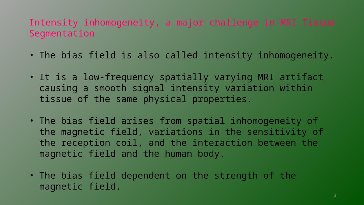
Intensity inhomogeneity, a major challenge in MRI Tissue Segmentation
• The bias field is also called intensity inhomogeneity.
• It is a low-frequency spatially varying MRI artifact causing a smooth signal intensity variation within tissue of the same physical properties.
• The bias field arises from spatial inhomogeneity of the magnetic field, variations in the sensitivity of the reception coil, and the interaction between the magnetic field and the human body.
• The bias field dependent on the strength of the magnetic field.
4
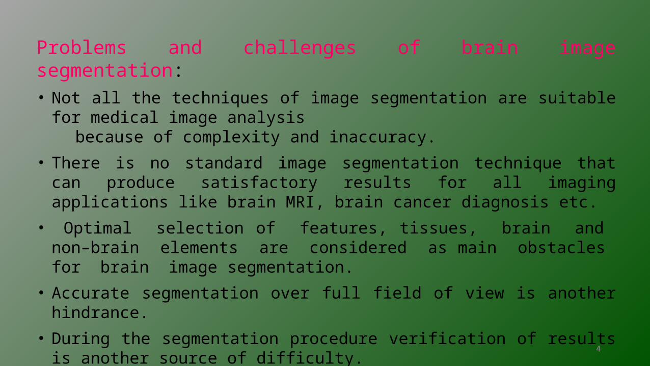
Problems and challenges of brain image segmentation:• Not all the techniques of image segmentation are suitable for medical image analysis because of complexity and inaccuracy.
• There is no standard image segmentation technique that can produce satisfactory results for all imaging applications like brain MRI, brain cancer diagnosis etc.
• Optimal selection of features, tissues, brain and non–brain elements are considered as main obstacles for brain image segmentation.
• Accurate segmentation over full field of view is another hindrance.
• During the segmentation procedure verification of results is another source of difficulty.
5
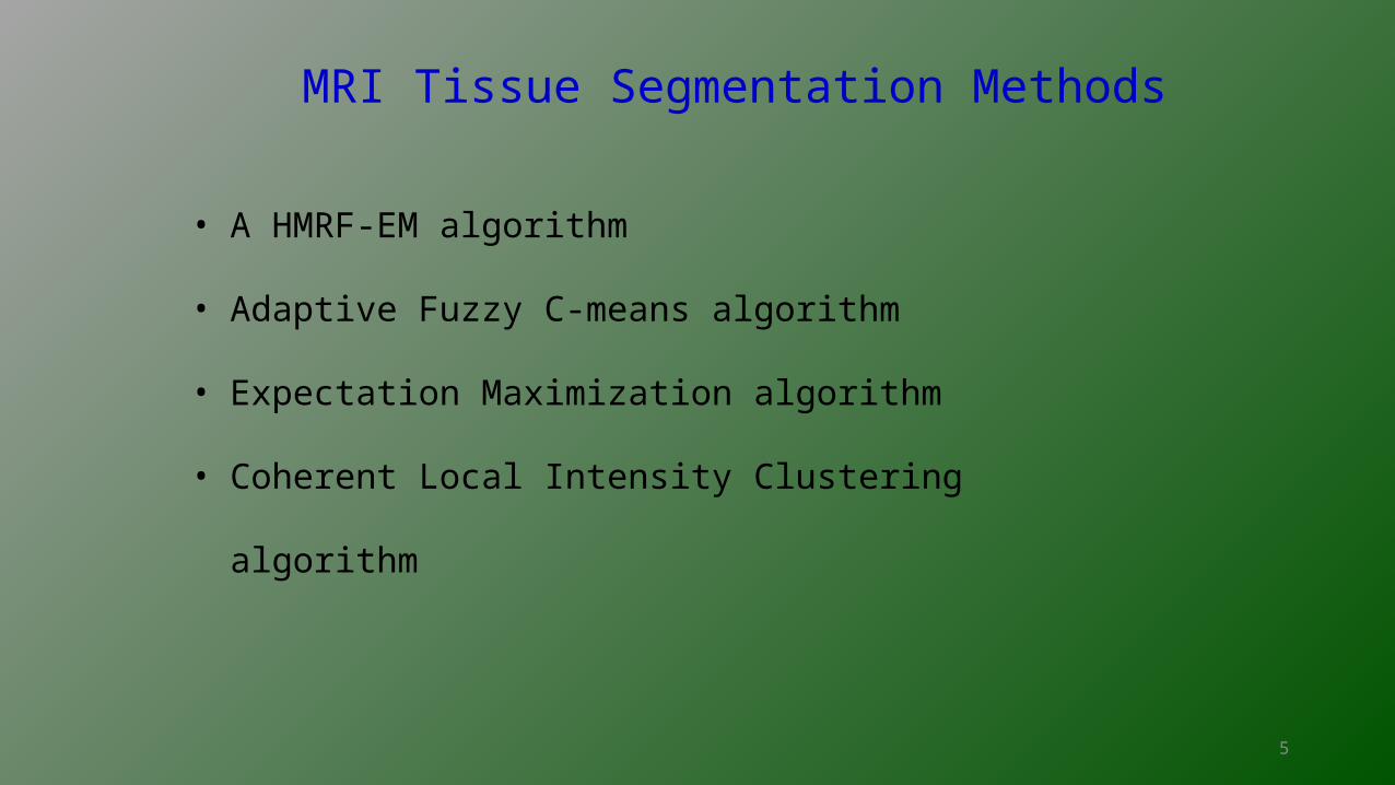
MRI Tissue Segmentation Methods
• A HMRF-EM algorithm
• Adaptive Fuzzy C-means algorithm
• Expectation Maximization algorithm
• Coherent Local Intensity Clustering algorithm
6
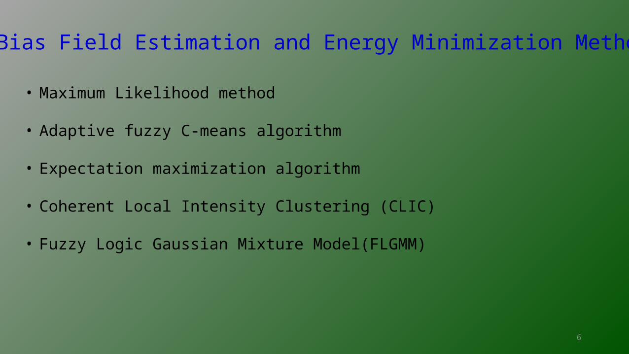
Bias Field Estimation and Energy Minimization Methods
• Maximum Likelihood method
• Adaptive fuzzy C-means algorithm
• Expectation maximization algorithm
• Coherent Local Intensity Clustering (CLIC)
• Fuzzy Logic Gaussian Mixture Model(FLGMM)
7
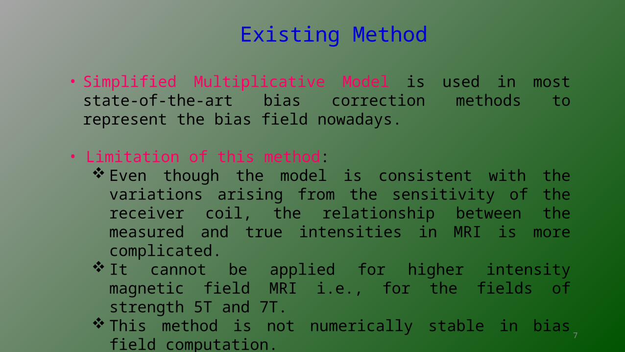
Existing Method
• Simplified Multiplicative Model is used in most state-of-the-art bias
correction methods to represent the bias field nowadays.
• Limitation of this method: Even though the model is consistent with the variations arising from the
sensitivity of the receiver coil, the relationship between the measured and true intensities in MRI is more complicated.
It cannot be applied for higher intensity magnetic field MRI i.e., for the fields of strength 5T and 7T.
This method is not numerically stable in bias field computation.
8
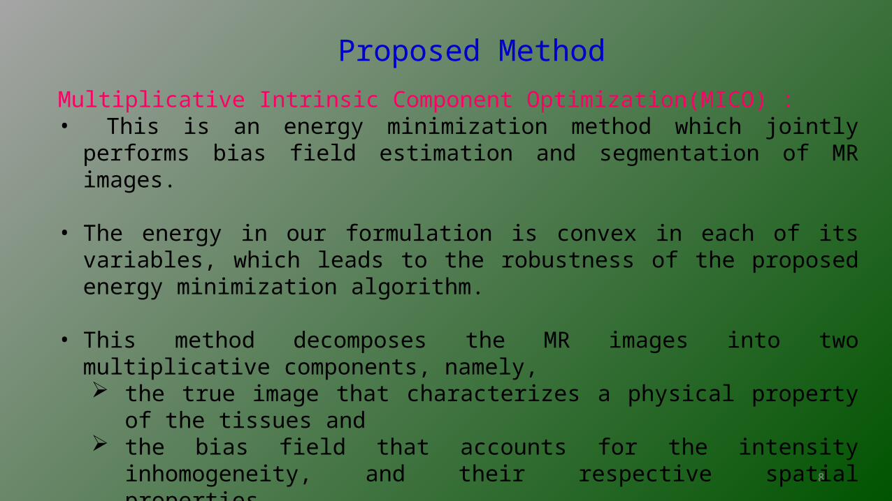
Proposed Method
Multiplicative Intrinsic Component Optimization(MICO) :• This is an energy minimization method which jointly performs bias field estimation
and segmentation of MR images.
• The energy in our formulation is convex in each of its variables, which leads to the robustness of the proposed energy minimization algorithm.
• This method decomposes the MR images into two multiplicative components, namely, the true image that characterizes a physical property of the tissues and the bias field that accounts for the intensity inhomogeneity, and their respective
spatial properties.
9
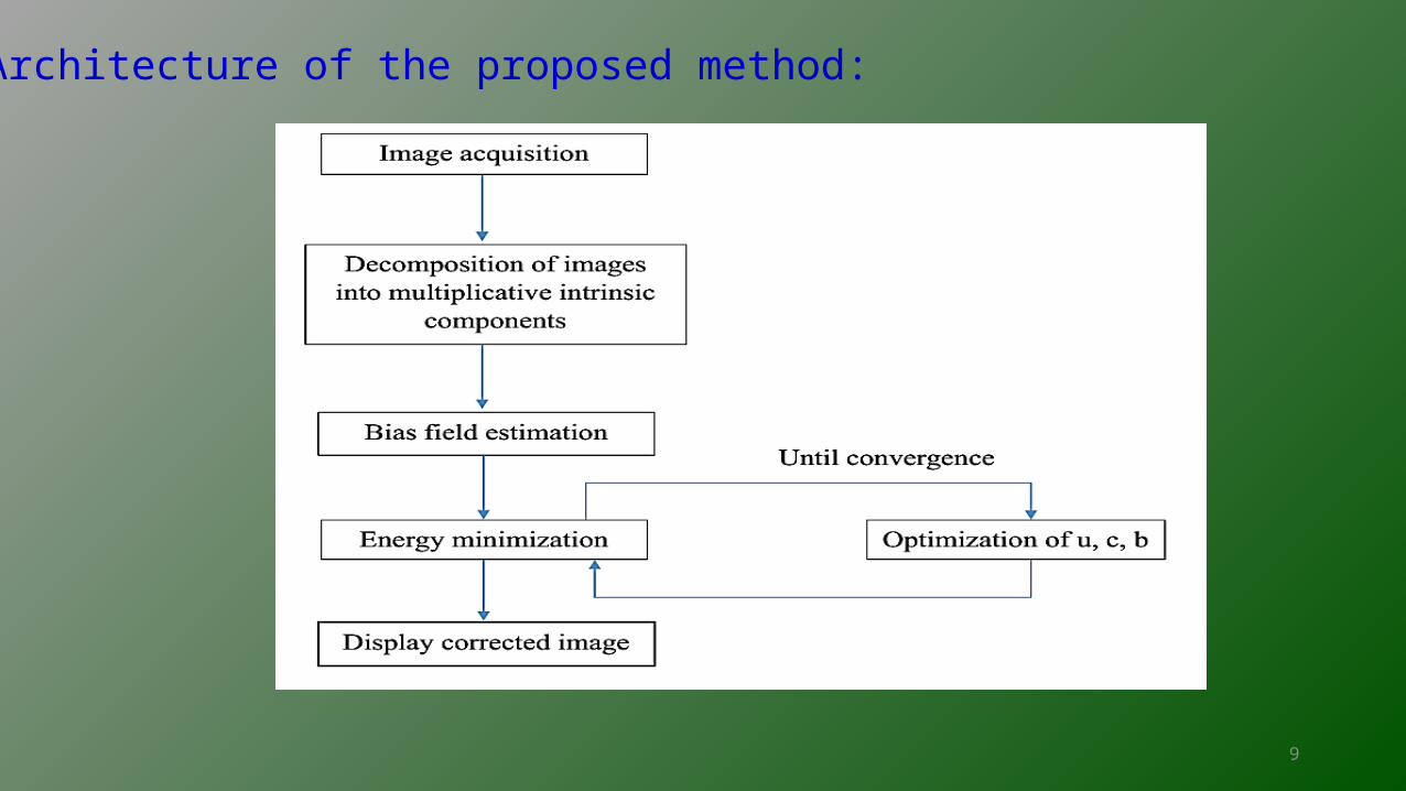
Architecture of the proposed method:
10
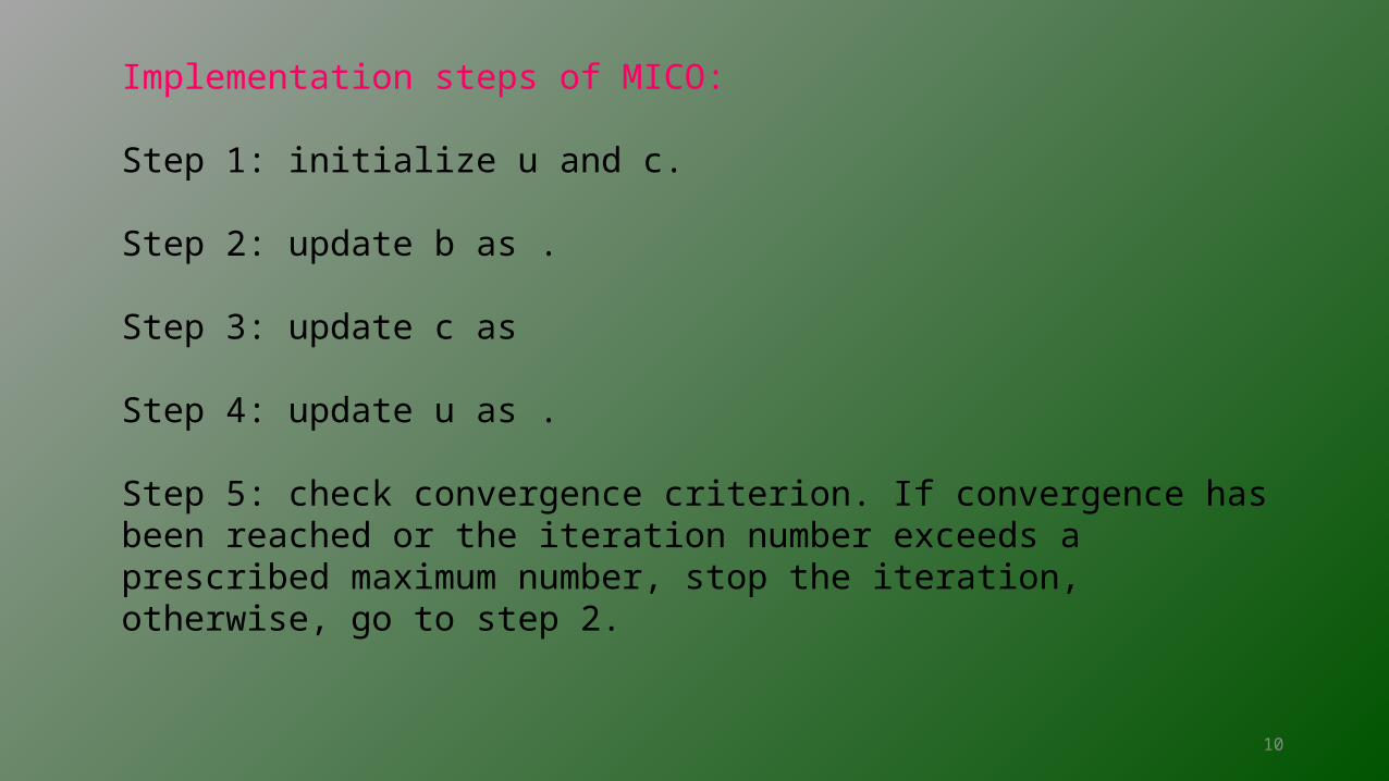
Implementation steps of MICO:
Step 1: initialize u and c.
Step 2: update b as .
Step 3: update c as
Step 4: update u as .
Step 5: check convergence criterion. If convergence has been reached or the iteration number exceeds a prescribed maximum number, stop the iteration, otherwise, go to step 2.
11
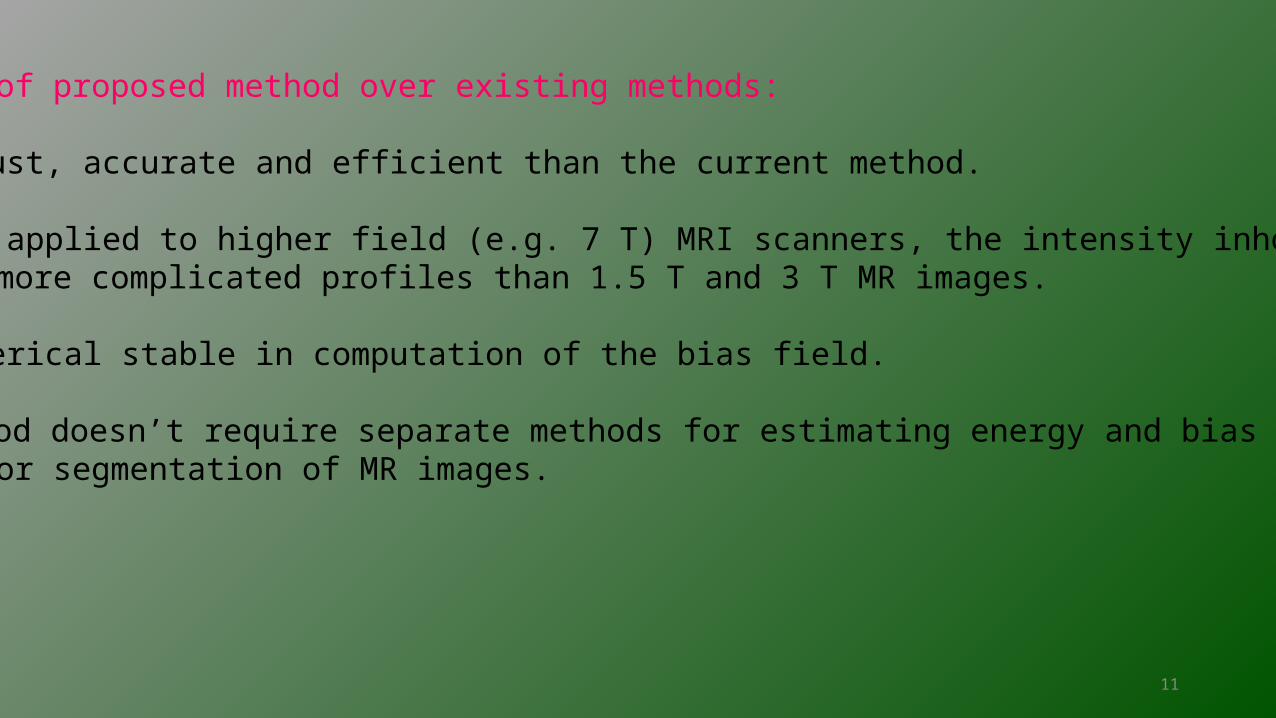
Advantages of proposed method over existing methods:
• It is robust, accurate and efficient than the current method.
• It can be applied to higher field (e.g. 7 T) MRI scanners, the intensity inhomogeneities have more complicated profiles than 1.5 T and 3 T MR images.
• It is numerical stable in computation of the bias field.
• This method doesn’t require separate methods for estimating energy and bias field, and also for segmentation of MR images.
12
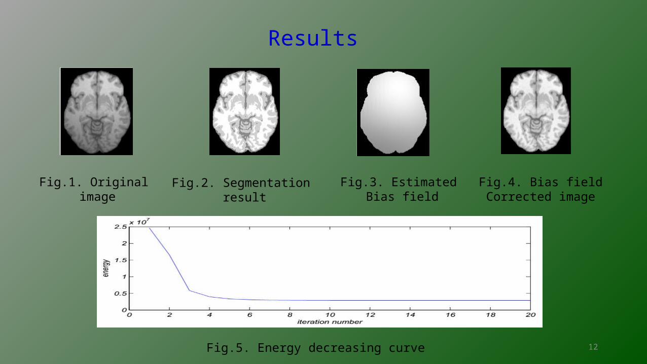
Results
Fig.1. Original image
Fig.2. Segmentation result
Fig.3. Estimated Bias field
Fig.4. Bias fieldCorrected image
Fig.5. Energy decreasing curve
13
Conclusion
The proposed method MICO, for bias field estimation and segmentation of MR images in a new energy minimization formulation is robust, efficient and accurate than the existing method. This method has been successfully applied to 1.5T and 3T MR images with desirable results.
14
Future Work
• The MICO formulation need to be extended to 3D/4D tissue segmentation with spatial/spatiotemporal regularization.
15
References
• Chunming Li, John C.Gore, Christos Davatzikos, ” Multiplicative Intrinsic component Optimization(MICO) for MRI bias field estimation and tissue segmentation”, Elsevier, Magnetic Resonance Imaging 32(2014) 913-923.

16
Thank you