Morphological and vascular variations of the left kidney ... · The renal vein and the ureter leave...
3
Anatomy Page 1 of 3 Compeng interests: none declared. Conflict of interests: none declared. All authors contributed to the concepon, design, and preparaon of the manuscript, as well as read and approved the final manuscript. All authors abide by the Associaon for Medical Ethics (AME) ethical rules of disclosure. For citation purposes: Satheesha Nayak B, Shetty SD, Rao Sirasanagandla S, Swamy Ravindra S, Kumar N, Jetti R, et al. Morphological and vascular variations of the left kidney: A case report. OA Case Reports 2013 Sep 10;2(10):98 Licensee OA Publishing London 2013. Creative Commons Attribution License (CC-BY) Case Report Compeng interests: none declared. Conflict of interests: none declared. All authors contributed to the concepon, design, and preparaon of the manuscript, as well as read and approved the final manuscript. All authors abide by the Associaon for Medical Ethics (AME) ethical rules of disclosure. Abstract Introduction The left kidney is supplied by the left renal artery, which is a branch of the abdominal aorta and drained by left renal vein, which is a tributary of the inferior vena cava. This article reports a case of morphological and vascular variations of the left kidney. Case report During dissection classes for medical undergraduates, we observed mor- phological and vascular variations of the left kidney in a male cadaver aged approximately 65 years. Hilum of the left kidney was situated on the medial half of its anterior surface. The left renal artery made a prominent down- ward curve at its origin and then coursed upward and to the left. It divided into two major branches before entering the kidney. The left renal vein had two divisions: an upper and a lower. The upper division was formed by a union of two veins, and the lower division was a single vein. The upper division and lower division had a crisscross arrangement before they united to form a single vein. Conclusion Knowledge of the morphological and vascular variations of the kidney reported here is of importance to urologists, radiologists, endocrinolo- gists and surgeons. Introduction The left kidney is situated at the posterior abdominal wall on the left Morphological and vascular variations of the left kidney: a case report B Satheesha Nayak *, SD Shetty, S Rao Sirasanagandla, S Swamy Ravindra, N Kumar, R Jetti, P Abhinitha side of the vertebral column. It has a hilum in the middle of its medial border and the branches of the renal artery enter the kidney through it. The renal vein and the ureter leave the kidney through the hilum. The left renal artery is a branch of the abdom- inal aorta and the left renal vein drains into the inferior vena cava. The left renal vein receives the left gonadal and suprarenal veins. It is very common to have vascular varia- tions of the kidney, and many studies have reported several variations of them. Left renal arteries may vary in number and position. They may arise from the aorta below or above the usual level of origin 1,2 . The entry point of renal arteries into the kidney is also liable to variations 3 . The left renal vein may also vary in its posi- tion, relations and number of tribu- taries 4–6 . Renal vascular variations might hinder the blood flow through the kidney and alter its functions. Knowledge of the variations is impor- tant for operating surgeons and radi- ologists. Here, we report left renal vascular variations and discuss their functional and clinical importance. Case report During dissection classes for medical undergraduates, we observed mor- phological and vascular variations of the left kidney in a male cadaver aged approximately 65 years. The right kidney was normal in its morphology and vascular supply. The left kidney had an oval hilum on the medial half of its anterior surface (Figures 1 and 2). The left renal artery made a prom- inent downward curve at its origin and then coursed upward and to the left (Figures 1 and 2). It divided into two major branches, which entered the kidney through the upper part of the hilum. The left renal vein had two divisions: an upper and a lower. The upper division was formed by the union of two veins, and the lower division was a single vein. The upper division and lower division had a crisscross arrangement before they united to form a single vein (Figures 1 and 2). At the point of union, the lower division exhibited an acute bend. The left testicular vein opened into the lower division and the left suprarenal vein opened into the main trunk of the left renal vein. Discussion The definitive kidneys develop from the metanephros. They start their development in the pelvis in front of the sacrum and ascend to their final level due to the differential growth of the body. By the ninth week of foetal life, the kidneys reach the suprarenal glands, and after that, their ascent stops. In the early stage, the hilum of the kidney faces anteriorly, but as the kidney ascends, the hilum rotates medially and comes to lie along the medial border 7 . Rarely, one or both the kidneys may fail to rotate or rotate abnormally during their ascent. Failure of rotation results in a hilum facing anteriorly. Chaudhary and Rao reported an abnormal rota- tion of kidney where the hilum faced posteriorly 8 , and such abnormal rota- tions of the kidney might cause uret- eropelvic obstructions 9 . In the current case, the hilum was situated on the medial half of the anterior surface and was directed anteromedially. *Corresponding author Email: [email protected] Melaka Manipal Medical College (Manipal Campus), Manipal University, Manipal 576104, Karnataka, India
Transcript of Morphological and vascular variations of the left kidney ... · The renal vein and the ureter leave...

Ana
tom
yPage 1 of 3
Com
petin
g in
tere
sts:
non
e de
clar
ed. C
onfli
ct o
f int
eres
ts: n
one
decl
ared
. A
ll au
thor
s co
ntrib
uted
to th
e co
ncep
tion,
des
ign,
and
pre
para
tion
of th
e m
anus
crip
t, a
s w
ell a
s re
ad a
nd a
ppro
ved
the
final
man
uscr
ipt.
A
ll au
thor
s ab
ide
by th
e A
ssoc
iatio
n fo
r Med
ical
Eth
ics
(AM
E) e
thic
al ru
les
of d
iscl
osur
e.
For citation purposes: Satheesha Nayak B, Shetty SD, Rao Sirasanagandla S, Swamy Ravindra S, Kumar N, Jetti R, et al. Morphological and vascular variations of the left kidney: A case report. OA Case Reports 2013 Sep 10;2(10):98
Licensee OA Publishing London 2013. Creative Commons Attribution License (CC-BY)
Case Report
Com
petin
g in
tere
sts:
non
e de
clar
ed. C
onfli
ct o
f int
eres
ts: n
one
decl
ared
. A
ll au
thor
s co
ntrib
uted
to th
e co
ncep
tion,
des
ign,
and
pre
para
tion
of th
e m
anus
crip
t, a
s w
ell a
s re
ad a
nd a
ppro
ved
the
final
man
uscr
ipt.
A
ll au
thor
s ab
ide
by th
e A
ssoc
iatio
n fo
r Med
ical
Eth
ics
(AM
E) e
thic
al ru
les
of d
iscl
osur
e.
AbstractIntroductionThe left kidney is supplied by the left renal artery, which is a branch of the abdominal aorta and drained by left renal vein, which is a tributary of the inferior vena cava. This article reports a case of morphological and vascular variations of the left kidney.Case reportDuring dissection classes for medical undergraduates, we observed mor-phological and vascular variations of the left kidney in a male cadaver aged approximately 65 years. Hilum of the left kidney was situated on the medial half of its anterior surface. The left renal artery made a prominent down-ward curve at its origin and then coursed upward and to the left. It divided into two major branches before entering the kidney. The left renal vein had two divisions: an upper and a lower. The upper division was formed by a union of two veins, and the lower division was a single vein. The upper division and lower division had a crisscross arrangement before they united to form a single vein. ConclusionKnowledge of the morphological and vascular variations of the kidney reported here is of importance to urologists, radiologists, endocrinolo-gists and surgeons.
IntroductionThe left kidney is situated at the posterior abdominal wall on the left
Morphological and vascular variations of the left kidney: a case report
B Satheesha Nayak *, SD Shetty, S Rao Sirasanagandla, S Swamy Ravindra, N Kumar, R Jetti, P Abhinitha
side of the vertebral column. It has a hilum in the middle of its medial border and the branches of the renal artery enter the kidney through it. The renal vein and the ureter leave the kidney through the hilum. The left renal artery is a branch of the abdom-inal aorta and the left renal vein drains into the inferior vena cava. The left renal vein receives the left gonadal and suprarenal veins. It is very common to have vascular varia-tions of the kidney, and many studies have reported several variations of them. Left renal arteries may vary in number and position. They may arise from the aorta below or above the usual level of origin1,2. The entry point of renal arteries into the kidney is also liable to variations3. The left renal vein may also vary in its posi-tion, relations and number of tribu-taries4–6. Renal vascular variations might hinder the blood flow through the kidney and alter its functions. Knowledge of the variations is impor-tant for operating surgeons and radi-ologists. Here, we report left renal vascular variations and discuss their functional and clinical importance.
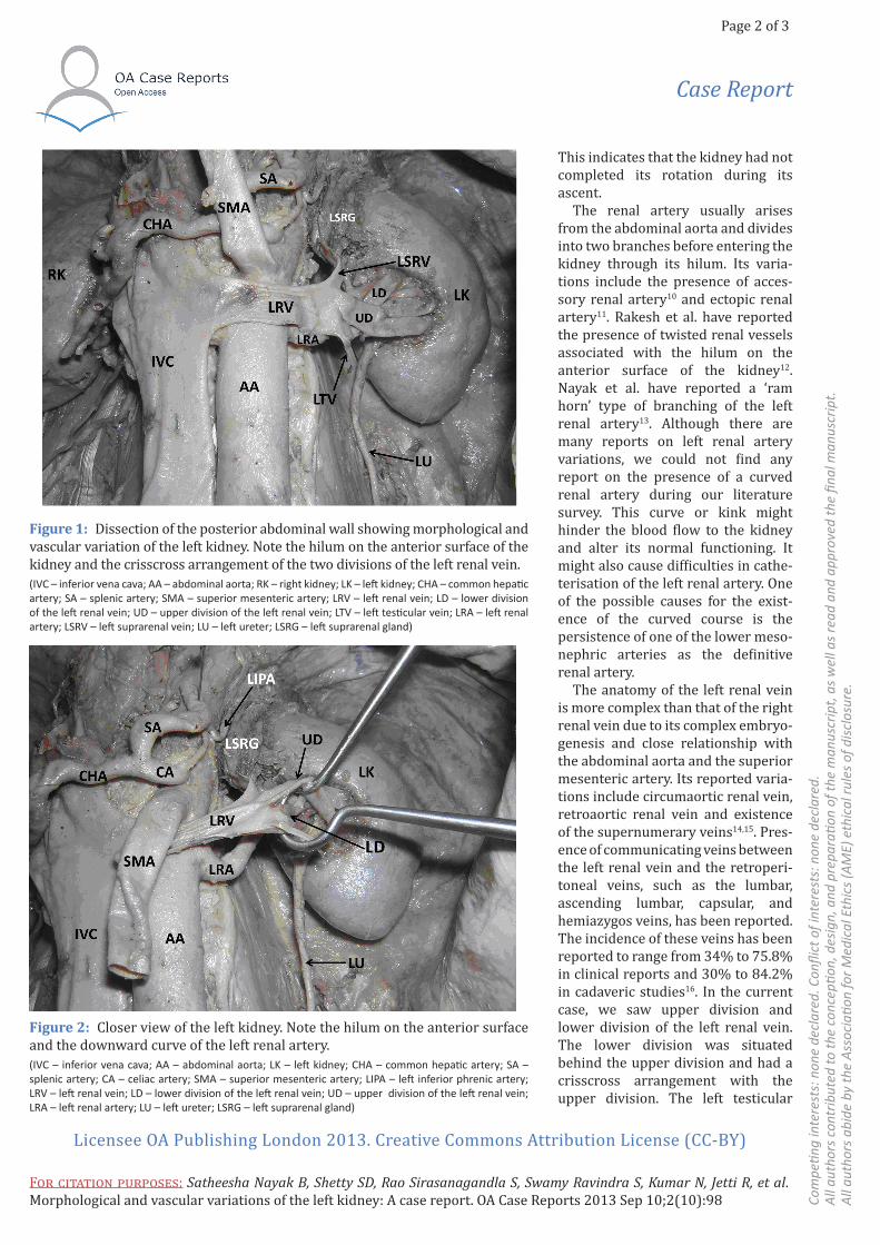
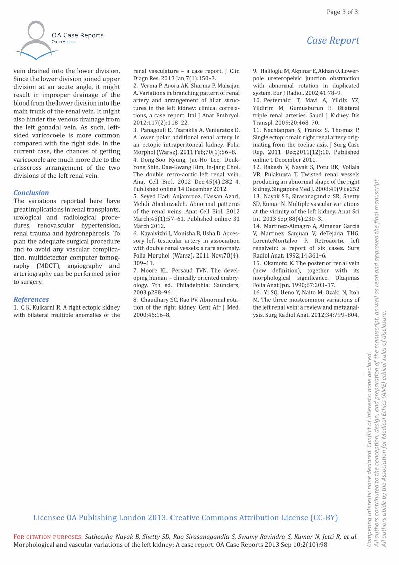
Case reportDuring dissection classes for medical undergraduates, we observed mor-phological and vascular variations of the left kidney in a male cadaver aged approximately 65 years. The right kidney was normal in its morphology and vascular supply. The left kidney had an oval hilum on the medial half of its anterior surface (Figures 1 and 2). The left renal artery made a prom-inent downward curve at its origin and then coursed upward and to the
left (Figures 1 and 2). It divided into two major branches, which entered the kidney through the upper part of the hilum. The left renal vein had two divisions: an upper and a lower. The upper division was formed by the union of two veins, and the lower division was a single vein. The upper division and lower division had a crisscross arrangement before they united to form a single vein (Figures 1 and 2). At the point of union, the lower division exhibited an acute bend. The left testicular vein opened into the lower division and the left suprarenal vein opened into the main trunk of the left renal vein.
DiscussionThe definitive kidneys develop from the metanephros. They start their development in the pelvis in front of the sacrum and ascend to their final level due to the differential growth of the body. By the ninth week of foetal life, the kidneys reach the suprarenal glands, and after that, their ascent stops. In the early stage, the hilum of the kidney faces anteriorly, but as the kidney ascends, the hilum rotates medially and comes to lie along the medial border7. Rarely, one or both the kidneys may fail to rotate or rotate abnormally during their ascent. Failure of rotation results in a hilum facing anteriorly. Chaudhary and Rao reported an abnormal rota-tion of kidney where the hilum faced posteriorly8, and such abnormal rota-tions of the kidney might cause uret-eropelvic obstructions9. In the current case, the hilum was situated on the medial half of the anterior surface and was directed anteromedially.
*Corresponding author Email: [email protected]
Melaka Manipal Medical College (Manipal Campus), Manipal University, Manipal 576104, Karnataka, India
Page 2 of 3
Com
petin
g in
tere
sts:
non
e de
clar
ed. C
onfli
ct o
f int
eres
ts: n
one
decl
ared
. A
ll au
thor
s co
ntrib
uted
to th
e co
ncep
tion,
des
ign,
and
pre
para
tion
of th
e m
anus
crip
t, a
s w
ell a
s re
ad a
nd a
ppro
ved
the
final
man
uscr
ipt.
A
ll au
thor
s ab
ide
by th
e A
ssoc
iatio
n fo
r Med
ical
Eth
ics
(AM
E) e
thic
al ru
les
of d
iscl
osur
e.
For citation purposes: Satheesha Nayak B, Shetty SD, Rao Sirasanagandla S, Swamy Ravindra S, Kumar N, Jetti R, et al. Morphological and vascular variations of the left kidney: A case report. OA Case Reports 2013 Sep 10;2(10):98
Licensee OA Publishing London 2013. Creative Commons Attribution License (CC-BY)
Case Report
This indicates that the kidney had not completed its rotation during its ascent.
The renal artery usually arises from the abdominal aorta and divides into two branches before entering the kidney through its hilum. Its varia-tions include the presence of acces-sory renal artery10 and ectopic renal artery11. Rakesh et al. have reported the presence of twisted renal vessels associated with the hilum on the anterior surface of the kidney12. Nayak et al. have reported a ‘ram horn’ type of branching of the left renal artery13. Although there are many reports on left renal artery variations, we could not find any report on the presence of a curved renal artery during our literature survey. This curve or kink might hinder the blood flow to the kidney and alter its normal functioning. It might also cause difficulties in cathe-terisation of the left renal artery. One of the possible causes for the exist-ence of the curved course is the persistence of one of the lower meso-nephric arteries as the definitive renal artery.
The anatomy of the left renal vein is more complex than that of the right renal vein due to its complex embryo-genesis and close relationship with the abdominal aorta and the superior mesenteric artery. Its reported varia-tions include circumaortic renal vein, retroaortic renal vein and existence of the supernumerary veins14,15. Pres-ence of communicating veins between the left renal vein and the retroperi-toneal veins, such as the lumbar, ascending lumbar, capsular, and hemiazygos veins, has been reported. The incidence of these veins has been reported to range from 34% to 75.8% in clinical reports and 30% to 84.2% in cadaveric studies16. In the current case, we saw upper division and lower division of the left renal vein. The lower division was situated behind the upper division and had a crisscross arrangement with the upper division. The left testicular
Figure 1: Dissection of the posterior abdominal wall showing morphological and vascular variation of the left kidney. Note the hilum on the anterior surface of the kidney and the crisscross arrangement of the two divisions of the left renal vein.(IVC – inferior vena cava; AA – abdominal aorta; RK – right kidney; LK – left kidney; CHA – common hepatic artery; SA – splenic artery; SMA – superior mesenteric artery; LRV – left renal vein; LD – lower division of the left renal vein; UD – upper division of the left renal vein; LTV – left testicular vein; LRA – left renal artery; LSRV – left suprarenal vein; LU – left ureter; LSRG – left suprarenal gland)
Figure 2: Closer view of the left kidney. Note the hilum on the anterior surface and the downward curve of the left renal artery.(IVC – inferior vena cava; AA – abdominal aorta; LK – left kidney; CHA – common hepatic artery; SA – splenic artery; CA – celiac artery; SMA – superior mesenteric artery; LIPA – left inferior phrenic artery; LRV – left renal vein; LD – lower division of the left renal vein; UD – upper division of the left renal vein; LRA – left renal artery; LU – left ureter; LSRG – left suprarenal gland)
Page 3 of 3
Com
petin
g in
tere
sts:
non
e de
clar
ed. C
onfli
ct o
f int
eres
ts: n
one
decl
ared
. A
ll au
thor
s co
ntrib
uted
to th
e co
ncep
tion,
des
ign,
and
pre
para
tion
of th
e m
anus
crip
t, a
s w
ell a
s re
ad a
nd a
ppro
ved
the
final
man
uscr
ipt.
A
ll au
thor
s ab
ide
by th
e A
ssoc
iatio
n fo
r Med
ical
Eth
ics
(AM
E) e
thic
al ru
les
of d
iscl
osur
e.
For citation purposes: Satheesha Nayak B, Shetty SD, Rao Sirasanagandla S, Swamy Ravindra S, Kumar N, Jetti R, et al. Morphological and vascular variations of the left kidney: A case report. OA Case Reports 2013 Sep 10;2(10):98
Licensee OA Publishing London 2013. Creative Commons Attribution License (CC-BY)
Case Report
vein drained into the lower division. Since the lower division joined upper division at an acute angle, it might result in improper drainage of the blood from the lower division into the main trunk of the renal vein. It might also hinder the venous drainage from the left gonadal vein. As such, left-sided varicocoele is more common compared with the right side. In the current case, the chances of getting varicocoele are much more due to the crisscross arrangement of the two divisions of the left renal vein.
ConclusionThe variations reported here have great implications in renal transplants, urological and radiological proce-dures, renovascular hypertension, renal trauma and hydronephrosis. To plan the adequate surgical procedure and to avoid any vascular complica-tion, multidetector computer tomog-raphy (MDCT), angiography and arteriography can be performed prior to surgery.
References1. C K, Kulkarni R. A right ectopic kidney with bilateral multiple anomalies of the
renal vasculature – a case report. J Clin Diagn Res. 2013 Jan;7(1):150–3. 2. Verma P, Arora AK, Sharma P, Mahajan A. Variations in branching pattern of renal artery and arrangement of hilar struc-tures in the left kidney: clinical correla-tions, a case report. Ital J Anat Embryol. 2012;117(2):118–22. 3. Panagouli E, Tsaraklis A, Venieratos D. A lower polar additional renal artery in an ectopic intraperitoneal kidney. Folia Morphol (Warsz). 2011 Feb;70(1):56–8.4. Dong-Soo Kyung, Jae-Ho Lee, Deuk-Yong Shin, Dae-Kwang Kim, In-Jang Choi. The double retro-aortic left renal vein. Anat Cell Biol. 2012 Dec;45(4):282–4. Published online 14 December 2012. 5. Seyed Hadi Anjamrooz, Hassan Azari, Mehdi Abedinzadeh. Abnormal patterns of the renal veins. Anat Cell Biol. 2012 March;45(1):57–61. Published online 31 March 2012. 6. Kayalvizhi I, Monisha B, Usha D. Acces-sory left testicular artery in association with double renal vessels: a rare anomaly. Folia Morphol (Warsz). 2011 Nov;70(4): 309–11.7. Moore KL, Persaud TVN. The devel-oping human – clinically oriented embry-ology. 7th ed. Philadelphia: Saunders; 2003.p288–96.8. Chaudhary SC, Rao PV. Abnormal rota-tion of the right kidney. Cent Afr J Med. 2000;46:16–8.
9. Haliloglu M, Akpinar E, Akhan O. Lower-pole ureteropelvic junction obstruction with abnormal rotation in duplicated system. Eur J Radiol. 2002;41:78–9.10. Pestemalci T, Mavi A, Yildiz YZ, Yildirim M, Gumusburun E. Bilateral triple renal arteries. Saudi J Kidney Dis Transpl. 2009;20:468–70.11. Nachiappan S, Franks S, Thomas P. Single ectopic main right renal artery orig-inating from the coeliac axis. J Surg Case Rep. 2011 Dec;2011(12):10. Published online 1 December 2011. 12. Rakesh V, Nayak S, Potu BK, Vollala VR, Pulakunta T. Twisted renal vessels producing an abnormal shape of the right kidney. Singapore Med J. 2008;49(9):e25213. Nayak SB, Sirasanagandla SR, Shetty SD, Kumar N. Multiple vascular variations at the vicinity of the left kidney. Anat Sci Int. 2013 Sep;88(4):230–3..14. Martinez-Almagro A, Almenar Garcia V, Martinez Sanjuan V, deTejada THG, LorenteMontalvo P. Retroaortic left renalvein: a report of six cases. Surg Radiol Anat. 1992;14:361–6.15. Okamoto K. The posterior renal vein (new definition), together with its morphological significance. Okajimas Folia Anat Jpn. 1990;67:203–17.16. Yi SQ, Ueno Y, Naito M, Ozaki N, Itoh M. The three mostcommon variations of the left renal vein: a review and metaanal-ysis. Surg Radiol Anat. 2012;34:799–804.