Chapter 25. Urinary System Two kidneys Two ureters Urethra urinary bladder.
Upload
annie-cheshireCategory
view
233download
2
The Urinary System
2
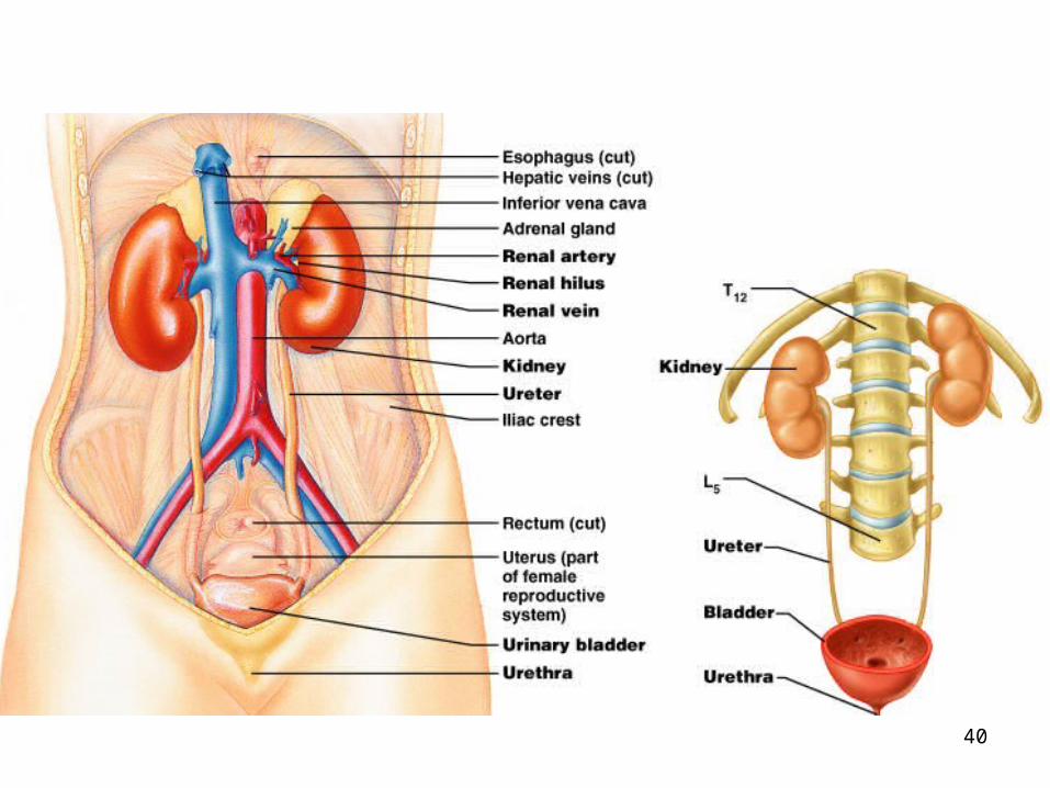
The Urinary System
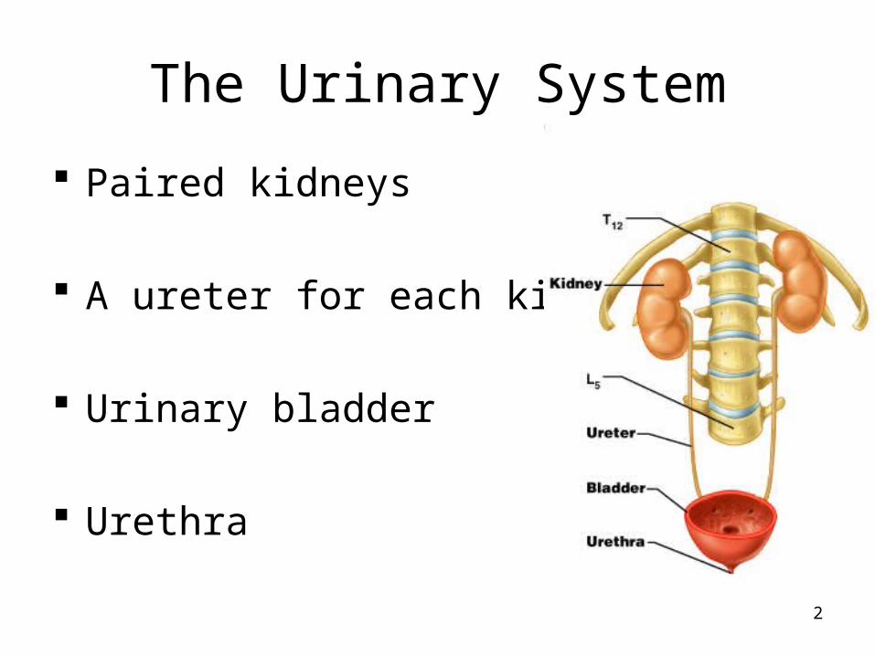
Paired kidneys
A ureter for each kidney
Urinary bladder
Urethra

3
Main Functions of Urinary System
Kidneys filter blood to keep it pure Toxins Metabolic wastes Excess water Excess ions
Dispose of nitrogenous wastes from blood Urea Uric acid Creatinine
Regulate the balance of water and electrolytes, acids and bases
4
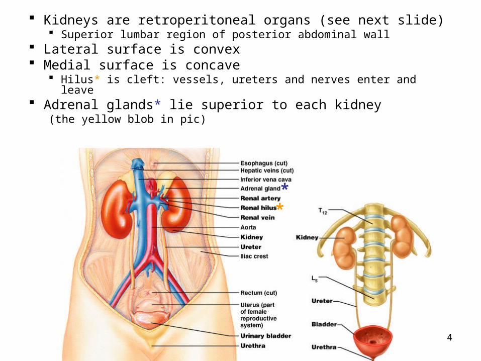
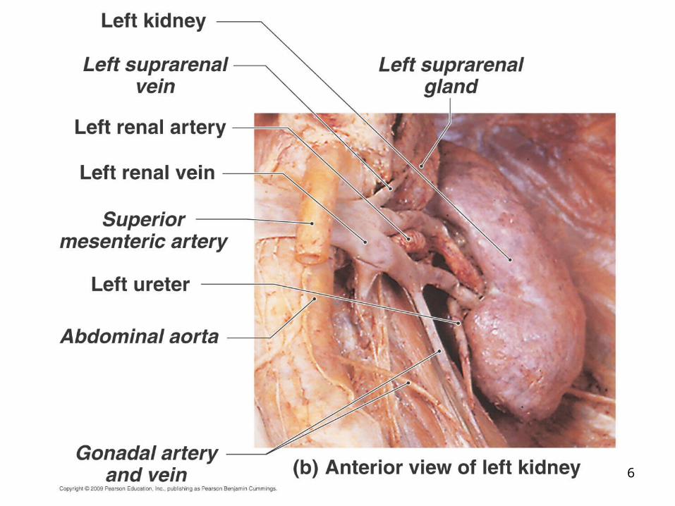
Kidneys are retroperitoneal organs (see next slide) Superior lumbar region of posterior abdominal wall
Lateral surface is convex Medial surface is concave
Hilus* is cleft: vessels, ureters and nerves enter and leave Adrenal glands* lie superior to each kidney
(the yellow blob in pic)
**

5
6

7
Transverse sections show retroperitoneal position of kidneys
Note also: liver, aorta muscles on CT
Note layers of adipose (fat), capsule, fascia
8

9
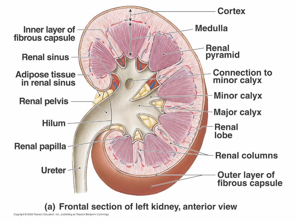
Kidney has two regions Cortex: outer
Columns of cortex divide medulla into “pyramids” Medulla: inner
Darker, cone-shaped medullary or renal pyramids Parallel bundles of urine-collecting tubules
10
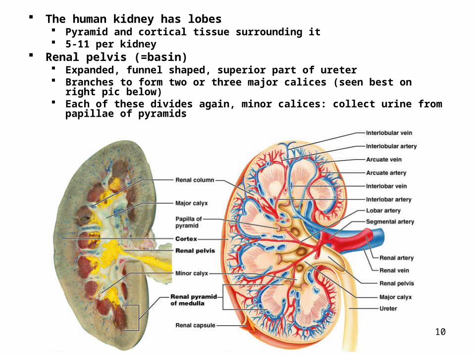
The human kidney has lobes Pyramid and cortical tissue surrounding it 5-11 per kidney
Renal pelvis (=basin) Expanded, funnel shaped, superior part of ureter Branches to form two or three major calices (seen best on right pic below) Each of these divides again, minor calices: collect urine from papillae of
pyramids
11
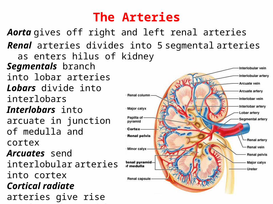
The ArteriesAorta gives off right and left renal arteries
Renal arteries divides into 5 segmental arteries as enters hilus of kidney
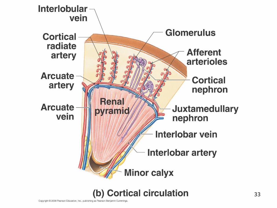
Segmentals branch into lobar arteriesLobars divide into interlobarsInterlobars into arcuate in junction of medulla and cortexArcuates send interlobular arteries into cortexCortical radiate arteries give rise to glomerular arterioles

12
Vasculature of the kidney
The glomerular capillary bed is unusual in having arterioles going both to it and away from it (afferent and efferent), instead of a vein going away as most
It is also unusual in having two capillary beds in series (one following the other)
13
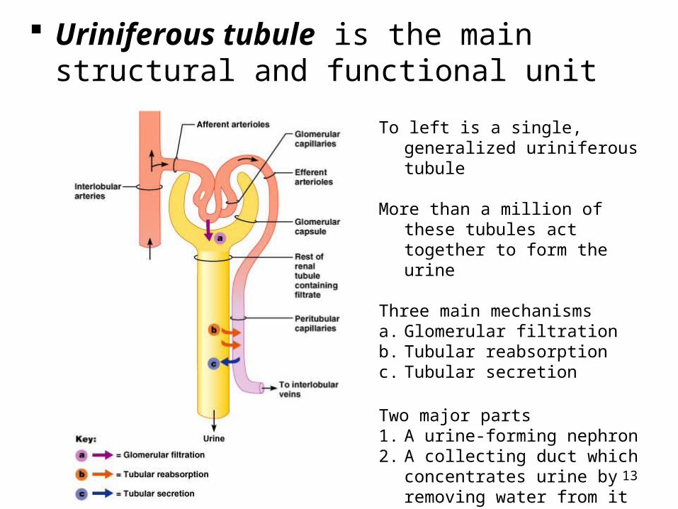
Uriniferous tubule is the main structural and functional unit
To left is a single, generalized uriniferous tubule
More than a million of these tubules act together to form the urine
Three main mechanismsa. Glomerular filtrationb. Tubular reabsorptionc. Tubular secretion
Two major parts1. A urine-forming nephron2. A collecting duct which
concentrates urine by removing water from it
14
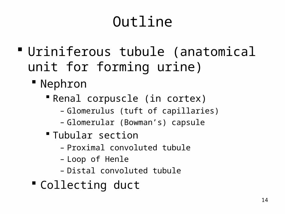
Outline
Uriniferous tubule (anatomical unit for forming urine) Nephron
Renal corpuscle (in cortex)– Glomerulus (tuft of capillaries)– Glomerular (Bowman’s) capsule
Tubular section– Proximal convoluted tubule– Loop of Henle– Distal convoluted tubule
Collecting duct
15
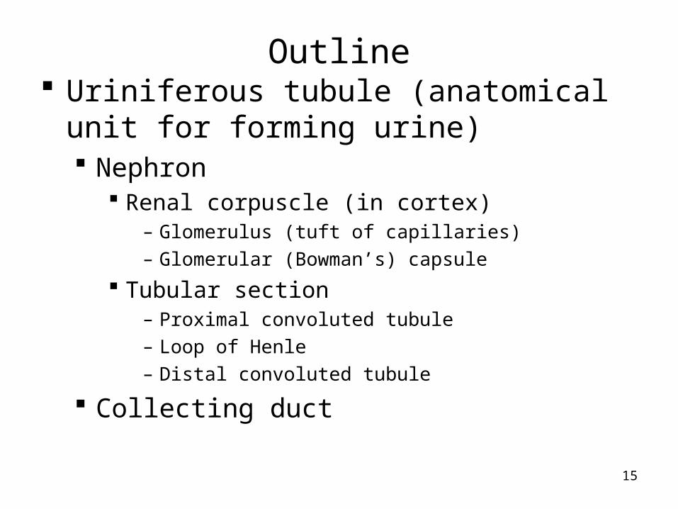
Outline Uriniferous tubule (anatomical unit for
forming urine) Nephron
Renal corpuscle (in cortex)– Glomerulus (tuft of capillaries)– Glomerular (Bowman’s) capsule
Tubular section– Proximal convoluted tubule– Loop of Henle– Distal convoluted tubule
Collecting duct

16
Understand at least this much:
Filtrationa. Fluid is squeezed out of
the glomerular capillary bed
Resorptionb. Most nutrients, water ad
essential ions are returned to the blood of the peritubular capillaries
Secretionc. Moves additional
undesirable molecules into tubule from blood of peritubular capillaries

17
Nephron Renal corpuscle Tubular section
Renal corpuscle: only in cortex Tuft of capillaries called
glomerulus Surrounded by cup-shaped,
hollow glomerular (Bowman’s) capsule
Uriniferous tubule (anatomical unit for forming urine) Nephron
Renal corpuscle (in cortex) Glomerulus (tuft of capillaries) Glomerular (Bowman’s) capsule
Tubular section Proximal convoluted tubule Loop of Henle Distal convoluted tubule
Collecting duct

18
(refer to this (refer to this pic as we go)pic as we go)
Visceral layer of capsule has podocytes Unusual branching
epithelial cells Foot processes with
slit processes between them
-------------------
19
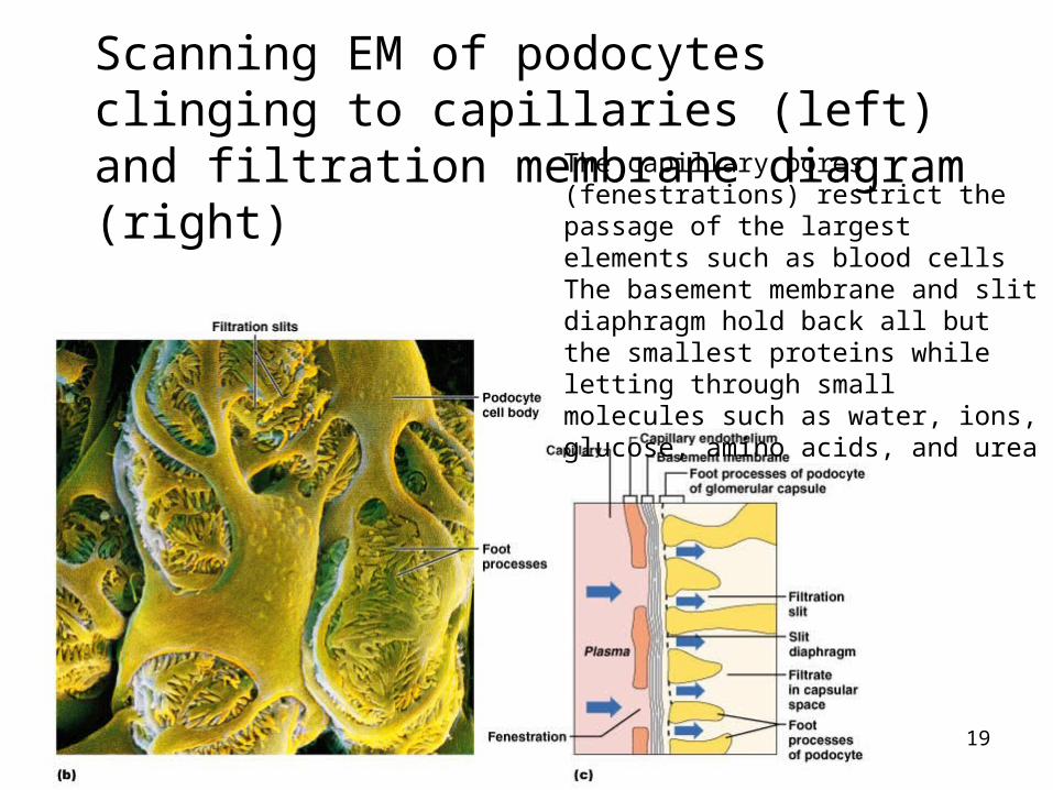
Scanning EM of podocytes clinging to capillaries (left) and filtration membrane diagram (right) The capillary pores (fenestrations)
restrict the passage of the largest elements such as blood cellsThe basement membrane and slit diaphragm hold back all but the smallest proteins while letting through small molecules such as water, ions, glucose, amino acids, and urea
20
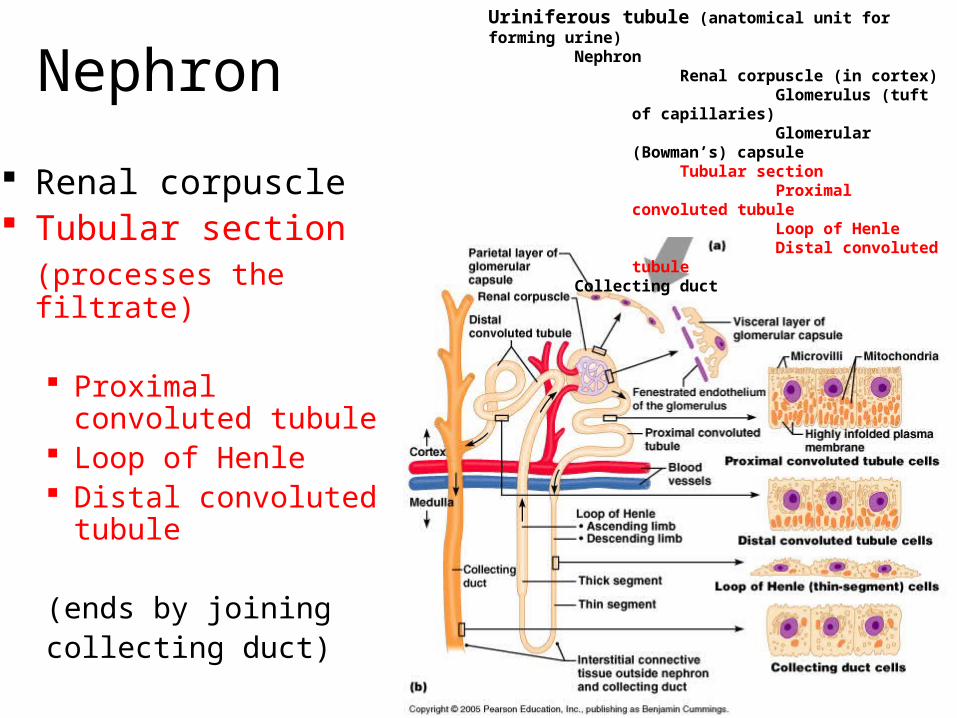
Nephron
Renal corpuscle Tubular section
(processes the filtrate)
Proximal convoluted tubule
Loop of Henle Distal convoluted
tubule
(ends by joiningcollecting duct)
Uriniferous tubule (anatomical unit for forming urine) Nephron
Renal corpuscle (in cortex) Glomerulus (tuft of capillaries) Glomerular (Bowman’s) capsule
Tubular section Proximal convoluted tubule Loop of Henle Distal convoluted tubule
Collecting duct

21
Proximal convoluted tubule
Confined to renal cortex
Cuboidal epithelial cells with long microvilli (fuzzy appearance in pics)
Resorption of water, ions and solutes
Uriniferous tubule (anatomical unit for forming urine) Nephron
Renal corpuscle (in cortex) Glomerulus (tuft of capillaries) Glomerular (Bowman’s) capsule
Tubular section Proximal convoluted tubule Loop of Henle Distal convoluted tubule
Collecting duct
*
22
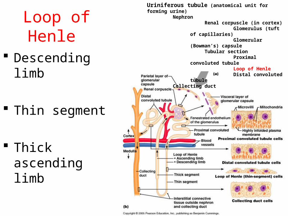
Loop of Henle
Descending limb
Thin segment
Thick ascending limb
Uriniferous tubule (anatomical unit for forming urine) Nephron
Renal corpuscle (in cortex) Glomerulus (tuft of capillaries) Glomerular (Bowman’s) capsule
Tubular section Proximal convoluted tubule Loop of Henle Distal convoluted tubule
Collecting duct
23
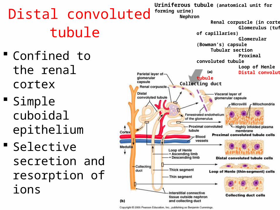
Distal convoluted tubule
Confined to the renal cortex
Simple cuboidal epithelium
Selective secretion and resorption of ions
Uriniferous tubule (anatomical unit for forming urine) Nephron
Renal corpuscle (in cortex) Glomerulus (tuft of capillaries) Glomerular (Bowman’s) capsule
Tubular section Proximal convoluted tubule Loop of Henle Distal convoluted tubule
Collecting duct
24
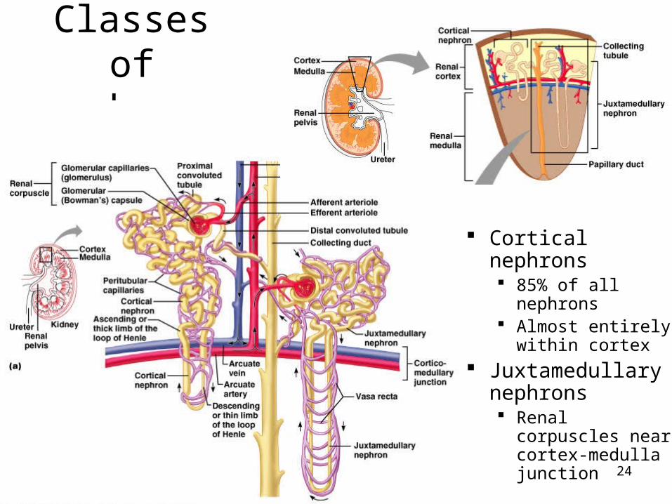
Classes of nephrons
Cortical nephrons 85% of all
nephrons Almost entirely
within cortex Juxtamedullary
nephrons Renal corpuscles
near cortex-medulla junction
25
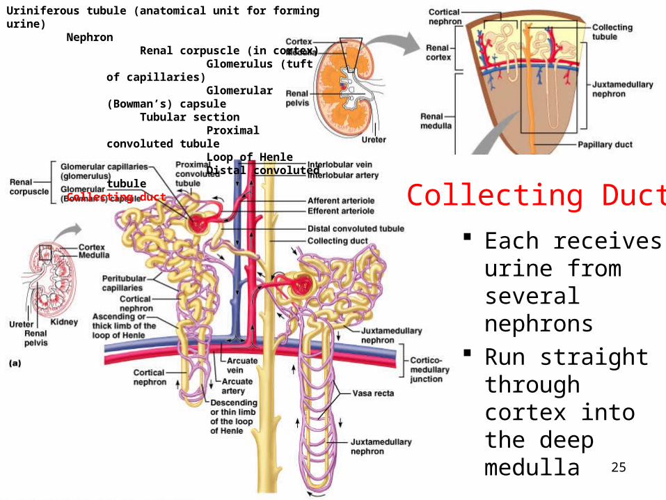
Collecting Ducts Each receives
urine from several nephrons
Run straight through cortex into the deep medulla
Uriniferous tubule (anatomical unit for forming urine) Nephron
Renal corpuscle (in cortex) Glomerulus (tuft of capillaries) Glomerular (Bowman’s) capsule
Tubular section Proximal convoluted tubule Loop of Henle Distal convoluted tubule
Collecting duct
Collecting Duct
26
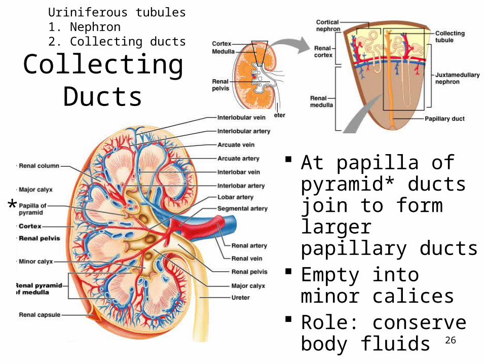
Collecting Ducts
At papilla of pyramid* ducts join to form larger papillary ducts
Empty into minor calices
Role: conserve body fluids
Uriniferous tubules1. Nephron 2. Collecting ducts
*

27
The collecting ducts
The most important role is to conserve body fluids
When the body must conserve water, the posterior pituitary gland secretes ADH (antidiuretic hormone)
ADH increases the permeability of the collecting tubules and distal tubules to water so more is reabsorbed
This decreases the total volume of urine Alcohol inhibits the release of ADH, so less
water is reabsorbed producing copious amounts of dilute urine (can cause dehydration)
28
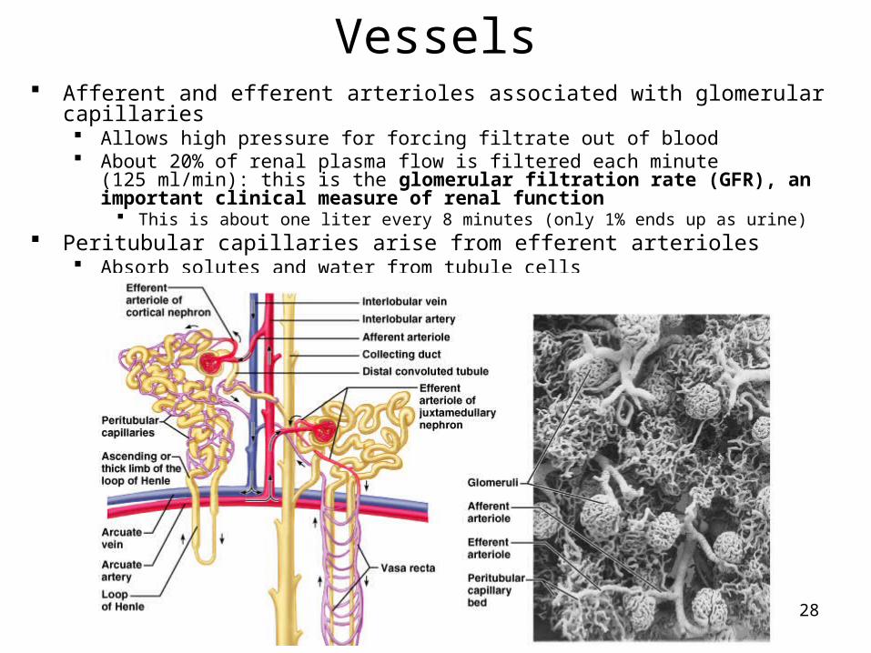
Vessels Afferent and efferent arterioles associated with glomerular capillaries
Allows high pressure for forcing filtrate out of blood About 20% of renal plasma flow is filtered each minute (125 ml/min): this is the
glomerular filtration rate (GFR), an important clinical measure of renal function This is about one liter every 8 minutes (only 1% ends up as urine)
Peritubular capillaries arise from efferent arterioles Absorb solutes and water from tubule cells
29
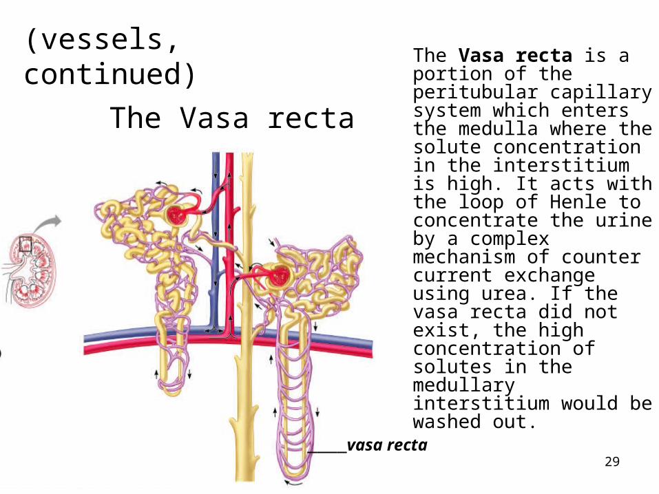
The Vasa recta is a portion of the peritubular capillary system which enters the medulla where the solute concentration in the interstitium is high. It acts with the loop of Henle to concentrate the urine by a complex mechanism of counter current exchange using urea. If the vasa recta did not exist, the high concentration of solutes in the medullary interstitium would be washed out.
____vasa recta
(vessels, continued)
The Vasa recta

30
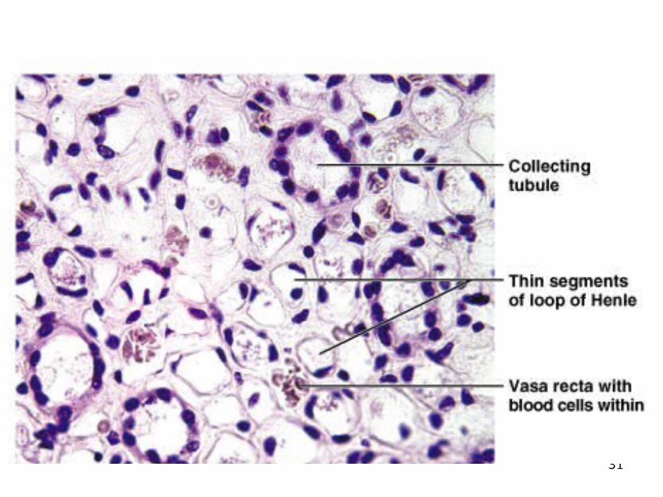
Histology
31
32
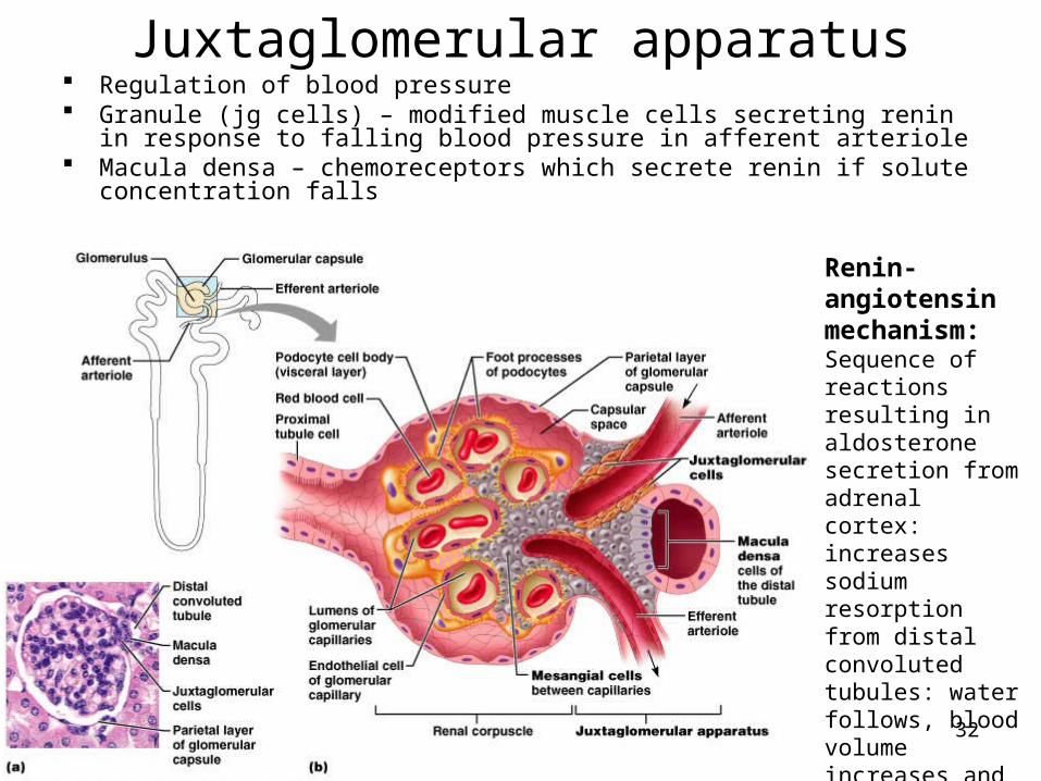
Juxtaglomerular apparatus Regulation of blood pressure Granule (jg cells) – modified muscle cells secreting renin in response to
falling blood pressure in afferent arteriole Macula densa – chemoreceptors which secrete renin if solute concentration
falls
Renin-angiotensin mechanism: Sequence of reactions resulting in aldosterone secretion from adrenal cortex: increases sodium resorption from distal convoluted tubules: water follows, blood volume increases and blood pressure increases
33
34
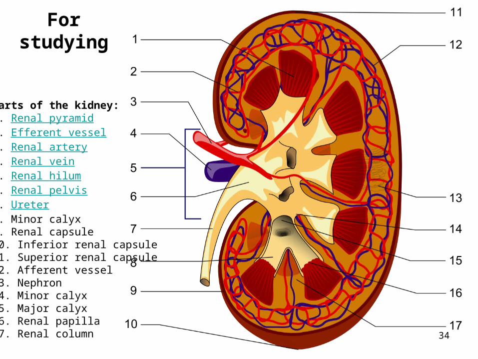
For studying
Parts of the kidney:1. Renal pyramid2. Efferent vessel3. Renal artery4. Renal vein5. Renal hilum6. Renal pelvis7. Ureter8. Minor calyx9. Renal capsule10. Inferior renal capsule11. Superior renal capsule12. Afferent vessel13. Nephron14. Minor calyx15. Major calyx16. Renal papilla17. Renal column

35
The Ureters Slender tubes about
25 cm (10 “) long leaving each renal pelvis
One for each kidney carrying urine to the bladder
Descend retroperitonealy and cross pelvic brim
Enter posterolateral corners of bladder
Run medially within posterior bladder wall before opening into interior
This oblique entry helps prevent backflow of urine
36
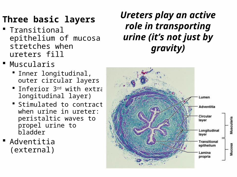
Ureters play an active role in transporting urine (it’s not just by gravity)
Three basic layers Transitional epithelium
of mucosa stretches when ureters fill
Muscularis Inner longitudinal, outer
circular layers Inferior 3rd with extra
longitudinal layer) Stimulated to contract
when urine in ureter: peristaltic waves to propel urine to bladder
Adventitia (external)

37
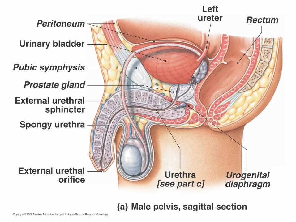
Urinary Bladder Collapsible muscular sac
Stores and expels urine
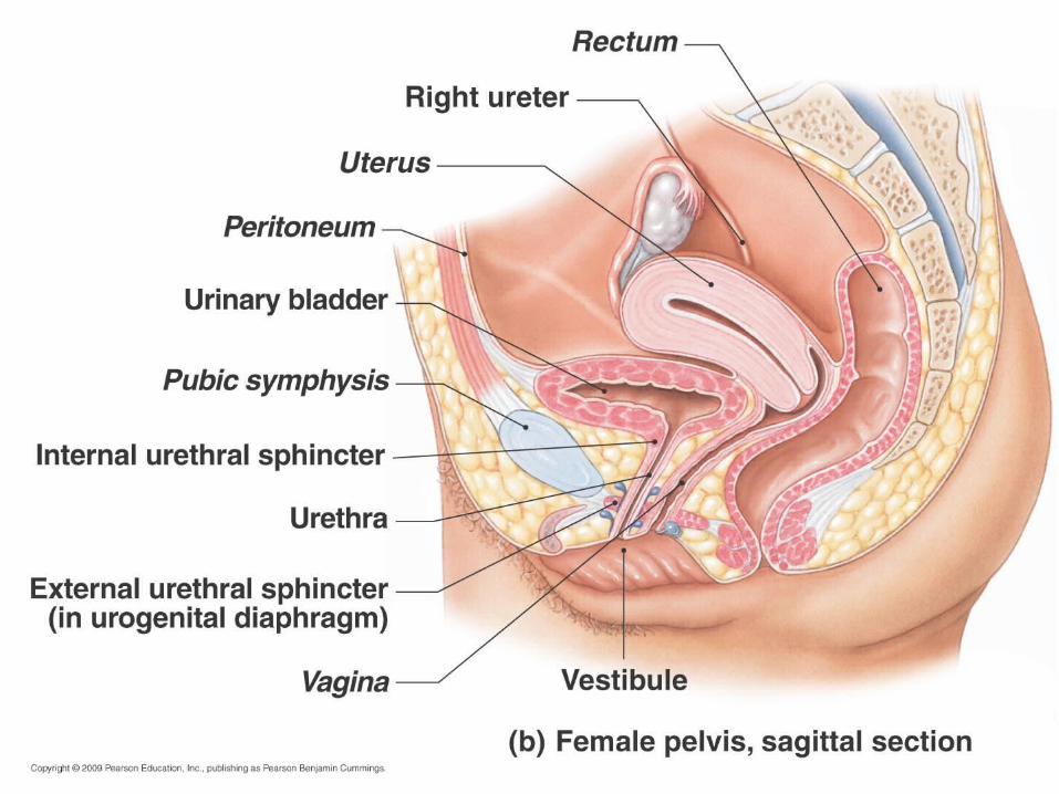
Lies on pelvic floor posterior to pubic symphysis Males: anterior to
rectum Females: just
anterior to the vagina and uterus
See also brief atlas
38
39
40

41
If full: bladder is spherical and extends into abdominal cavity (holds about 500 ml or 1 pt)
If empty: bladder lies entirely within pelvis with shape like upside-down pyramid
Urine exits via the urethra Trigone is inside area between ureters and
urethra: prone to infection (see slide 38)
42
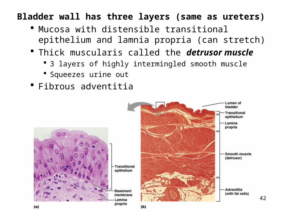
Bladder wall has three layers (same as ureters) Mucosa with distensible transitional epithelium and
lamnia propria (can stretch) Thick muscularis called the detrusor muscle
3 layers of highly intermingled smooth muscle Squeezes urine out
Fibrous adventitia
43
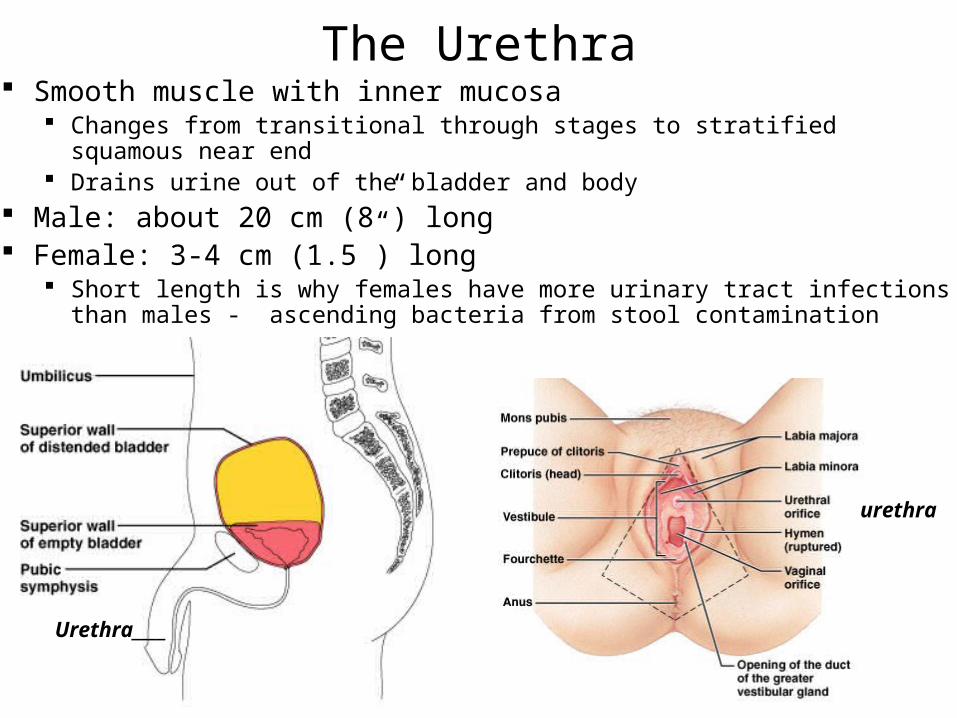
The Urethra Smooth muscle with inner mucosa
Changes from transitional through stages to stratified squamous near end Drains urine out of the bladder and body
Male: about 20 cm (8”) long Female: 3-4 cm (1.5”) long
Short length is why females have more urinary tract infections than males - ascending bacteria from stool contamination
Urethra____
urethra
44
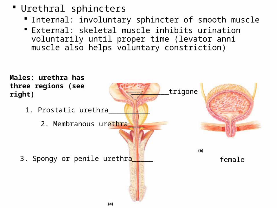
Urethral sphincters Internal: involuntary sphincter of smooth muscle External: skeletal muscle inhibits urination voluntarily
until proper time (levator anni muscle also helps voluntary constriction)
Males: urethra has three regions (see right)
1. Prostatic urethra__________
2. Membranous urethra____
3. Spongy or penile urethra_____
_________trigone
female

45
With all the labels
46
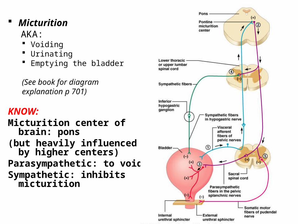
Micturition AKA: Voiding Urinating Emptying the bladder
(See book for diagramexplanation p 701)
KNOW:Micturition center of brain:
pons(but heavily influenced by
higher centers) Parasympathetic: to voidSympathetic: inhibits
micturition