BENIGN TUMORS OF KIDNEY URETER & BLADDER
76
KUB: Benign neoplasms Dr arif khan s
-
Upload
arif-s -
Category
Healthcare
-
view
144 -
download
0
Transcript of BENIGN TUMORS OF KIDNEY URETER & BLADDER

KUB:Benign neoplasms
Dr arif khan s

INTRODUCTION
URINARY SYSTEM
KIDNEYS
URETER
BLADDER
RENAL NEOPLASM
Adult
Paediatric
WHO classification categorises neoplasms on basis of cell of origin and histopathology.
Most renal tumors arise from the parenchyma, refered to as renal cell tumors
Small number from mesenchyme and collecting system.
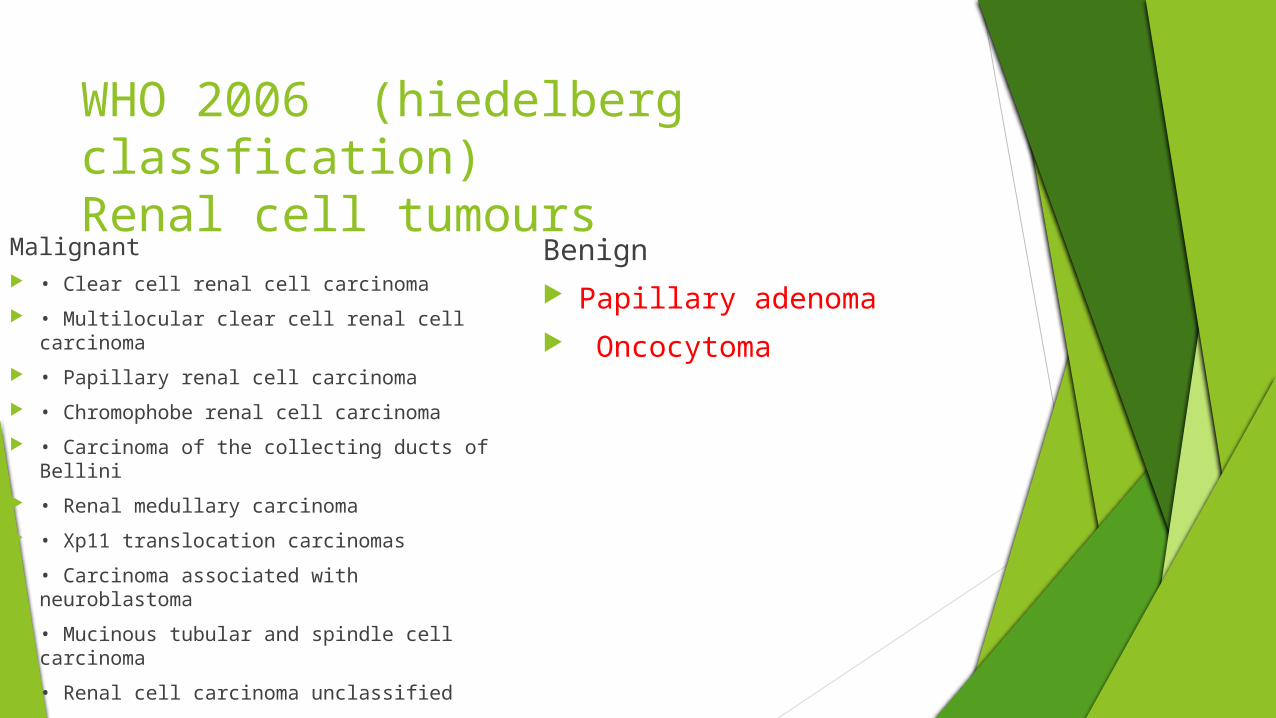
WHO 2006 (hiedelberg classfication)Renal cell tumours
Malignant • Clear cell renal cell carcinoma
• Multilocular clear cell renal cell carcinoma
• Papillary renal cell carcinoma
• Chromophobe renal cell carcinoma
• Carcinoma of the collecting ducts of Bellini
• Renal medullary carcinoma
• Xp11 translocation carcinomas
• Carcinoma associated with neuroblastoma
• Mucinous tubular and spindle cell carcinoma
• Renal cell carcinoma unclassified
Benign Papillary adenoma Oncocytoma
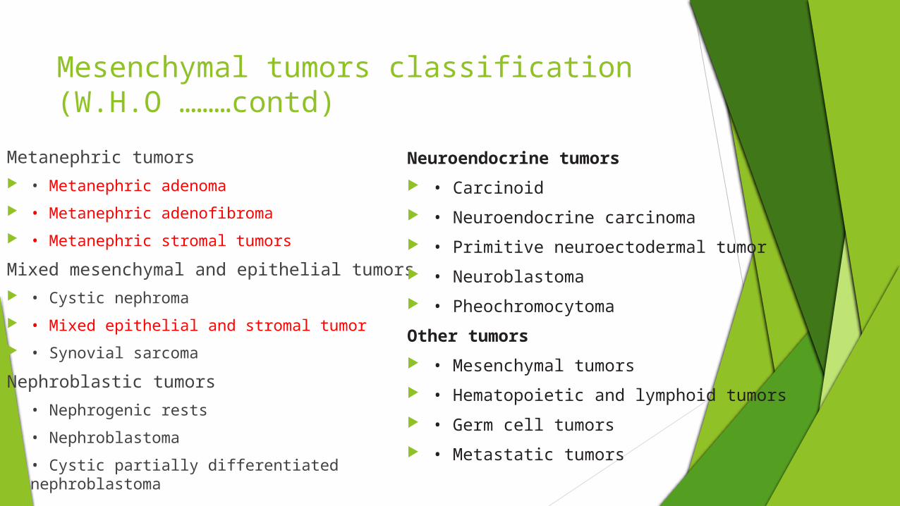
Mesenchymal tumors classification(W.H.O ………contd)
Metanephric tumors • Metanephric adenoma
• Metanephric adenofibroma
• Metanephric stromal tumors
Mixed mesenchymal and epithelial tumors • Cystic nephroma
• Mixed epithelial and stromal tumor
• Synovial sarcoma
Nephroblastic tumors • Nephrogenic rests
• Nephroblastoma
• Cystic partially differentiated nephroblastoma
Neuroendocrine tumors
• Carcinoid
• Neuroendocrine carcinoma
• Primitive neuroectodermal tumor
• Neuroblastoma
• Pheochromocytoma
Other tumors
• Mesenchymal tumors
• Hematopoietic and lymphoid tumors
• Germ cell tumors
• Metastatic tumors

Balls v/s beans concept By Harman and ros
BALL TYPE
Most by expansion appears as exophytic balls from the poles
Eg: cysts, renal cell carcinomas, angiomyolipoma, metastasis, oncocytoma
These masses are shaped similarly to spheres or balls and they displace and compress, rather that invade normal structure
BEAN TYPE
grow along the latticework of normal renal parenchyma
Infiltrating lesions swell the area of involved parenchyma,
do not deform the shape. The kidney retains the ‘bean’ shape.
These are difficult to detect radiologically since little mass effect
is associated.
Eg;
transitional cell carcinoma, squamous cell, lymphoma,
renal medullary carcinoma

Imaging modalities
IVU Excretory urography lacks specificity for accurately
characterizing a lesion.
Expansile masses cause contour abnormalities, calcyeal splaying, stretching and draping.
Infiltrating renal mass produce little parenchymal mass effect and maybe seen as a filling defect in the collecting system.

Ultrasound
Ultrasound is very useful for detection of renal masses and characterizing them as solid or cystic.
Ultrasound may also be useful when CT
pseudoenhancement is noted, leading to a simple renal cyst being mischaracterized as solid mass.
Color Doppler sonography can evaluate renal vein and IVC for presence of thrombus
The vascular flow within a renal mass, identified by color and power Doppler is strongly associated with clear cell carcinoma.
Peripheral or mixed peripheral and penetrating patterns are seen.

CT CT is accurate in detection, characterization and staging of renal
masses. MDCT allows fast, multiphase and high resolution imaging
For characterization of a renal mass, the examination is to performed before and after administration of IV contrast
the different phases of renal enhancement arterial (15-20sec delay) corticomedullary (35-80 sec), nephrographic (85-180 sec) and excretory (3 min or more) need to be evaluated.
The nephrographic phase is ideal for detection of masses as there is maximal andhomogenous parenchymal enhancement

In corticomedullary phase, small renal masses may be indistinguishable from renal medulla.
excretory phase, masses may be same attenuation as the parenchyma which has de-enhanced.
Cortico-medullary phase may help in characterizing pseudo-tumors such as hypertrophy of column of Bertin
Careful attention is to be given to enhancement that is identified as unequivocal and not due to pseudoenhancement which is elevated HU measurement of cyst due to image reconstruction algorithm
It may be useful to adopt gallbladder or a simple cyst (when present) as internal reference standard for Hounsfield unit measurement
An increase in attenuation of 10 HU or more in a lesion measuring at least 2 cm in diameter indicates enhancement

As enhancement is transient, washout of contrast is also useful.
If an area of enhancement decreases subjectively or quantitatively (at least 10 HU), neoplasm is suspected. This is called “de enhancement”.
CT urography is beginning to replace IVU as the imaging examination of choice to detect upper urinary tract tumors.
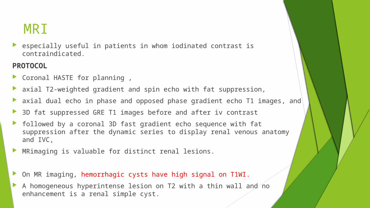
MRI especially useful in patients in whom iodinated contrast is contraindicated.
PROTOCOL Coronal HASTE for planning ,
axial T2-weighted gradient and spin echo with fat suppression,
axial dual echo in phase and opposed phase gradient echo T1 images, and
3D fat suppressed GRE T1 images before and after iv contrast
followed by a coronal 3D fast gradient echo sequence with fat suppression after the dynamic series to display renal venous anatomy and IVC,
MRimaging is valuable for distinct renal lesions.
On MR imaging, hemorrhagic cysts have high signal on T1WI.
A homogeneous hyperintense lesion on T2 with a thin wall and no enhancement is a renal simple cyst.
All 3 phases of renal excretion are obtained after contrast and subtraction images may be obtained to determine, percentage of enhancement
In and opposed phase images help in detecting intratumoral fat.
MR urogram is indicated in patients with suspected collecting system disease
Oncocytoma
solid non-fat containing renal mass,
Arising from proximal tubule with many characteristics of RCCs
Most oncocytomas are asymptomatic (80%), but few may present with hematuria pain or mass
The peak age of incidence is in the seventh decade with a male pre-ponderance.
IMAGING
as well demarcated, unencapsulated, fairly homogeneous renal cortical tumors.
Bilateral multiple oncocytomas are seen in hereditary syndromes of renal oncocytosis

USG may show a central stellate scar within a solid hematogenous mass.
Generally tend be large well demarcated tumours at presentation.
Non contrast
if less than 3 cm - homogenous attenuation
if more than 3 cm - heterogenous attenuation
perinephric fat-stranding may be present due to oedema
calcification may be present
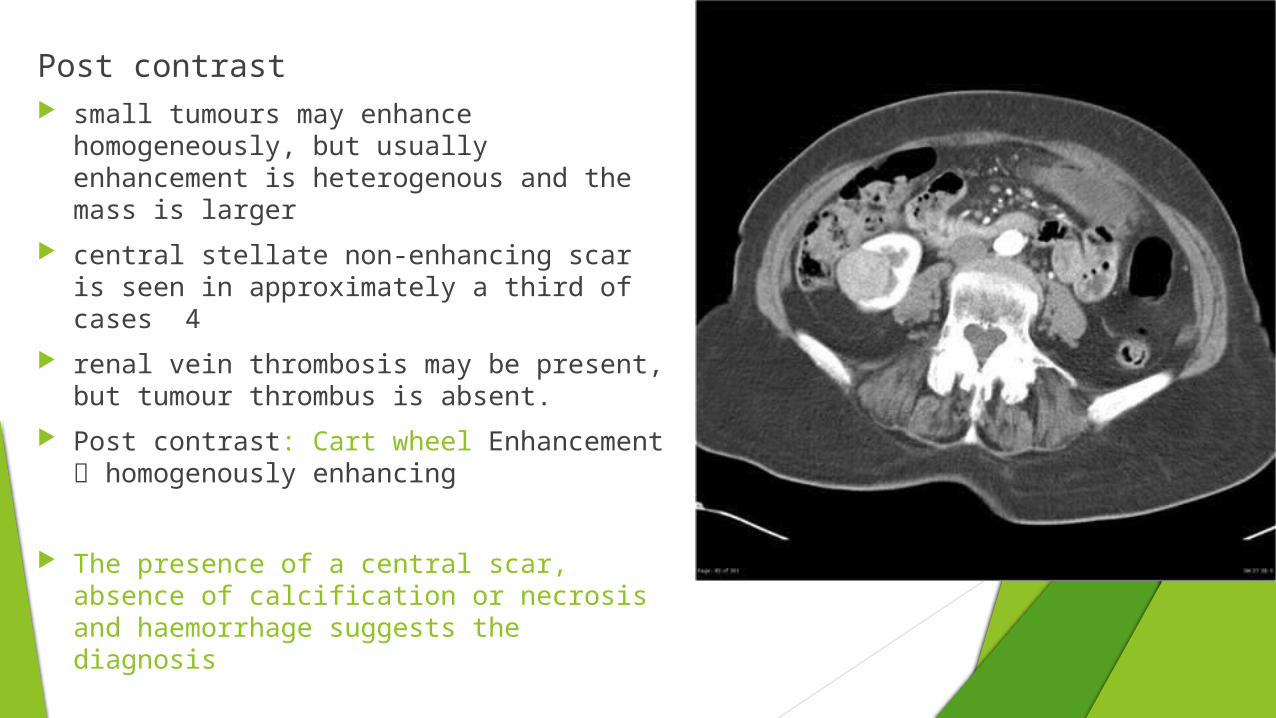
Post contrast small tumours may enhance
homogeneously, but usually enhancement is heterogenous and the mass is larger
central stellate non-enhancing scar is seen in approximately a third of cases 4
renal vein thrombosis may be present, but tumour thrombus is absent.
Post contrast: Cart wheel Enhancement homogenously enhancing
The presence of a central scar, absence of calcification or necrosis and haemorrhage suggests the diagnosis

Surgery is usually performed for these and HPE is indicated to confirm both.
MRI: T1W – Hypo to iso intense ; T2W hyperintense,
The central scar, if present is hypointense on T2
Post contrast show heterogenous enhancement

Renal Adenomas
Papillary adenomas are the most common renal epithelial neoplasm
According to autopsy series, approximately 40 percent of patients above the age of 70 harbor renal adenoma.
They are indistinguishable from RCCs. papillary adenomas measure 5 mm or less peak age in 5th-6th decades
a welldefined, unencapsulated, solitary solid mass. Metanephric adenomas display hypovascularity
and delayed, minimal enhancement

Metanephric adenoma
asymptomatic in many patients few with pain and hematuria.
IMAGING difficult to differentiate from other renal tumours such as renal cell
carcinomas typically appears as a well-defined expansile hypoechoic or
hyperechoic. , unencapsulated, solitary solid mass on sonography. appears as a hyper attenuating mass on unenhanced CT; large tumors appear as heterogeneous, hypovascular masses with
frequent foci of hemorrhage and necrosis. Calcification is seen in 20% of cases
MRI: T1 hypointense , T2 hyper intense ;few hypointense
ANGIOMYOLIPOMA
These benign lesions are mesenchymal neoplasms A type of hamartomas Represents Excess growth of fat , smooth muscle and thick
walled blood vessels Amount of Each component is variable Histologically proliferation of perivascular Epitheliod cells
(PEComas) Tuberous sclerosis is the most commonly associated
syndrome. Multiple and bilateral angiomyolipomas should suggest the
diagnosis of TS Other assosciations include NF-1 , ADPKD, vHL

IMAGING
large, heterogeneous masses with varying amount of macroscopic fat
intralesional aneurysms and hypervascular soft tissues seen.
Angiomyolipomas are composed of thick walled inelastic blood vessels,
Risk of intratumoral and perinephric hemorrhage is higher in lesion > 4 cm diameter
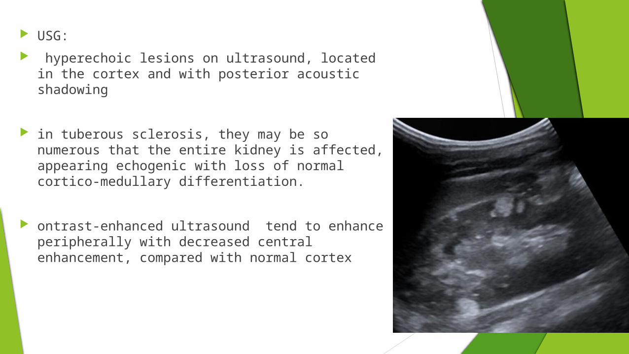
USG:
hyperechoic lesions on ultrasound, located in the cortex and with posterior acoustic shadowing
in tuberous sclerosis, they may be so numerous that the entire kidney is affected, appearing echogenic with loss of normal cortico-medullary differentiation.
ontrast-enhanced ultrasound tend to enhance peripherally with decreased central enhancement, compared with normal cortex

CT
Most lesions involve the cortex and demonstrate macroscopic fat (less than -20 HU)
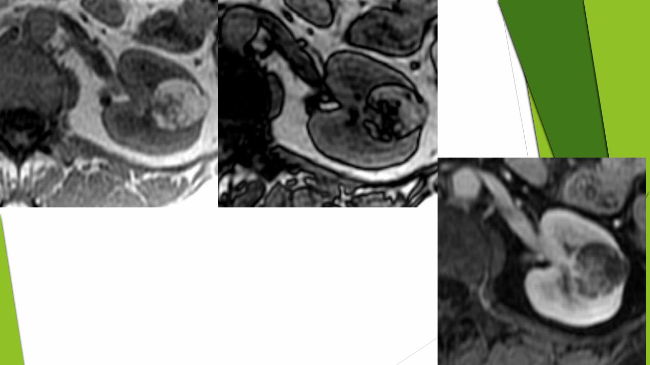
MRI
excellent at evaluating fat containing lesions
Appear hyper in T1 wt images ,
In RCC also mascroscopic fat is seen. But lack of uniformity and capsular breach all be there
macroscopic fat in RCC almost always occurs in the presence of ossification/calcification,
absence of ossification/calcification on imaging is in favour of AML.

DSA - angiography
Angiomyolipomas are hypervascular lesions demonstrating often characteristic features:
micro or macro aneurysms
sharply marginated
dense early arterial network
late whorled appearance
absent AV shunting

Renal lymphangiectasia
is a rare disorder
dilatation of perirenal, peripelvic and intrarenal lymphatics.
aspiration of chylous fluid is usually confirmatory.
Haematuria, flank pain and abdominal pain
Ascites, renal mass, pyelonephritis and renal insufficiency have also been described
Subscapular fluid accumulation relative ischemia Secondary Hypertension.
Renal or perinephric inflammation, blocking the lymphatics

IMAGING
USG:
perirenal collection and peripelvic cysts
retroperitoneal fluid collection
ascites
poor corticomedullary differentiation and diffuse echogenic renal parenchyma
CT:
perinephric fluid attenuation collection of HU usually 0-10.
Fluid collection in peritoneal or retroperitoneum compartments
Perinephric collection may give a page kidney like appearance.


MIXED EPITHELIAL AND STROMALTUMORS (MESTs)
This comprises of tumors previously referred to as hamartoma, adult mesoblastic nephroma.’
seen almost exclusively in perimenopausal women, most patients receiving estrogen therapy
imaging patterns including complex cysts, mixed solid cystic masses
show delayed and heterogeneous enhancement. On MR imaging the degree of delayed enhancement and T2
hypointense signal

It is typically seen as a multiloculated cystic renal mass with a variable proportion of solid and cystic components and containing internal septa. The latter may demonstrate heterogeneous and delayed contrast material enhancement

CYSTIC NEPHROMA
These are rare benign neoplasms that also show a female preponderance. Most patients are asymptomatic
predominantly unilateral, As well circumscribed cystic lesions with thin
septations. Hemorrhage or urinary obstruction may be caused by
the prolapse of the cystic mass into the renal pelvis.

Renal Cysts and Bosniack classification The Bosniak classification system of renal cystic
masses divides renal cystic masses into five groups.
based on imaging characteristics on contrast-enhanced CT
Useful in predicting risk of malignanacy and follow up
Classification
Bosniak 1 simple cyst, imperceptible wall, rounded
work-up: nil
percentage malignant: ~0%

Bosniak 2
minimally complex, a few thin (<1 mm) septa, thin calcifications;
non-enhancing high-attenuation (due to to proteinaceous or haemorrhagic fluid)
renal lesions of less than 3 cm are also included in this category;
these lesions are generally well marginated
work-up: nil percentage malignant: ~0%

Bosniak 3
indeterminate, thick or multiple septations, mural nodule, hyperdense on CT (see 2F)
treatment/work-up: partial nephrectomy or radiofrequency ablation in elderly or poor surgical candidates
percentage malignant: ~54%

Bosniak 4
clearly malignant, solid mass with large cystic or necrotic component
treatment: partial or total nephrectomy
percentage malignant: ~100

Incidental Solid Renal mass If a large >3 cm solid mass is discovered, renal cell carcinoma
(provided there is no fat on CT/MR protocols) is the most probable diagnosis and surgery is recommended. If
the tumor is 1-3 cm in size, RCC is most likely though percutaneous biopsy may be required.
Very small lesions < 1 cm are more likely to be benign; thin sections (< 3 mm) used .
Obsevation and follow up CT/MR at 3-6months, then at 12 months and then yearly may be done.
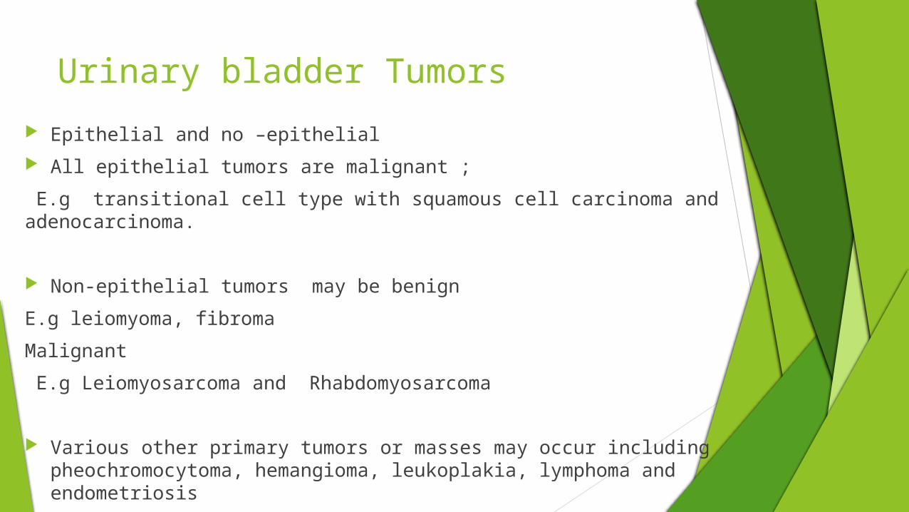
Urinary bladder Tumors
Epithelial and no –epithelial
All epithelial tumors are malignant ;
E.g transitional cell type with squamous cell carcinoma and adenocarcinoma.
Non-epithelial tumors may be benign
E.g leiomyoma, fibroma
Malignant
E.g Leiomyosarcoma and Rhabdomyosarcoma
Various other primary tumors or masses may occur including pheochromocytoma, hemangioma, leukoplakia, lymphoma and endometriosis

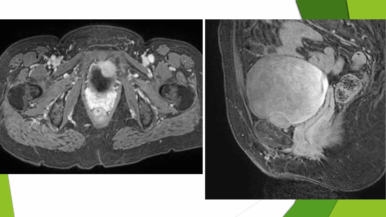
Urinarybladder leiomyoma the most common mesenchymal tumor
accounts for only 0.43% of bladder tumors.
Leiomyomas occur equally in men and women with a wide age range of 22–78 years
Most are small and asymptomatic and are discovered incidentally.
However, large tumors manifest with symptoms such as hesitancy, frequency, dribbling, hematuria, pressure from mass effect, or urinary obstruction.
Leiomyomas arise in the submucosa,
but growth may be submucosal (7%), intravesical (63%), or extravesical (30%)
USG:
smooth-walled homogeneous hypoechoic solid mass in the bladder with thin echogenic surface
Can determine the nature od spread
reveal smooth-walled solid lesion with homogeneous echogenicity

CT is accurate in detection and localization of these lesions, by presenting it as hypodense mass
contrast-enhanced CT scan the lesion is shown as a moderately enhan cing mass
MRI
Better demonstrating sub mucosal origin of the tumour
the preservation of the muscle layer
The imaging characteristics are similar to uterine leiomyomas:
T1: intermediate signal intensity
T2: low signal intensity
degenerated leiomyomas have more heterogeneous signal characteristics; cystic areas have high signal intensity
T1 C+ (Gad): contrast enhancement is variable, degenerated areas lack enhancement

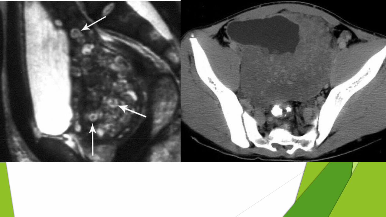
Neurofibroma
Neurofibromas of the bladder are rare,
The bladder is the most common genitourinary site of a neurofibroma.
They may be isolated or occur in association with neurofibromatosis type 1
Patients most commonly present with urinary tract infections. Other symptoms include hematuria, urinary frequency and urgency, mass, or obstruction

Characteristic CT and MR imaging features are diffuse, nodular bladder wall thickening with masses extending to the pelvic side wall .
MR imaging features are more specific Low signal intensity on T1-weighted images a target sign on T2-weighted images. which consists of low-signal-intensity fibrosis surrounded by
high-signal-intensity myxoid stroma

Bladder Hemangiomas
half manifest in childhood
the mean age is 58 years (range, 19–76 years) and they are more common in males in a ratio of 3.7:1
Painless gross hematuria is the most common presentation
a median size of 0.7 cm (range, 0.2–3 cm)
. Cavernous hemangioma is the most common type, accounting for 78% of cases,
capillary and arteriovenous types having equal rates of 10%

Most were single, broad-based, sessile bladder masses on the posterolateral walls
Hemangiomas are hypervascular masses at US, CT, and MR imaging
a focal, lobular, intramural mass OR diffuse bladder wall thickening
MRI
low to intermediate signal intensity on T1-weighted images
markedly high signal intensity on T2-weighted images
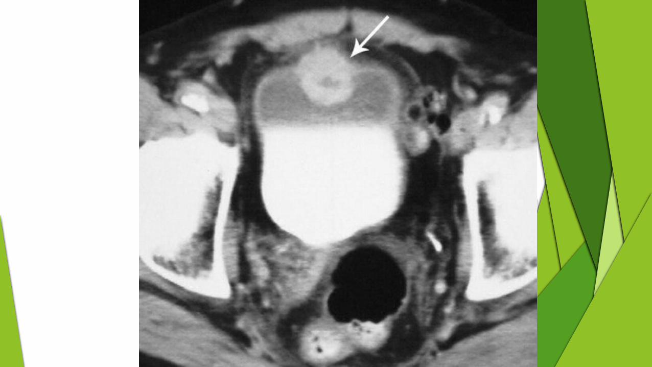

Paraganglioma
preferred term for pheochromocytomas arising outside the adrenal gland.
may rarely manifest as a bladder mass.
They account for 0.1% of all bladder tumors and 1% of all pheochromocytomas.
The age range is wide at 10–78 years, and there is a female preponderance
A characteristic clinical syndrome of catecholamine release during micturition, “micturition attack,” occurs in 50% of patients.
Nonfunctioning pheochromocytomas are also reported.
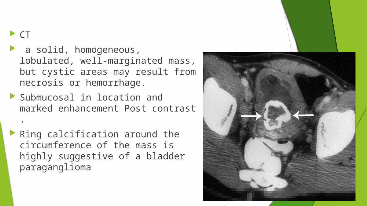
CT a solid, homogeneous, lobulated,
well-marginated mass, but cystic areas may result from necrosis or hemorrhage.
Submucosal in location and marked enhancement Post contrast .
Ring calcification around the circumference of the mass is highly suggestive of a bladder paraganglioma
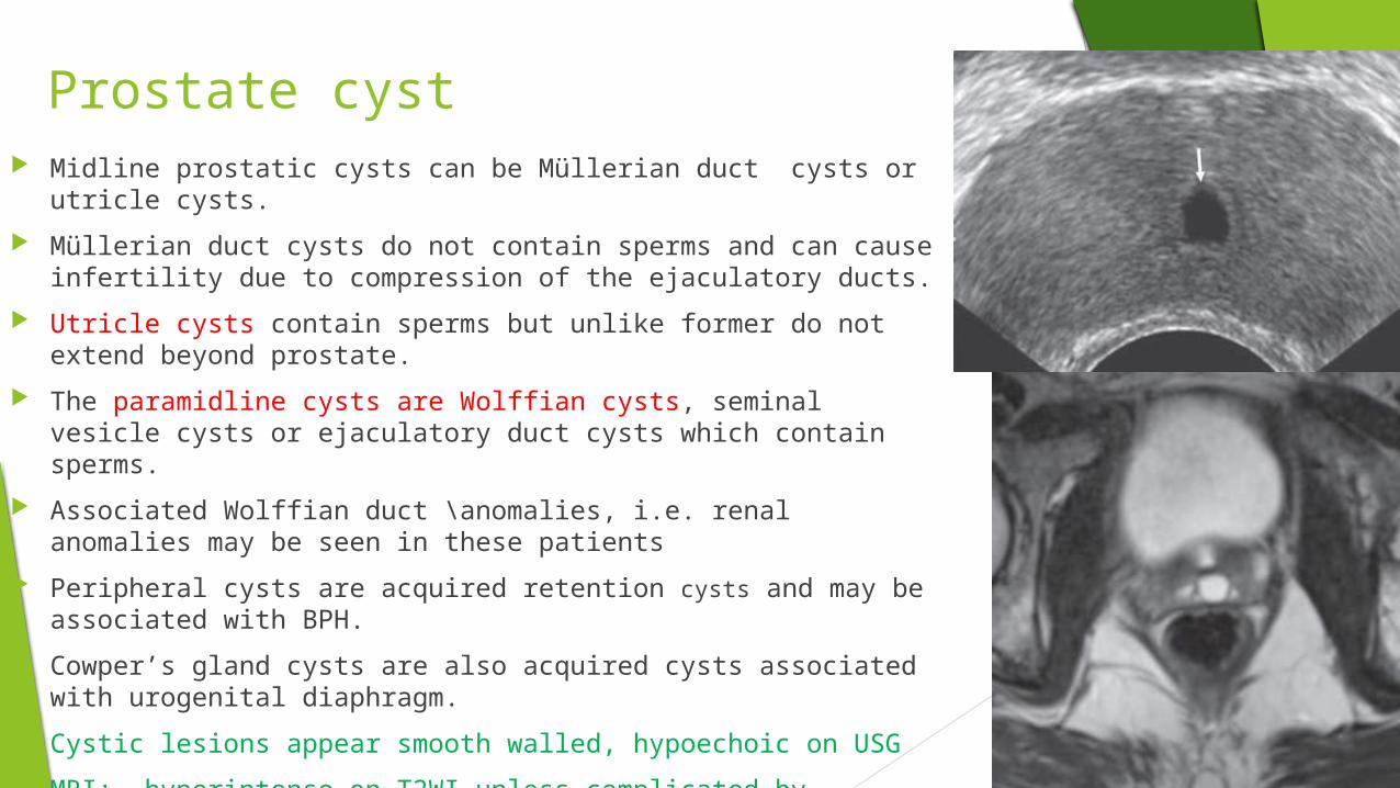
Prostate cyst Midline prostatic cysts can be Müllerian duct cysts or utricle cysts.
Müllerian duct cysts do not contain sperms and can cause infertility due to compression of the ejaculatory ducts.
Utricle cysts contain sperms but unlike former do not extend beyond prostate.
The paramidline cysts are Wolffian cysts, seminal vesicle cysts or ejaculatory duct cysts which contain sperms.
Associated Wolffian duct \anomalies, i.e. renal anomalies may be seen in these patients
Peripheral cysts are acquired retention cysts and may be associated with BPH.
Cowper’s gland cysts are also acquired cysts associated with urogenital diaphragm.
Cystic lesions appear smooth walled, hypoechoic on USG
MRI: hyperintense on T2WI unless complicated by infection or hemorrhage

Adrenal Tumors
Pheochromocytoma catecholamine producing tumors which arise from ganglion cells.
Arise from anywhere in the autonomic nervous system.
Ninety percent of pheochromocytomas originate in the adrenal medulla,
10 percent are extra-adrenal,
the common sites being paravertebral sympathetic ganglia, organ of Zuckerkandl, urinary bladder, neck or mediastinum.
10 percent are extra-adrenal, Ten percent of pheochromocytomas are bilateral and 10 percent are malignant..
Clinical features of hypertension, paroxysmal attacks of palpitation, headache, sweating
biochemical tests of plasma catecholamine levels and 24 hours urine vanillylmandelic acid level can provide the diagnosis
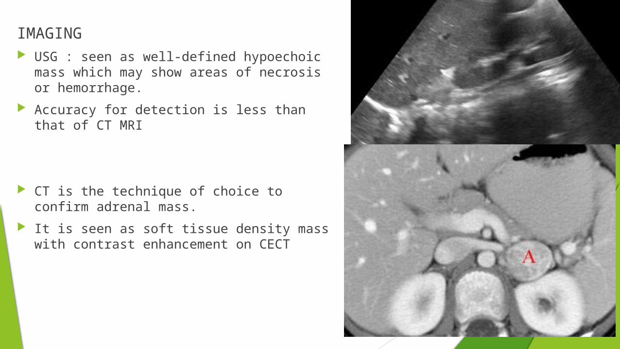
IMAGING USG : seen as well-defined hypoechoic mass
which may show areas of necrosis or hemorrhage.
Accuracy for detection is less than that of CT MRI
CT is the technique of choice to confirm adrenal mass.
It is seen as soft tissue density mass with contrast enhancement on CECT

MRI MRI is the most sensitive modality for identification of pheochromocytomas,
and is particularly useful in cases of extra-adrenal location. The overall sensitivity is said to be 98%
T1
slightly hypo-intense to the remainder of the adrenal
if necrotic and/or haemorrhagic then signal will be more heterogeneous
T2 markedly hyper-intense
areas of necrosis/haemorrhage/calcification will alter signal
T1 C+ (Gd) heterogenous enhancement
enhancement is prolonged, persisting for as long as 50 minutes

Adrenal adenomas Represent nonneoplastic overgrowth of adrenocortical cells of zona fasciculata.
Consist of cholesterol laden clear cells and contribute little to steroid production
Functional and non functional types are seen
Imaging wise both appear the same.
They account for almost 90 percent of all incidentalomas
CT: Low attenuation well defgined mass (HU values rarely exceed 10HU).
Contrast study shows enhancement with washout in delayed phase

Mri

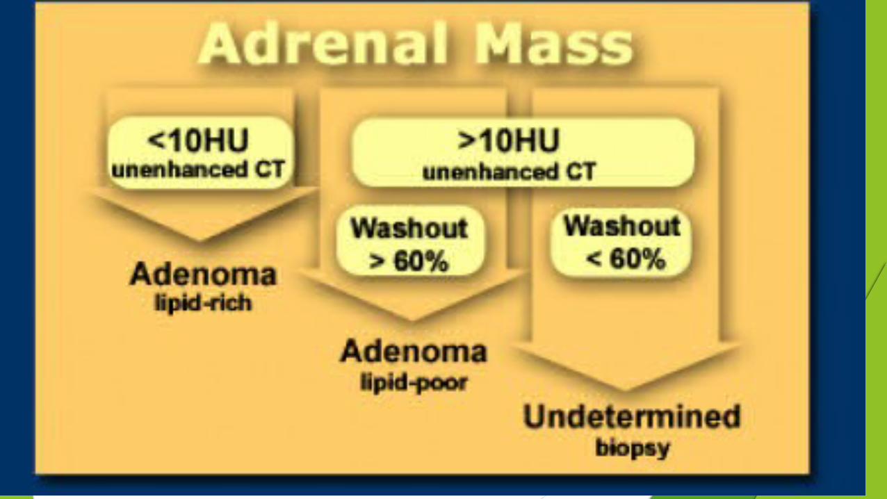
Adrenal Adenoma V/s Metastasis Adrenal adenomas have two properties that differentiate
them from non-adenomas.
. 70% of adenomas contain high intracellular fat (lipid-rich adenomas) and will be of low attenuation on unenhanced CT.
2. Adenomas rapidly wash out contrast.
Unenhanced CT : HU values less than 10HU Adenoma
If > 10HU then Contrast CT
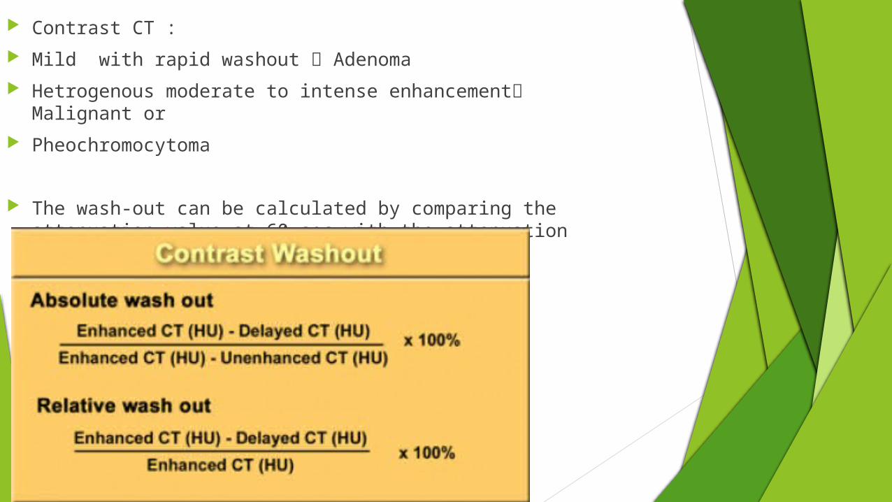
Contrast CT :
Mild with rapid washout Adenoma
Hetrogenous moderate to intense enhancement Malignant or
Pheochromocytoma
The wash-out can be calculated by comparing the attenuation value at 60 sec with the attenuation value on a delayed scan at 15 minutes.

Adrenal Cyst Adrenal cysts are rare and usually seen in females.
derived from endothelium in 45 percent of patients
the rest are epithelial, parasitic or pseudocysts from prior hemorrhage.
CT shows characteristically well-defined round low attenuating lesion suggesting fluid with rim enhancement
Rim calcification may be noted
The attenuation values may be mixed in the presence of debris or hemorrhage
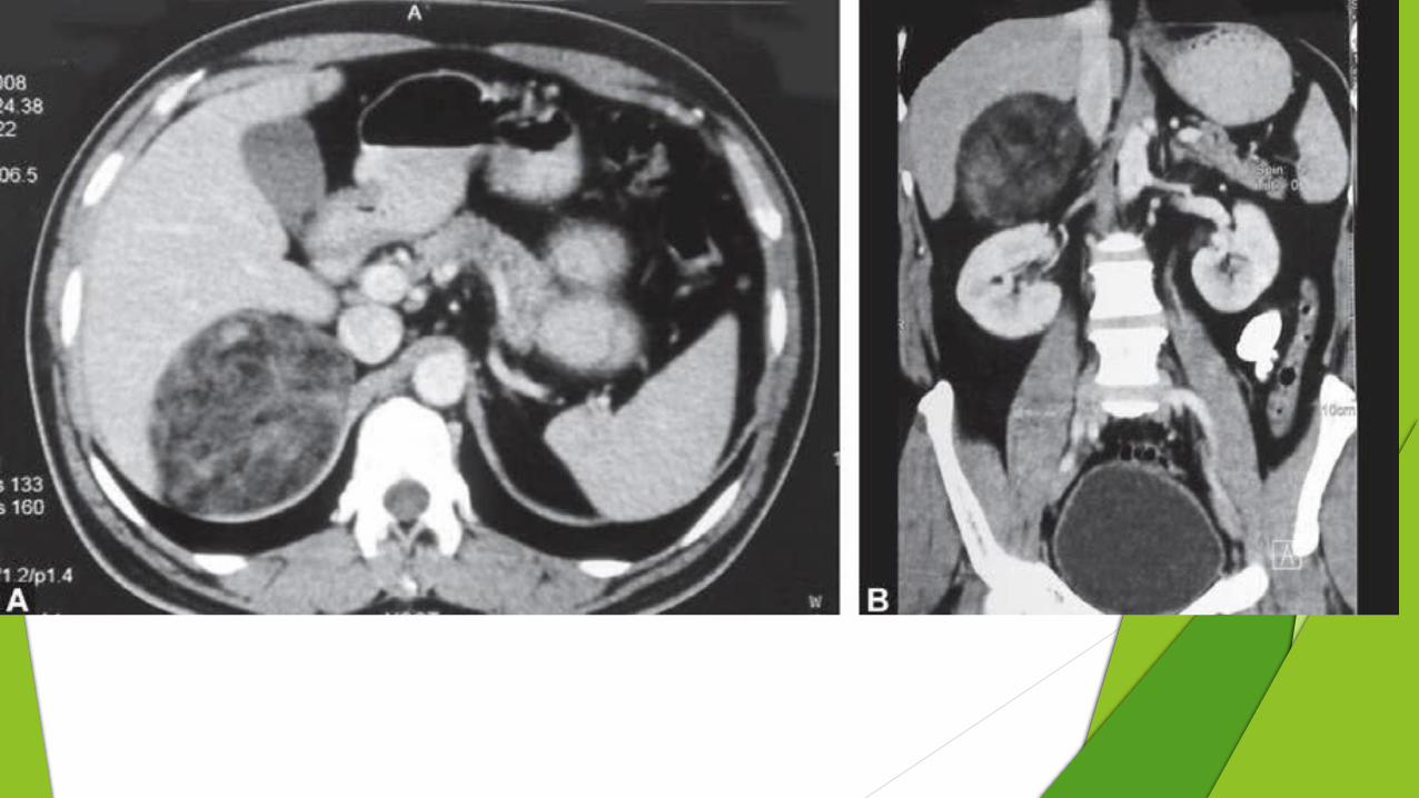
Adrenal Myelolipoma benign tumor of the cortex, comprised of mature fat and hematopoietic
elements
It accounts for about 7-15 percent of all incidentalomas.
Imaging appearance may vary depending on which histological component is dominant
USG : well-defined echogenic or heterogeneous adrenal mass,
may be associated with apparent posterior displacement of the diaphragm.
CT:
confirms presence of intratumoral fat.
Punctate calcification may be seen in 20 percent of cases
large myelolipomas may mimic retroperitoneal lipoma or liposarcoma.

GRE T1W in-phase (A) and opposed phase (B) image show a hyperintenseright adrenal mass which shows no drop in signal intensity but is suppressed on T2W fat suppressed image

Spotters


Double bubble sign


PINCER TYPE IMPINGEMENT
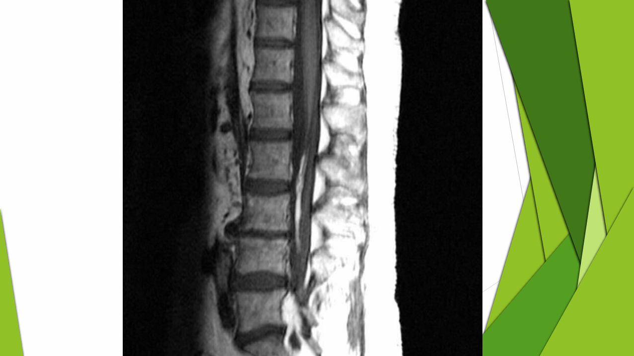

Lipoma of the filum terminale
is a relatively common finding on imaging of the lumbar spine, and in most cases is an incidental finding of no clinical concern.
In some patients however it may be associated with signs and symptoms of tethered cord syndrome. In such cases it is usually associated with a thickened filum and a low lying conus


Hamburger’s sign

THANK YOU