Renal Pelvis, Ureter, Bladder and Other Urinary · tumor(s) in both the right ureter and tumor(s)...
52
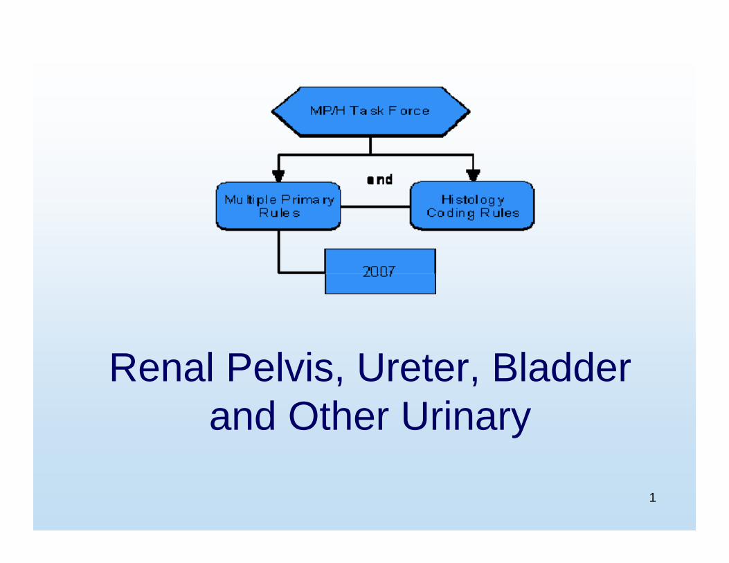
1 Renal Pelvis, Ureter, Bladder and Other Urinary
-
Upload
truongdang -
Category
Documents
-
view
219 -
download
0
Transcript of Renal Pelvis, Ureter, Bladder and Other Urinary · tumor(s) in both the right ureter and tumor(s)...
1
Renal Pelvis, Ureter, Bladder and Other Urinary

2
Equivalent Terms, Definitions,Tables and Illustrations

3
Introduction
• Change in groupings– Previous: Kidney, ureter, renal pelvis
• Bladder, ureter, renal pelvis– Lower urinary tract– Lined by transitional epithelium /
urothelium

4
Urothelium
• Frequent multiple or multifocal tumors– Field effect: Widespread change in urothelium– Implantation: Cells washed along in urine

5
Flat Carcinoma In Situ
• Direct spread within the epithelium • Direct extension• Field effect• Implantation

6
Squamous Cell Carcinoma
Pure squamous cell carcinoma has a poor prognosis
See histology coding rules H5 and H13

7
Most Invasive - Bladder
• Mucosa• Lamina propria (some pathologists equate
this to submucosa)• Muscularis mucosae (this layer not always
present, may not be mentioned)• Submucosa• Muscular layer (muscularis propria,
detrusor muscle)• Serosa, adventitia
8
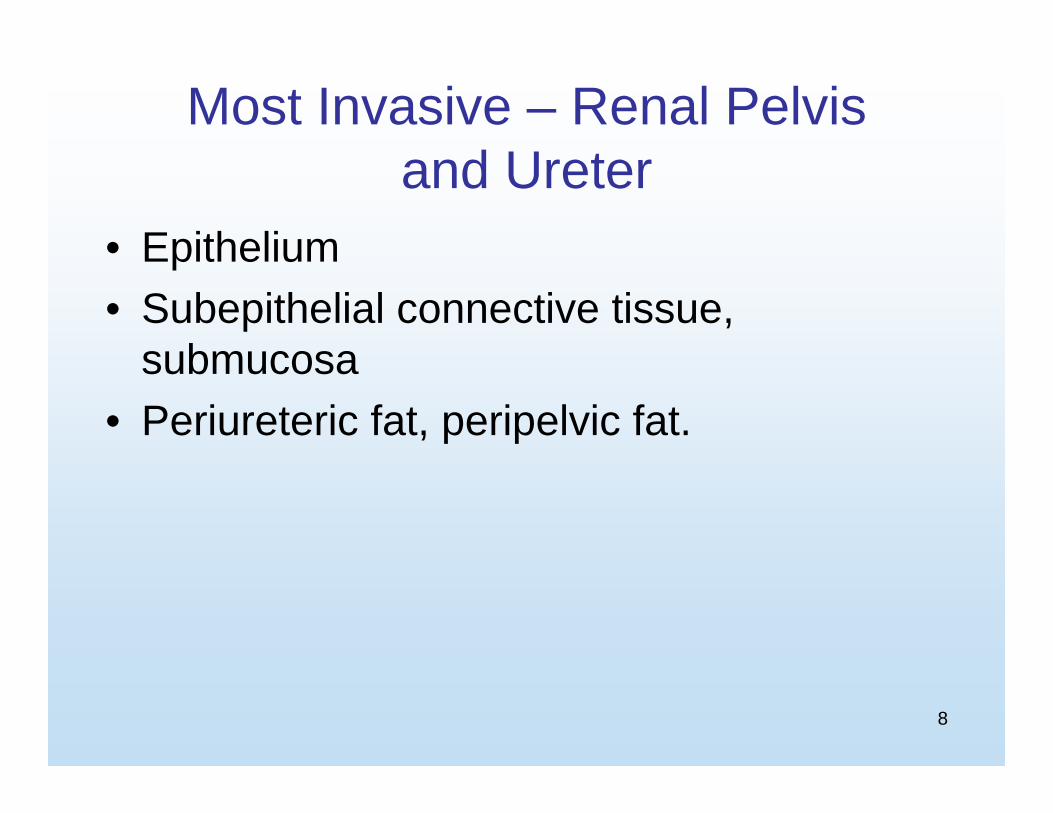
Most Invasive – Renal Pelvis and Ureter
• Epithelium• Subepithelial connective tissue,
submucosa• Periureteric fat, peripelvic fat.

9
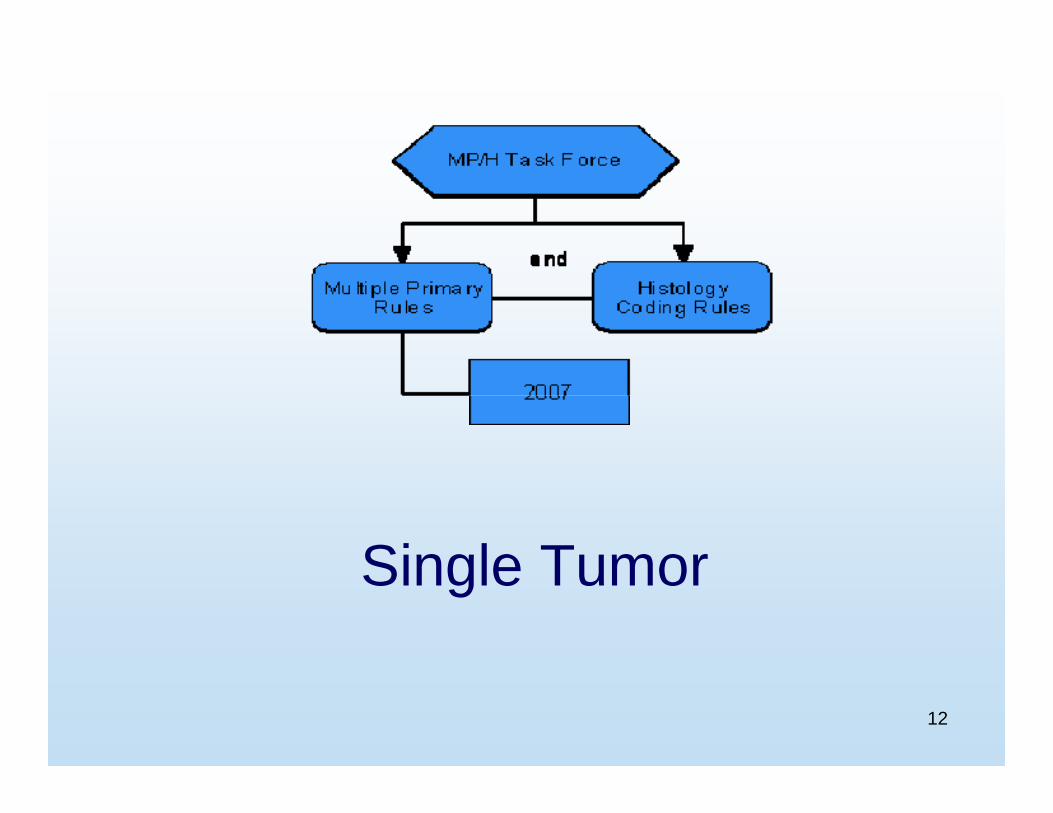
Multiple Primary Rules

10
Unknown if Single or Multiple Tumors
11
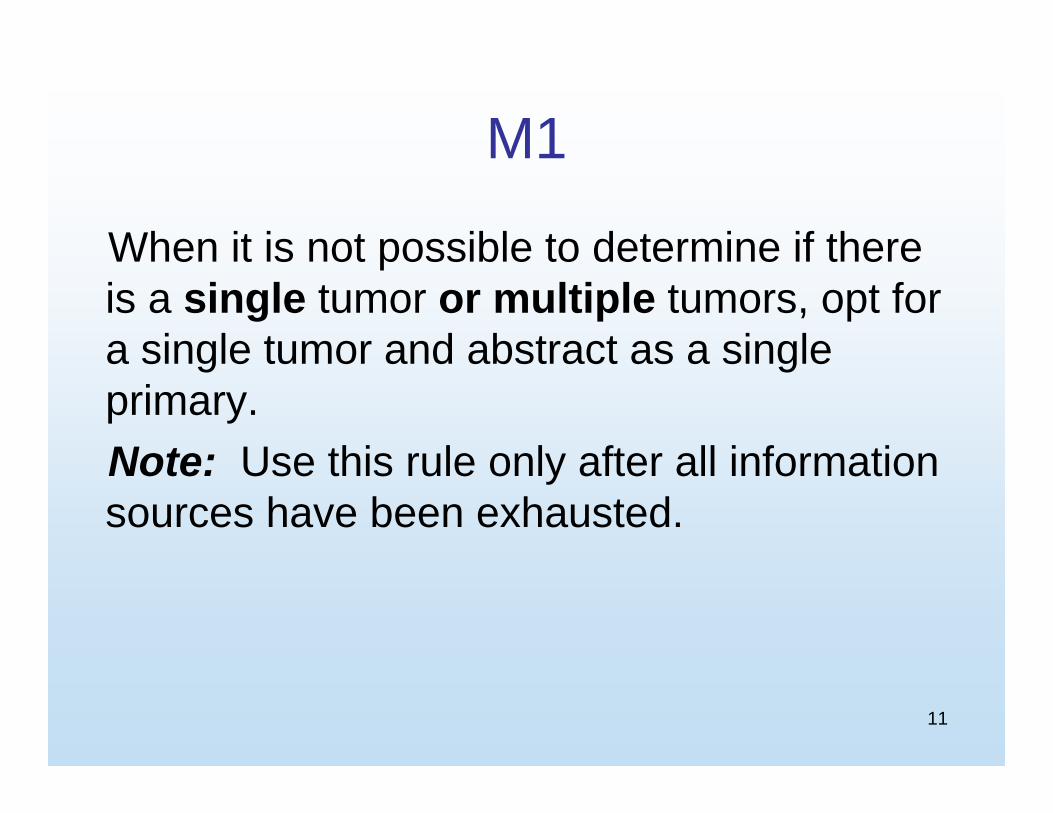
M1
When it is not possible to determine if there is a single tumor or multiple tumors, opt for a single tumor and abstract as a singleprimary.Note: Use this rule only after all information sources have been exhausted.
12
Single Tumor

13
M2
A single tumor is always a single primary.
Note: The tumor may overlap onto or extend into adjacent/contiguous site or subsite.

14
Multiple Tumors

15
M3
When no other urinary sites are involved, tumor(s) in both the right renal pelvis andtumor(s) in the left renal pelvis are multiple primaries.
Note: Use this rule and abstract as a multiple primary unless documented to be metastatic.

16
M4
When no other urinary sites are involved, tumor(s) in both the right ureter andtumor(s) in the left ureter are multiple primaries.
Note: Use this rule and abstract as a multiple primary unless documented to be metastatic.

17
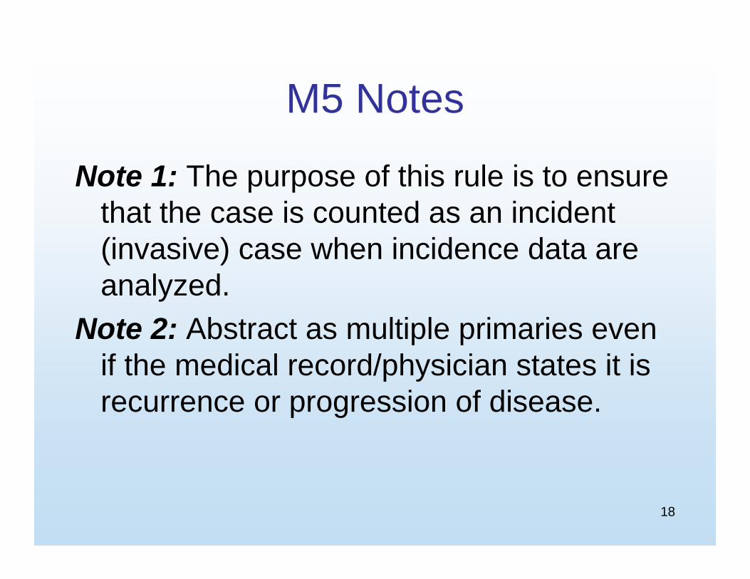
M5
An invasive tumor following a non-invasive or in situ tumor more than 60 days after diagnosis is a multiple primary.
18
M5 Notes
Note 1: The purpose of this rule is to ensure that the case is counted as an incident (invasive) case when incidence data are analyzed.
Note 2: Abstract as multiple primaries even if the medical record/physician states it is recurrence or progression of disease.

19
M6
Bladder tumors with any combination of the following histologies: papillary carcinoma(8050), transitional cell carcinoma (8120-8124), or papillary transitional cell carcinoma (8130-8131), are a single primary.

20
M7
Tumors diagnosed more than three (3) years apart are multiple primaries.
21
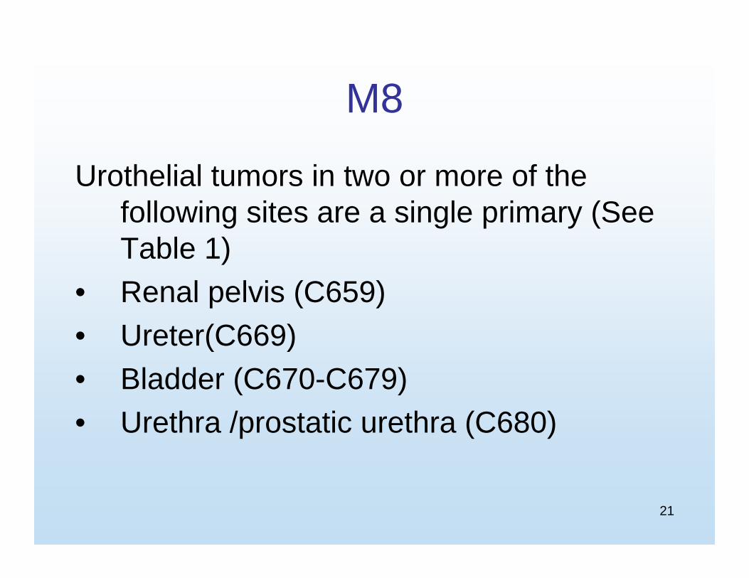
M8
Urothelial tumors in two or more of the following sites are a single primary (See Table 1)
• Renal pelvis (C659) • Ureter(C669)• Bladder (C670-C679) • Urethra /prostatic urethra (C680)

22
M9
Tumors with ICD-O-3 histology codes that are different at the first (xxxx), second (xxxx) or third (xxxx) number are multiple primaries.
23
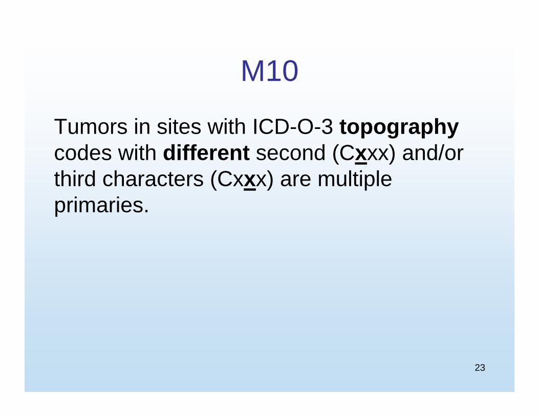
M10
Tumors in sites with ICD-O-3 topographycodes with different second (Cxxx) and/or third characters (Cxxx) are multiple primaries.

24
M11
Tumors that do not meet any of the above criteria are a single primary.
Note: When an invasive tumor follows an in situ tumor within 60 days, abstract as a single primary.

25
Histology Rules

26
Single Tumor

27
H1
Code the histology documented by the physician when there is no pathology/cytology specimen or the pathology/cytology report is not available.

28
H1 Notes
Note 1: Priority for using documents to code the histology
– Documentation in the medical record that refers to pathologic or cytologic findings
– Physician’s reference to type of cancer (histology) in the medical record
– CT or MRI scans

29
H1 Notes
Note 2: Code the specific histology when documented.
Note 3: Code the histology to 8000 (cancer/malignant neoplasm) or 8010 (carcinoma, NOS) as stated by the physician when nothing more specific is documented.

30
H2
Code the histology from the metastatic site when there is no pathology/cytology specimen from the primary site.
Note: Code the behavior /3

31
H3
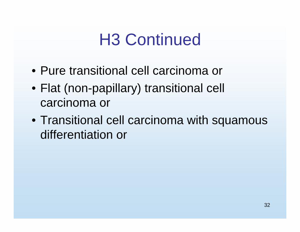
Code 8120 (transitional cell/urothelialcarcinoma) (Table 1 - Code 8120) when there is:
32
H3 Continued
• Pure transitional cell carcinoma or• Flat (non-papillary) transitional cell
carcinoma or• Transitional cell carcinoma with squamous
differentiation or
33
H3 Continued
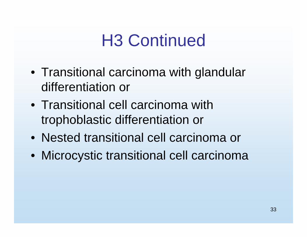
• Transitional carcinoma with glandular differentiation or
• Transitional cell carcinoma with trophoblastic differentiation or
• Nested transitional cell carcinoma or• Microcystic transitional cell carcinoma
34
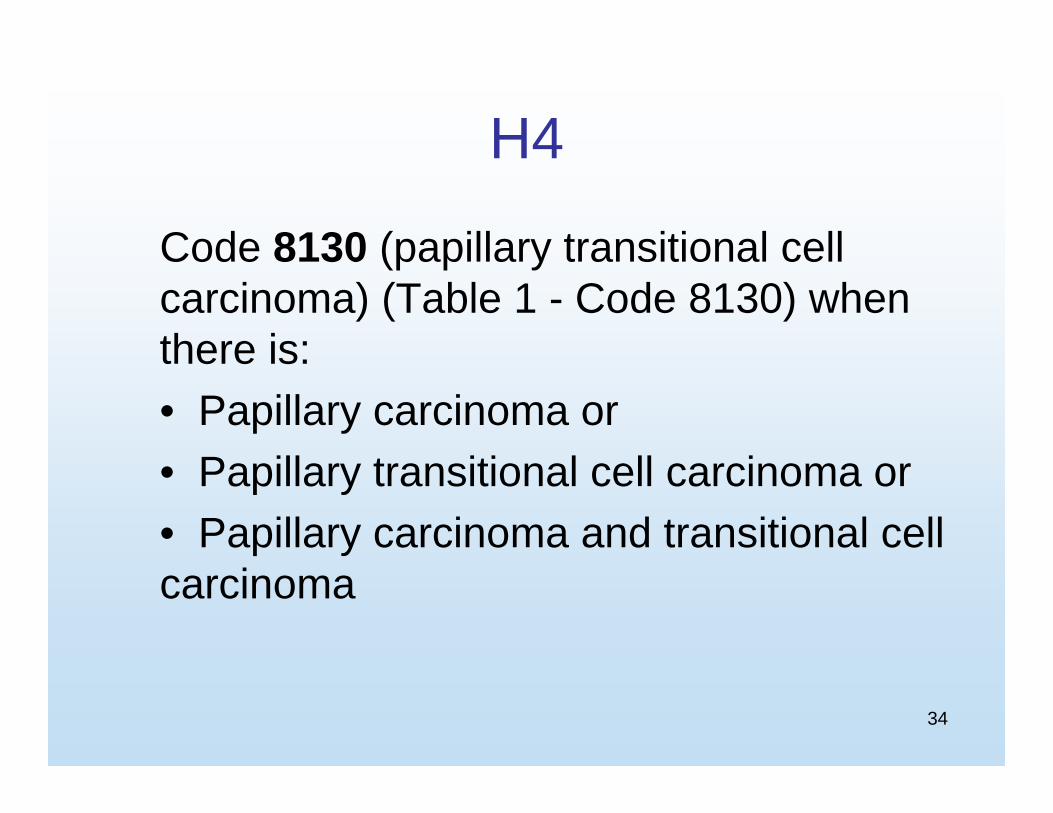
H4
Code 8130 (papillary transitional cell carcinoma) (Table 1 - Code 8130) when there is:• Papillary carcinoma or• Papillary transitional cell carcinoma or• Papillary carcinoma and transitional cell carcinoma
35
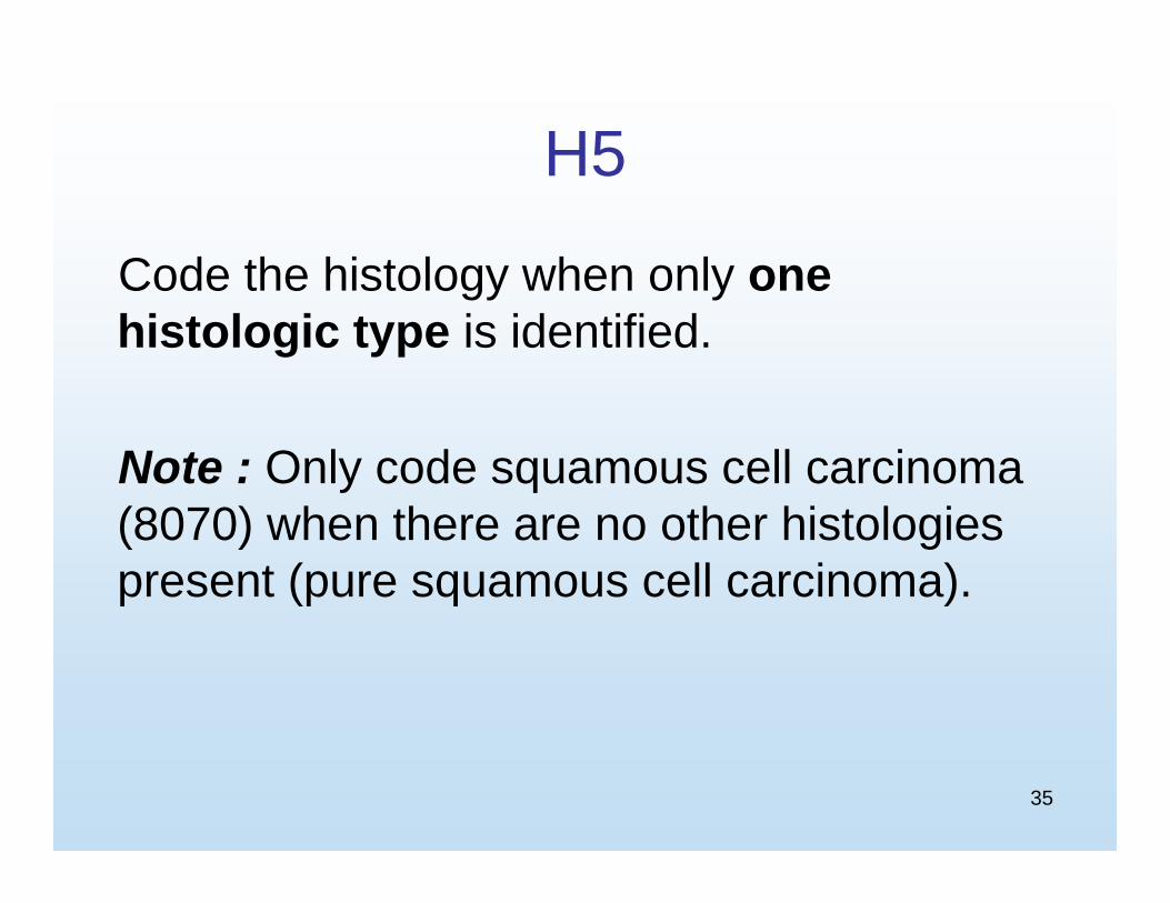
H5
Code the histology when only one histologic type is identified.
Note : Only code squamous cell carcinoma (8070) when there are no other histologies present (pure squamous cell carcinoma).
36
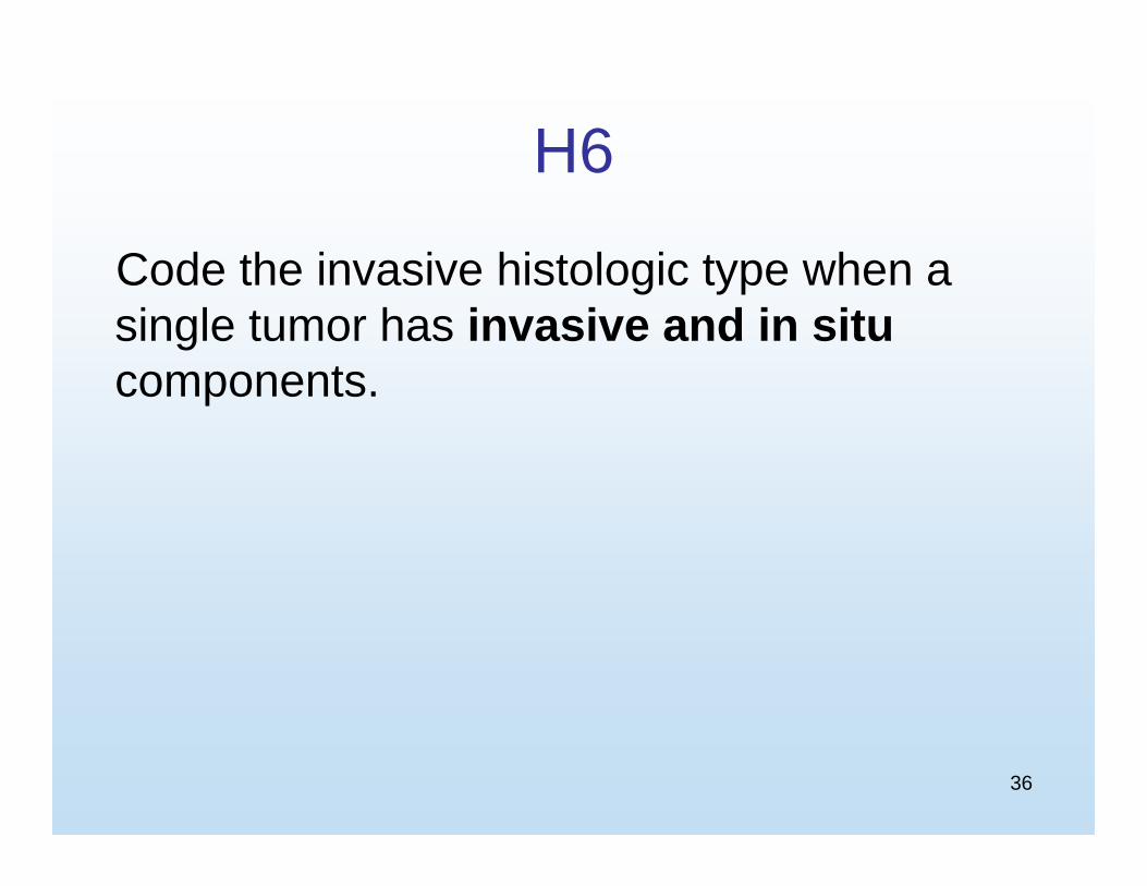
H6
Code the invasive histologic type when a single tumor has invasive and in situ components.

37
H7
Code the most specific histologic term.Examples• Cancer/malignant neoplasm, NOS
(8000) and a more specific histology or• Carcinoma, NOS (8010) and a more
specific carcinoma or• Sarcoma, NOS (8800) and a more
specific sarcoma (invasive only)

38
H7 Notes
Note 1: The specific histology for in situtumors may be identified as pattern, architecture, type, subtype, predominantly, with features of, major, or with ____differentiation
Note 2: The specific histology for invasive tumors may be identified as type, subtype, predominantly, with features of, major, or with ____differentiation

39
H8
Code the histology with the numerically higher ICD-O-3 code.

40
Multiple Tumors Abstracted as a Single Primary

41
H9
Code the histology documented by the physician when there is no pathology/cytology specimen or the pathology/cytology report is not available.

42
H9 Notes
Note 1: Priority for using documents to code the histology– Documentation in the medical record that
refers to pathologic or cytologic findings– Physician’s reference to type of cancer
(histology) in the medical record– CT or MRI scans
43
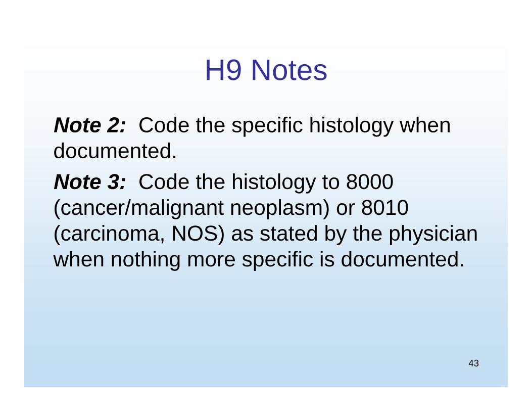
H9 Notes
Note 2: Code the specific histology when documented.Note 3: Code the histology to 8000 (cancer/malignant neoplasm) or 8010 (carcinoma, NOS) as stated by the physician when nothing more specific is documented.
44
H10
Code the histology from the metastatic site when there is no pathology/cytology specimen from the primary site.
Note: Code the behavior /3

45
H11
Code 8120 (transitional cell/urothelialcarcinoma) (Table 1 – Code 8120) when there is:
• Pure transitional cell carcinoma or• Flat (non-papillary) transitional cell
carcinoma or

46
H11 Continued
• Transitional cell carcinoma with squamous differentiation or
• Transitional cell carcinoma with glandular differentiation or
• Transitional cell carcinoma with trophoblastic differentiation or
• Nested transitional cell carcinoma or• Microcystic transitional cell carcinoma

47
H12
Code 8130 (papillary transitional cell carcinoma) (Table 1 – Code 8130) when there is:
• Papillary carcinoma or• Papillary transitional cell carcinoma or• Papillary carcinoma and transitional
cell carcinoma

48
H13
Code the histology when only one histologic type is identified.
Note: Only code squamous cell carcinoma (8070) when there are no other histologies present (pure squamous cell carcinoma).

49
H14
Code the histology of the most invasivetumor.
Note: See the Renal Pelvis, Ureter, Bladder and Other Urinary Equivalent Terms, Definitions, Tables and Illustrations for the definition of most invasive.

50
H14 Continued
• If one tumor is in situ and one is invasive, code the histology from the invasive tumor.
• If both/all histologies are invasive, code the histology of the most invasive tumor.

51
H15
Code the histology with the numerically higher ICD-O-3 code.

52
MP/H Task Force