mirracle: An Augmented Reality Magic Mirror System for...
2
mirracle: An Augmented Reality Magic Mirror System for Anatomy Education Tobias Blum * Valerie Kleeberger Christoph Bichlmeier Nassir Navab Computer Aided Medical Procedures & Augmented Reality (CAMP), Technische Universit¨ at M ¨ unchen, Munich, Germany ABSTRACT We present an augmented reality magic mirror for teaching anatomy. The system uses a depth camera to track the pose of a user standing in front of a large display. A volume visualization of a CT dataset is augmented onto the user, creating the illusion that the user can look into his body. Using gestures, different slices from the CT and a photographic dataset can be selected for visual- ization. In addition, the system can show 3D models of organs, text information and images about anatomy. For interaction with this data we present a new interaction metaphor that makes use of the depth camera. The visibility of hands and body is modified based on the distance to a virtual interaction plane. This helps the user to understand the spatial relations between his body and the virtual interaction plane. Index Terms: H.5.1 [Information Interfaces and Presentation]: Artificial, augmented, and virtual realities—; 1 I NTRODUCTION Knowledge about human anatomy is an important part of education, especially for medical professionals. Teaching anatomy is difficult and often a large amount of effort is expended, e.g., when doctors present demonstrations in dissection courses, when creating illus- trations and plastic models of anatomy or by utilizing 3D computer graphics. We present a novel way of intuitively teaching anatomy using an augmented reality (AR) magic mirror system displaying anatomical structures on the user’s body. Furthermore we present a new concept for gesture-based interaction. Figure 1: The system consists of a display device, a color camera and a depth camera. The user is tracked and a CT dataset is aug- mented onto the user. 2 METHODS The mirracle system, which is presented in this paper, has mainly been developed for education of anatomy in classrooms, museums or exhibitions. It focuses on bones and a small number of important * e-mail: [email protected] organs of the abdomen, namely liver, lungs, pancreas, stomach and small intestine. Figure 1 shows a photo of the mirracle hardware setup. The first component of our system is a display device. The second component is a color camera mounted next to the display surface. The third component is a depth camera which is placed next to the color camera. Our system uses the Microsoft Kinect which is sold as an add-on for the Xbox 360 video game console to enable games using gestures and body movement as input. It consists of a color and a depth camera. 2.1 AR In-situ visualization of human anatomy For an intuitive visualization of organs we use the concept of a magic mirror. The camera image is flipped horizontally and shown on the screen such that the user has the impression of standing in front of a mirror. Virtual objects can be added to the image of the real scene. The magic mirror concept has been used previously to augment virtual shoes [4], shirts [3] or knight’s armors [5] onto the user. For tracking the user, previous systems have been using markers [5, 3] or required the user to wear a shirt with a rectangular highly textured region [6]. While previous systems have augmented objects onto the user, our system extends the magic mirror concept for training of anatomy. It creates the illusion that the user can look inside her body. We augment a volume visualization of a CT dataset onto the user. To allow a correct augmentation of the CT, the pose of the user has to be tracked. This is done based on the depth image using the NITE skeleton tracking 1 software. Our system could use a CT scan of the user. However a CT of the user is usually not available. Therefore we use the Visible Korean Human dataset (VKH) [8]. This dataset consists of a CT scan, a MR volume and a photographic volume which has been acquired by stacking up cryosections. The CT volume is scaled to the size of the user and augmented onto the user. For visualization of the bones a transfer function is used as bones can be distinguished easily in the CT volume based on the voxel intensities. For the organs a segmentation of the VKH is used. The augmentation uses contextual in-situ visualization [2] such that the virtual objects are only shown through a circular win- dow as is shown in figure 1. This leads to a better perception of depth, compared to a simple augmentation of the whole CT. 2.2 Gesture based interaction for slices Medical volumes are usually visualized by showing slices that are aligned with the axes of the volume. A volume can be seen as a stack of sagittal slices starting from the left side and going to the right. When the system is in sagittal slice mode the user selects the current sagittal slice by moving the right hand from left to right. To switch to a transverse slice mode the user has to move the hand up or down, respectively front or back for the coronal slice mode. The current slice is shown on the right part of the monitor while a green rectangle is augmented onto the user. This rectangle visualizes the pose of the current slice. The system can switch between slices from the CT or the photographic volume. 1 www.openni.org 115 IEEE Virtual Reality 2012 4-8 March, Orange County, CA, USA 978-1-4673-1246-2/12/$31.00 ©2012 IEEE
Transcript of mirracle: An Augmented Reality Magic Mirror System for...

mirracle: An Augmented Reality Magic Mirror System for AnatomyEducation
Tobias Blum∗ Valerie Kleeberger Christoph Bichlmeier Nassir Navab
Computer Aided Medical Procedures & Augmented Reality (CAMP), Technische Universitat Munchen, Munich, Germany
ABSTRACT
We present an augmented reality magic mirror for teachinganatomy. The system uses a depth camera to track the pose of auser standing in front of a large display. A volume visualizationof a CT dataset is augmented onto the user, creating the illusionthat the user can look into his body. Using gestures, different slicesfrom the CT and a photographic dataset can be selected for visual-ization. In addition, the system can show 3D models of organs, textinformation and images about anatomy. For interaction with thisdata we present a new interaction metaphor that makes use of thedepth camera. The visibility of hands and body is modified basedon the distance to a virtual interaction plane. This helps the userto understand the spatial relations between his body and the virtualinteraction plane.
Index Terms: H.5.1 [Information Interfaces and Presentation]:Artificial, augmented, and virtual realities—;
1 INTRODUCTION
Knowledge about human anatomy is an important part of education,especially for medical professionals. Teaching anatomy is difficultand often a large amount of effort is expended, e.g., when doctorspresent demonstrations in dissection courses, when creating illus-trations and plastic models of anatomy or by utilizing 3D computergraphics. We present a novel way of intuitively teaching anatomyusing an augmented reality (AR) magic mirror system displayinganatomical structures on the user’s body. Furthermore we present anew concept for gesture-based interaction.
Figure 1: The system consists of a display device, a color cameraand a depth camera. The user is tracked and a CT dataset is aug-mented onto the user.
2 METHODS
The mirracle system, which is presented in this paper, has mainlybeen developed for education of anatomy in classrooms, museumsor exhibitions. It focuses on bones and a small number of important
∗e-mail: [email protected]
organs of the abdomen, namely liver, lungs, pancreas, stomach andsmall intestine. Figure 1 shows a photo of the mirracle hardwaresetup. The first component of our system is a display device. Thesecond component is a color camera mounted next to the displaysurface. The third component is a depth camera which is placednext to the color camera. Our system uses the Microsoft Kinectwhich is sold as an add-on for the Xbox 360 video game consoleto enable games using gestures and body movement as input. Itconsists of a color and a depth camera.
2.1 AR In-situ visualization of human anatomy
For an intuitive visualization of organs we use the concept of amagic mirror. The camera image is flipped horizontally and shownon the screen such that the user has the impression of standing infront of a mirror. Virtual objects can be added to the image of thereal scene. The magic mirror concept has been used previouslyto augment virtual shoes [4], shirts [3] or knight’s armors [5] ontothe user. For tracking the user, previous systems have been usingmarkers [5, 3] or required the user to wear a shirt with a rectangularhighly textured region [6].
While previous systems have augmented objects onto the user,our system extends the magic mirror concept for training ofanatomy. It creates the illusion that the user can look inside herbody. We augment a volume visualization of a CT dataset onto theuser. To allow a correct augmentation of the CT, the pose of theuser has to be tracked. This is done based on the depth image usingthe NITE skeleton tracking 1 software. Our system could use a CTscan of the user. However a CT of the user is usually not available.Therefore we use the Visible Korean Human dataset (VKH) [8].This dataset consists of a CT scan, a MR volume and a photographicvolume which has been acquired by stacking up cryosections. TheCT volume is scaled to the size of the user and augmented ontothe user. For visualization of the bones a transfer function is usedas bones can be distinguished easily in the CT volume based onthe voxel intensities. For the organs a segmentation of the VKHis used. The augmentation uses contextual in-situ visualization [2]such that the virtual objects are only shown through a circular win-dow as is shown in figure 1. This leads to a better perception ofdepth, compared to a simple augmentation of the whole CT.
2.2 Gesture based interaction for slices
Medical volumes are usually visualized by showing slices that arealigned with the axes of the volume. A volume can be seen as astack of sagittal slices starting from the left side and going to theright. When the system is in sagittal slice mode the user selects thecurrent sagittal slice by moving the right hand from left to right. Toswitch to a transverse slice mode the user has to move the hand upor down, respectively front or back for the coronal slice mode. Thecurrent slice is shown on the right part of the monitor while a greenrectangle is augmented onto the user. This rectangle visualizes thepose of the current slice. The system can switch between slicesfrom the CT or the photographic volume.
1www.openni.org
115
IEEE Virtual Reality 20124-8 March, Orange County, CA, USA978-1-4673-1246-2/12/$31.00 ©2012 IEEE
2.3 Frosted glass interaction metaphor
In addition to the AR in-situ visualization of anatomy we want todisplay text, images and 3D models to provide more informationabout anatomical structures. To do this, our system switches to amode, where no magic mirror visualization is used, but the wholescreen is used to display additional information. In this mode theinteraction metaphor of frosted glass is used which is described be-low. Over the last years, multi-touch surfaces have become verypopular. As they are used in many mobile phones people have be-come familiar with this kind of interaction. Gestures, like zoomingby framing a target area by two fingers and moving these fingersare known to most people. At the first glance it seems as interac-tion using depth cameras would enable the same kind of gestures ina touch-free setup. However, there is an important difference.
One example is the zoom gesture. The first step is selecting thearea to zoom. This step does not yet involve touching the surfacebut the user only decides which area to zoom and moves the fin-gers towards the points framing this area. Directing the fingers tothese points is easy as the image and the fingers are in the same 3Dspace. The second step is confirming the selection and starting tozoom. This is done by touching the surface with both fingers. As aphysical surface is touched the user receives haptic feedback aboutthis action. The zooming itself is performed by moving both fingersand the zoom interaction is ended by taking the fingers away fromthe display.
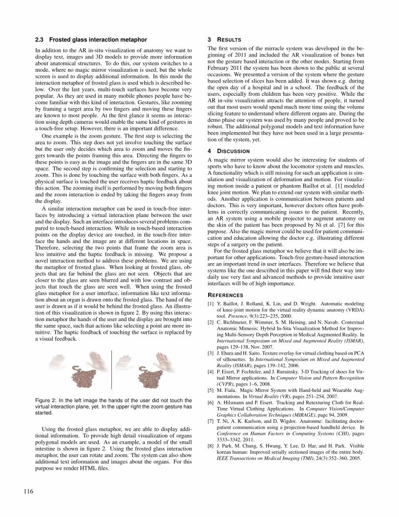
A similar interaction metaphor can be used in touch-free inter-faces by introducing a virtual interaction plane between the userand the display. Such an interface introduces several problems com-pared to touch-based interaction. While in touch-based interactionpoints on the display device are touched, in the touch-free inter-face the hands and the image are at different locations in space.Therefore, selecting the two points that frame the zoom area isless intuitive and the haptic feedback is missing. We propose anovel interaction method to address these problems. We are usingthe metaphor of frosted glass. When looking at frosted glass, ob-jects that are far behind the glass are not seen. Objects that arecloser to the glass are seen blurred and with low contrast and ob-jects that touch the glass are seen well. When using the frostedglass metaphor for a user interface, information like text informa-tion about an organ is drawn onto the frosted glass. The hand of theuser is drawn as if it would be behind the frosted glass. An illustra-tion of this visualization is shown in figure 2. By using this interac-tion metaphor the hands of the user and the display are brought intothe same space, such that actions like selecting a point are more in-tuitive. The haptic feedback of touching the surface is replaced bya visual feedback.
Figure 2: In the left image the hands of the user did not touch thevirtual interaction plane, yet. In the upper right the zoom gesture hasstarted.
Using the frosted glass metaphor, we are able to display addi-tional information. To provide high detail visualization of organspolygonal models are used. As an example, a model of the smallintestine is shown in figure 2. Using the frosted glass interactionmetaphor, the user can rotate and zoom. The system can also showadditional text information and images about the organs. For thispurpose we render HTML files.
3 RESULTS
The first version of the mirracle system was developed in the be-ginning of 2011 and included the AR visualization of bones butnot the gesture based interaction or the other modes. Starting fromFebruary 2011 the system has been shown to the public at severaloccasions. We presented a version of the system where the gesturebased selection of slices has been added. It was shown e.g. duringthe open day of a hospital and in a school. The feedback of theusers, especially from children has been very positive. While theAR in-situ visualization attracts the attention of people, it turnedout that most users would spend much more time using the volumeslicing feature to understand where different organs are. During thedemo phase our system was used by many people and proved to berobust. The additional polygonal models and text information havebeen implemented but they have not been used in a large presenta-tion of the system, yet.
4 DISCUSSION
A magic mirror system would also be interesting for students ofsports who have to know about the locomotor system and muscles.A functionality which is still missing for such an application is sim-ulation and visualization of deformation and motion. For visualiz-ing motion inside a patient or phantom Baillot et al. [1] modeledknee joint motion. We plan to extend our system with similar meth-ods. Another application is communication between patients anddoctors. This is very important, however doctors often have prob-lems in correctly communicating issues to the patient. Recently,an AR system using a mobile projector to augment anatomy onthe skin of the patient has been proposed by Ni et al. [7] for thispurpose. Also the magic mirror could be used for patient communi-cation and education allowing the doctor e.g. illustrating differentsteps of a surgery on the patient.
For the frosted glass metaphor we believe that it will also be im-portant for other applications. Touch-free gesture-based interactionare an important trend in user interfaces. Therefore we believe thatsystems like the one described in this paper will find their way intodaily use very fast and advanced methods to provide intuitive userinterfaces will be of high importance.
REFERENCES
[1] Y. Baillot, J. Rolland, K. Lin, and D. Wright. Automatic modelingof knee-joint motion for the virtual reality dynamic anatomy (VRDA)tool. Presence, 9(3):223–235, 2000.
[2] C. Bichlmeier, F. Wimmer, S. M. Heining, and N. Navab. ContextualAnatomic Mimesis: Hybrid In-Situ Visualization Method for Improv-ing Multi-Sensory Depth Perception in Medical Augmented Reality. InInternational Symposium on Mixed and Augmented Reality (ISMAR),pages 129–138, Nov. 2007.
[3] J. Ehara and H. Saito. Texture overlay for virtual clothing based on PCAof silhouettes. In International Symposium on Mixed and AugmentedReality (ISMAR), pages 139–142, 2006.
[4] P. Eisert, P. Fechteler, and J. Rurainsky. 3-D Tracking of shoes for Vir-tual Mirror applications. In Computer Vision and Pattern Recognition(CVPR), pages 1–6, 2008.
[5] M. Fiala. Magic Mirror System with Hand-held and Wearable Aug-mentations. In Virtual Reality (VR), pages 251–254, 2007.
[6] A. Hilsmann and P. Eisert. Tracking and Retexturing Cloth for Real-Time Virtual Clothing Applications. In Computer Vision/ComputerGraphics Collaboration Techniques (MIRAGE), page 94, 2009.
[7] T. Ni, A. K. Karlson, and D. Wigdor. Anatonme: facilitating doctor-patient communication using a projection-based handheld device. InConference on Human Factors in Computing Systems (CHI), pages3333–3342, 2011.
[8] J. Park, M. Chung, S. Hwang, Y. Lee, D. Har, and H. Park. Visiblekorean human: Improved serially sectioned images of the entire body.IEEE Transactions on Medical Imaging (TMI), 24(3):352–360, 2005.
116