Measuring Steroid Hormones in Avian Eggs - Top 100 ... · Measuring Steroid Hormones in Avian Eggs...
13
University of Groningen Measuring Steroid Hormones in Avian Eggs Engelhardt, Nikolaus von; Groothuis, Ton G.G. Published in: Annals of the New York Academy of Scieces DOI: 10.1196/annals.1343.015 IMPORTANT NOTE: You are advised to consult the publisher's version (publisher's PDF) if you wish to cite from it. Please check the document version below. Document Version Publisher's PDF, also known as Version of record Publication date: 2005 Link to publication in University of Groningen/UMCG research database Citation for published version (APA): Engelhardt, N. V., & Groothuis, T. G. G. (2005). Measuring Steroid Hormones in Avian Eggs. Annals of the New York Academy of Scieces, 1046(1), 181-192. https://doi.org/10.1196/annals.1343.015 Copyright Other than for strictly personal use, it is not permitted to download or to forward/distribute the text or part of it without the consent of the author(s) and/or copyright holder(s), unless the work is under an open content license (like Creative Commons). Take-down policy If you believe that this document breaches copyright please contact us providing details, and we will remove access to the work immediately and investigate your claim. Downloaded from the University of Groningen/UMCG research database (Pure): http://www.rug.nl/research/portal. For technical reasons the number of authors shown on this cover page is limited to 10 maximum. Download date: 04-05-2019
Transcript of Measuring Steroid Hormones in Avian Eggs - Top 100 ... · Measuring Steroid Hormones in Avian Eggs...

University of Groningen
Measuring Steroid Hormones in Avian EggsEngelhardt, Nikolaus von; Groothuis, Ton G.G.
Published in:Annals of the New York Academy of Scieces
DOI:10.1196/annals.1343.015
IMPORTANT NOTE: You are advised to consult the publisher's version (publisher's PDF) if you wish to cite fromit. Please check the document version below.
Document VersionPublisher's PDF, also known as Version of record
Publication date:2005
Link to publication in University of Groningen/UMCG research database
Citation for published version (APA):Engelhardt, N. V., & Groothuis, T. G. G. (2005). Measuring Steroid Hormones in Avian Eggs. Annals of theNew York Academy of Scieces, 1046(1), 181-192. https://doi.org/10.1196/annals.1343.015
CopyrightOther than for strictly personal use, it is not permitted to download or to forward/distribute the text or part of it without the consent of theauthor(s) and/or copyright holder(s), unless the work is under an open content license (like Creative Commons).
Take-down policyIf you believe that this document breaches copyright please contact us providing details, and we will remove access to the work immediatelyand investigate your claim.
Downloaded from the University of Groningen/UMCG research database (Pure): http://www.rug.nl/research/portal. For technical reasons thenumber of authors shown on this cover page is limited to 10 maximum.
Download date: 04-05-2019
181
Ann. N.Y. Acad. Sci. 1046: 181–192 (2005). © 2005 New York Academy of Sciences.doi: 10.1196/annals.1343.015
Measuring Steroid Hormones in Avian Eggs
NIKOLAUS VON ENGELHARDT AND TON G. G. GROOTHUIS
Research Group Animal Behaviour, University of Groningen, 9750 AA Haren,the Netherlands
ABSTRACT: Avian eggs contain substantial levels of various hormones of ma-ternal origin and have recently received a lot of interest, mainly from behavior-al ecologists. These studies strongly depend on the measurement of egghormone levels, but the method of measuring these levels has received little at-tention. This paper describes the sampling, extraction, and assay of hormonesin avian eggs and related methodological problems. The method of sampling isimportant because hormones are not homogeneously distributed within theegg, and after onset of embryonic development their levels may decrease andincrease due to changes in egg structure and secretion or uptake of hormonesby the embryo. The extraction of hormones from the yolk and chromatograph-ic separation of different hormones for immunoassays can strongly influencethe results because such procedures remove interfering substances such as pro-teins, lipids, and other hormones and their metabolites, which can cross-reactwith the antiserum used. Finally, the assay itself needs more validation thanmany studies report, especially with respect to the accuracy and specificity ofthe hormone measurements. We conclude that the addressed issues need moreattention for the correct interpretation of differences in hormone levels withinand between studies.
KEYWORDS: maternal hormones; steroids; androgens; testosterone; valida-tion; immunoassay; egg; yolk; albumen
INTRODUCTION
The discovery of hormones of maternal origin—such as androgens, estrogens,progesterone, corticosterone, and thyroid hormones—in avian eggs and their impacton offspring development1–8 has stimulated many studies measuring and manipulat-ing hormones in avian eggs (for recent reviews, see Refs. 9 and 10). Most studiesfocus on the intriguing question of whether deposition of maternal hormones reflectsan adaptive maternal effect. For example, can a female bird influence offspring de-velopment by varying the deposition of hormones in her eggs, and in this way adjustoffspring development to prevailing environmental conditions? Other questions in-clude the assessment of maternal endocrine state using eggs instead of more invasiveplasma samples,11,12 the assessment of environmental contamination with steroids
Address for correspondence: N. von Engelhardt, Research Group Animal Behaviour, Univer-sity of Groningen, P.O. Box 14, 9750 AA Haren, the Netherlands. Voice: +31-503637850; fax:+31-50-3632148.
182 ANNALS NEW YORK ACADEMY OF SCIENCES
or endocrine disruptors,13 and the assessment of offspring endocrine state using hor-mones in allantoic fluid.14,15
This article deals with the methodology of hormone measurements in avian eggs.It is concerned primarily with (sex) steroid hormones of maternal origin in the yolkof eggs, but the general principles hold for the measurement of hormones in eggwhite and allantoic fluid of other origin. The paper complements others in this issueon conceptual questions in the study of maternal hormones,16 the measurement ofsteroid hormones in feces,17 the transfer of corticosteroids from the maternal circu-lation into the egg,12 and steroid hormones in allantoic fluid.15
It has been known for a long time that avian egg yolk has hormone-like activity,18
but only since Hubert Schwabl1 demonstrated that steroid hormones are present inavian eggs, did they begin to attract a lot of attention, especially from behavioralecologists interested in adaptive maternal effects. These studies use immunoassaysto determine egg levels of hormones, both for describing patterns of maternal hor-mone deposition in relation to, for example, environmental factors, and for estimat-ing dosages for experimental manipulation of egg hormone levels within the naturalrange. Clearly, such studies rely heavily on the proper use and validation of hormonequantification methods used, but these methods have not yet received the attentionthey deserve.
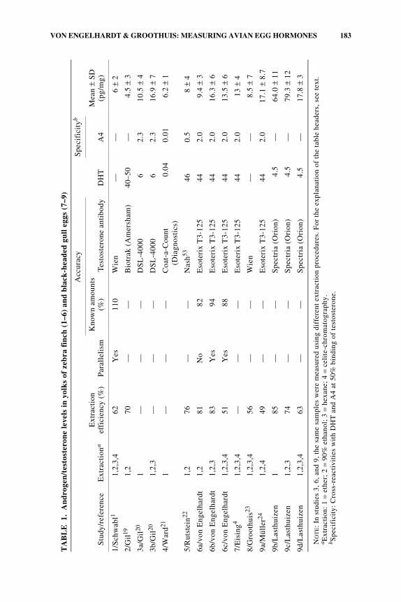
A comparison of nine different studies measuring androgens in egg yolks of zebrafinches and black-headed gulls shows both the wide variation in methods used andthe wide variation in the measured levels (TABLE 1; studies 6 and 9 show our unpub-lished data (N. von Engelhardt, and M. Lasthuizen, B. de Vries, and T. Groothuis,respectively), and part of the values of the other studies are calculated from the datain the original papers).1,4,19–24 The differences in levels may partly be due to truedifferences in androgen content of eggs of different populations, years, or experi-mental conditions. This cannot be the case for three studies (3, 6, 9), in which dif-ferences of 150%–400% were found when assaying the same samples usingsimplified extraction with ether, additional purification with ethanol and hexane toremove proteins and lipids, or chromatographic purification and separation. Thepresence of proteins and lipids25 or of steroids that cross-react with the antiserumused are the most important causes of the diverging results.
This example indicates the problems that can be encountered when measuringegg hormone levels. The qualitative and quantitative measurements of chemical an-alytes using indirect methods such as immunoassays are usually validated by deter-mining their accuracy (closeness to “true” value), specificity (the degree to whichthe method also measures other compounds), precision (variability of repeated mea-sures), and sensitivity (an indication of the smallest amount of analyte that can bedetected with the method).
Unreliable estimates of hormone levels are a special concern for studies that usepublished data to compare absolute egg hormone levels or relative differences be-tween species or populations. They also can be a problem for studies that intend todraw quantitative conclusions regarding causes or consequences of variation in egghormones. In addition, they are problematic if one wants to accurately manipulateyolk hormone levels within the ranges that can be encountered under normal condi-tions in the species studied.
The goal of this paper is to give an overview of the currently used methods tosample, extract, and assay egg hormones; to point out which methodological issues
183VON ENGELHARDT & GROOTHUIS: MEASURING AVIAN EGG HORMONES
TA
BL
E1.
An
dro
gen
/tes
tost
eron
e le
vels
in y
olk
s of
zeb
ra f
inch
(1–
6) a
nd b
lack
-hea
ded
gull
egg
s (7
–9)
NO
TE:
In s
tudi
es 3
, 6, a
nd 9
, the
sam
e sa
mpl
es w
ere
mea
sure
d us
ing
diff
eren
t ex
trac
tion
pro
cedu
res.
For
the
exp
lana
tion
of
the
tabl
e he
ader
s, s
ee t
ext.
a Ext
ract
ion:
1 =
eth
er;
2 =
90%
eth
anol
; 3
= he
xane
; 4
= ce
lite
-chr
omat
ogra
phy.
b Spe
cifi
city
: C
ross
-rea
ctiv
itie
s w
ith
DH
T a
nd A
4 at
50%
bin
ding
of
test
oste
rone
.
Stu
dy/r
efer
ence
Ext
ract
iona
Ext
ract
ion
effi
cien
cy (
%)
Par
alle
lism
Acc
urac
y
DH
T
Spe
cifi
city
b
Mea
n ±
SD
(pg/
mg)
Kno
wn
amou
nts
(%)
Tes
tost
eron
e an
tibo
dyA
4
1/S
chw
abl1
1,2,
3,4
62Y
es11
0W
ien
——
6 ±
2
2/G
il19
1,2
70—
—B
iotr
ak (
Am
ersh
am)
40–5
0—
4.5
± 3
3a/G
il20
1—
——
DS
L-4
000
62.
310
.5 ±
43b
/Gil
201,
2,3
——
—D
SL
-400
06
2.3
16.9
± 7
4/W
ard21
1—
——
Coa
t-a-
Cou
nt
(Dia
gnos
tics
)0.
040.
016.
2 ±
1
5/R
utst
ein22
1,2
76—
—N
ash53
460.
58
± 4
6a/v
on E
ngel
hard
t1,
281
No
82E
sote
rix
T3-
125
442.
09.
4 ±
3
6b/v
on E
ngel
hard
t1,
2,3
83Y
es94
Eso
teri
x T
3-12
544
2.0
16.3
± 6
6c/v
on E
ngel
hard
t1,
2,3,
451
Yes
88E
sote
rix
T3-
125
442.
013
.5 ±
67/
Eis
ing4
1,2,
3,4
——
—E
sote
rix
T3-
125
442.
013
± 4
8/G
root
huis
231,
2,3,
456
——
Wie
n—
—8.
5 ±
7
9a/M
ülle
r241,
2,4
49—
—E
sote
rix
T3-
125
442.
017
.1 ±
8.7
9b/L
asth
uize
n1
85—
—S
pect
ria
(Ori
on)
4.5
—64
.0 ±
11
9c/L
asth
uize
n1,
2,3
74—
—S
pect
ria
(Ori
on)
4.5
—79
.3 ±
12
9d/L
asth
uize
n1,
2,3,
463
——
Spe
ctri
a (O
rion
) 4
.5—
17.8
± 3

184 ANNALS NEW YORK ACADEMY OF SCIENCES
require particular attention and how problems can be tackled; and to demonstratehow important validation studies are.
SAMPLING
Inside the yolk of freshly laid eggs, hormones are not distributed homogeneously;rather, there is a radial gradient reflecting the concentric layering of yolk.7,12,26–28
Hormones in the peripheral layers of the yolk may be taken up at different stages ofdevelopment by the embryo than hormones in the center of the yolk, and therby servedifferent functions. Therefore, measurement of hormone levels in a random biopt orfrom a whole homogenized yolk would miss potentially valuable information. Ac-cording to our own experience it may, however, in practice be very difficult—if notimpossible—to sample yolk from specific locations of a live egg, because the yolkmoves within the egg when touched by the needle used for taking the biopsy speci-men.
The levels of different hormones in the yolk change rapidly after the onset of in-cubation, although the cause of this change is unknown.28–30 Currently, there is littleinformation concerning changes in yolk structure during embryonic development,the changes in the distribution of yolk hormones within the egg over time, and thestages at which different parts of the yolk are taken up by the embryo. There is aninflux of water from the albumen in the first days after incubation, which dilutes andenlarges the yolk.31,32 A decrease in yolk hormone concentrations is therefore ex-pected, and hormones might also diffuse within the yolk and to albumen and allan-tois. Egg hormone levels may also decrease or increase during embryonicdevelopment due to the uptake, secretion, and metabolism of hormones by the devel-oping embryo (see also Ref. 16). Because yolk hormone levels do not remain con-stant during development, one must be careful when drawing conclusions regardingmaternal hormones when measuring hormone levels after the onset of embryonicdevelopment.
It is therefore advisable to assess hormone levels immediately after eggs are laid.In field research, this may not be possible, so at the very least, the developmentalstage at which a sample has been taken should be assessed by, for example, measur-ing yolk and embryo size. The obvious problem here is that yolk hormones canaccelerate or delay embryonic development,4,5,33 so embryonic size may not indi-cate developmental stage independently of the hormone concentration in that egg.
Finally, a question that has hardly been addressed is the presence of hormones inthe albumen—almost all studies focus on yolk hormones. This focus is based on theassumption that the yolk contains most of the hormone in the egg due to the lipo-philic nature of steroid hormones and that the hormone is deposited during yolk for-mation. However, hormone levels in albumen can be similar to those in the yolk11,32
and require further study because they can certainly also be relevant for offspring de-velopment. It is conceivable that hormone in the albumen is partly derived from thefollicular wall (via diffusion from the yolk after ovulation) and partly deposited dur-ing albumen production in the magnum and/or when more water is added to the eggin the shell gland. Albumen is produced in about a day, whereas yolk deposition
185VON ENGELHARDT & GROOTHUIS: MEASURING AVIAN EGG HORMONES
takes several days. Therefore, the levels of hormones in the albumen may better re-flect short-term changes in the plasma levels of hormones than those in the yolk.
EXTRACTION
Hormones cannot be measured directly in the yolk, and extraction and purifica-tion is necessary to remove interfering substances. So-called matrix effects, causedby substances such as proteins and lipids that can bind hormones or interfere withthe binding of the hormone to antibody and charcoal, can strongly influence the re-sults of immunoassays.25 The removal of lipids requires particular attention becausemany factors can influence both maternal hormones and the presence of lipids in avi-an eggs. Therefore, apparent differences in hormone levels might rather be due todifferences in lipid content.
Most studies on avian eggs follow the extraction protocol published by Schwabl.1
First, samples are extracted twice with 3 mL of petroleum ether/diethyl ether, 30:70(vol:vol), a combination of solvents that extracts many different steroids from eggs.Samples are then dissolved in 90% ethanol and frozen overnight to precipitate pro-teins and neutral lipids. Finally, more lipids are removed by washing samples withhexane. Diethylether alone or 80% methanol has also been used for extraction of ste-roid hormones from the egg matrix.6,7
The physiochemical properties of different solvents determine the extent to whichthey extract different hormones and interfering substances (e.g., petroleum ether ex-tracts nonpolar steroids such as progesterone, but not the polar corticosteroids)34 andcan be a cause of disagreement between studies in absolute levels of measured ste-roids. One should therefore choose an adequate extraction procedure according tothe literature and test the extraction efficiency for the hormones of interest before-hand by adding radioactively labeled steroid hormones to yolk before extraction andassessing the amount of label recovered during each extraction step. This validationis also important because egg composition may differ between species, so differentextraction methods may be optimal or required.
For quantitative measurements of absolute hormone levels, it is advisable to de-termine recovery for each sample, because the percentage of recovery can fluctuatesubstantially.
CHROMATOGRAPHY
Chromatography is used primarily to separate the various hormones present in thesample, but also leads to further sample purification. Chromatography is not neces-sary if no cross-reacting steroids are present in the sample and if samples are suffi-ciently clean that no other interfering substances are present.
When chromatographic separation is used, the presence of several hormones canbe determined in a single sample, which is an advantage especially when samples(e.g., biopts) are too small and hormone levels too low to allow splitting of the sam-ple for several immunoassays. Chromatography also allows the use of nonspecificantibodies, which can be used to measure more than one hormone. Testosterone anddihydrotestosterone are frequently assayed after chromatographic separation using

186 ANNALS NEW YORK ACADEMY OF SCIENCES
an antiserum that cross-reacts with both hormones. The advantage of specific antis-era is obviously that the relatively time-consuming chromatography may not be nec-essary, but these antisera are often more expensive and removal of interferingsubstances such as lipids is still necessary.
Avian endocrinologists use mostly celite column chromatography for the separa-tion of steroid hormones.1,34,35 Other methods using commercially available col-umns can be found in the literature but have not yet been used for hormones ineggs.36–39
The packing of the columns and the extraction, especially for the separation ofseveral hormones, requires practice and careful validation following the publishedprotocols.1,34,35 The absolute volumes, mixtures, and succession of the eluting sol-vents (e.g., increasing polarities) influence the elution profile of the hormones. Also,the speed of elution and the temperature influence the quality of the separation andshould remain constant.34 It must first be established that the hormone of interest iseluted primarily in the fraction used for assaying this hormone; second, one must becertain that other hormones elute in this fraction only to a minor extent. This is im-portant for two reasons. First, elution of hormones in other fractions will lead to anoverestimation of recovery when using more than one labeled hormone at the sametime, because the amount of radioactivity recovered in the eluate is used to calculaterecovery and different hormones cannot be distinguished unless different labels (3H,14C) are used. However, 14C labels have a low specific activity (low radioactivity rel-ative to the mass of the molecule), so the large amount of label required for recoveryestimation can interfere with the assay. Second, elution of several hormones in thesame fraction that cross-react with the antibody will lead to inflated hormone levels.The degree of potential interference can be estimated from the amount of hormonethat is present in the sample, the percentage of cross-reactivity with the antibodyused, and the proportion present in the eluate. A hormone that has a high cross-reactivity and elutes in the same fraction may not interfere with the assay if it is hard-ly present in the sample, whereas a hormone with low cross-reactivity present inmuch larger amounts than the hormone of interest may cause substantial.
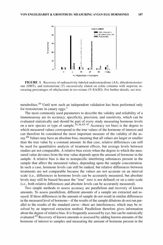
Therefore, the quality of the separation has to be tested beforehand by adding ra-dioactively labeled steroids—singly and in combination—to yolk samples and mea-suring recovery and separation in the different fractions. FIGURE 1 shows an exampleof our own validations for separating androstenedione, dihydrotestosterone, and tes-tosterone on celite columns.
IMMUNOASSAY
Currently, steroid hormones in avian eggs are measured using immunoassays,mostly radioimmunoassays, which are readily available, cheap, fast, and very sensi-tive. They cannot, however, be used to biochemically identify a hormone because an-tisera cross-react to some extent with other hormones or metabolites. They aretherefore an indirect method to measure amounts of a certain hormone, and for anaccurate measurement they must be validated using an independent method such asa combination of gas chromatography and mass spectrometry (GC–MS), which canbiochemically identify and quantify the hormone of interest.40–43 Even these meth-ods may have difficulty in distinguishing very similar analytes, such as isomeric
187VON ENGELHARDT & GROOTHUIS: MEASURING AVIAN EGG HORMONES
metabolites.44 Until now such an independent validation has been performed onlyfor testosterone in canary eggs.1
The most commonly used parameters to describe the validity and reliability of aimmunoassay are its accuracy, specificity, precision, and sensitivity, which can beevaluated statistically and should be part of every study measuring hormone levelson a new species or type of sample.34,36,45–47 Accuracy (or bias) is the degree towhich measured values correspond to the true values of the hormone of interest andcan therefore be considered the most important measure of the validity of the as-say.48 Values may have an absolute bias, meaning that all values are larger or smallerthan the true value by a constant amount. In that case, relative differences can stillbe used for quantitative analysis of treatment effects, but average levels betweenstudies are not comparable. A relative bias exists when the degree to which the mea-sured value deviates from the true value depends upon the amount of hormone in thesample. A relative bias is due to nonspecific interfering substances present in thesample that affect the measured values, depending upon the sample concentration.In such a case, hormone levels can still be ranked, but relative differences betweentreatments are not comparable because the values are not accurate on an intervalscale (i.e., differences in hormone levels can be accurately measured, but absolutelevels may still be biased because the “true” zero is not defined) or on a ratio scale(i.e., both relative differences and absolute levels can be accurately measured).
Two simple methods to assess accuracy are parallelism and recovery of knownamounts. To assess parallelism, different amounts of a sample are extracted and as-sayed. If these differences in the amount of sample do not result in similar differencesin the measured level of hormone—if the results of the sample dilutions do not run par-allel to the results of the standard curve—there are interferences, which may be re-solved by an improved extraction method. Parallelism therefore gives informationabout the degree of relative bias. It is frequently assessed by eye, but can be statisticallyevaluated.45 Recovery of known amounts is assessed by adding known amounts of thehormone of interest to samples and measuring the amount of hormone present in the
FIGURE 1. Recovery of radioactivity-labeled androstenedione (A4), dihydrotestoster-one (DHT), and testosterone (T) successively eluted on celite columns with stepwise in-creasing percentages of ethylacetate in iso-octane (% EA/IO). For further details, see text.
188 ANNALS NEW YORK ACADEMY OF SCIENCES
sample before and after addition of the hormone. If the difference is not equal to theamount of hormone added, then differences between samples do not accurately reflecttrue differences in endogenous hormone levels, most likely due to the presence of non-specific interferences, such as proteins or lipids, which bind steroid hormones. Thisgives an indication of both absolute bias and relative bias, but an absolute bias of thehormone levels can ultimately be detected only by validation with an accepted refer-ence method such as GC–MS.40–43 Another relatively easy method to evaluate accu-racy and specificity (see below) is to measure both the amount of radioactive label andthe level of hormone in several fractions of a chromatographic separation.49 A constantratio of the amount of radioactivity and the amount of hormone measured in differentfractions indicates that the assay measures the hormone of interest accurately andspecifically.
Specificity indicates the extent to which the assay measures only one specifichormone or also other hormones or metabolites. For example, a nonspecific assayfor testosterone may in fact measure several androgens, and conclusions are thenlimited to general conclusions regarding androgens.50 Furthermore, no quantitativeconclusions can be drawn because the different hormones bind with different affin-ities to the antibody. For example, a certain hormone level measured with an antise-rum that binds testosterone twice as well as dihydrotestosterone can reflect that levelof testosterone or twice as much dihydrotestosterone.
Increased specificity can be obtained by a chromatographic separation of the cross-reacting steroids or by finding an antiserum that has lower cross-reactivities with theinterfering hormones. Even cross-reactivities below 1% may cause a problem for spe-cific measurement if the cross-reacting steroid is present in much larger amounts thanthe hormone of interest. For example, yolk levels of androstenedione in gulls are 30-fold higher than testosterone,23 and progesterone levels in starlings are 500 times high-er than testosterone levels.51 Unfortunately, studies with radiolabeled hormone injec-tion in the mother, a standard validation technique in fecal hormone analyses,17 aredifficult for identification of reproductive hormones (and their metabolites) in the egg,because these steroids are produced in the follicles surrounding the egg. They thereforeprobably enter the yolk directly from the follicular wall,10,16 and only a very small per-centage of the injected hormone will reach the egg via the maternal circulation.27 Ra-diometabolism studies will require in vitro experiments with follicles using labeledhormones and their precursors. Radiometabolism studies are much easier to performand more valid for hormones, such as corticosteroids and thyroid hormones, producedin extragonadal glands.
Precision is the variability of the measures (either within or between assays)when a sample is assayed repeatedly. It is reported as the coefficient of variation[CV = (standard deviation/mean) × 100]. Precision is highest when samples aremeasured in a single assay, so many studies attempt to measure all samples of thestudy within a single assay. If samples must be measured in several assays, it is ob-viously very important that one does not measure samples of different experimentalgroups in different assays, because it would then be impossible to disentangle inter-assay effects from the effects of interest in the study.
Sensitivity or limit of detection gives an indication of the smallest amount of hor-mone that can be reliably detected. Determining sensitivity is methodologically andstatistically nontrival, and there is no general agreement regarding its calculation.46
Sensitivity is frequently defined as two or three standard deviations above the blank
189VON ENGELHARDT & GROOTHUIS: MEASURING AVIAN EGG HORMONES
value—the apparent amount of hormone in a sample that is identical to the samplesused in the assay but does not contain the analyte of interest. There is no optimal wayof producing such a sample, because the method for removing the analyte may itselfintroduce error (e.g., removal of endogenous hormones with charcoal). Becausemost hormones are present in large amounts in avian eggs, samples can and shouldbe measured at a concentration for which assay sensitivity is not an important issue,but some hormones, especially estradiol, are present in such low amounts that theyare difficult to detect, and assay sensitivity must be assessed. Most current studieson yolk hormones report the precision and sensitivity of their method, but do not re-port or assess specificity and accuracy. Accuracy can be assessed to a certain extentrelatively easily by demonstrating parallelism and recovery of a known amount ofhormone added to samples. We recommend that this should become common prac-tice for yolk hormone assays, as has been recommended for hormone assays in gen-eral.48,52 If parallelism cannot be demonstrated and if known amounts are notrecovered accurately, a given method is clearly not valid for quantitative compari-sons of relative or absolute differences of hormone levels, but the measurements maystill reflect the ranking and qualitative differences between samples.
CONCLUSION
Hormones in avian eggs vary in relation to various factors and can have strongeffects on offspring development. However, contradictory results have been found,10
and because these may partly be due to the methods used to measure yolk hormonesit is important to validate and standardize the way yolk hormones are measured.
Accurate measurement of yolk hormones usually requires extraction and chro-matographic purification and separation of samples because of the presence of sub-stances that can interfere with the assay. Validation of the method is thereforenecessary when setting up an assay for hormones in avian eggs in a new laboratory,for a different species, or for a different type of sample. Most endocrinologists agreethat a minimal validation should include an assessment of the accuracy, specificity,precision, and sensitivity of the method.34,46–48 The possibility of species-specificmetabolites requires a validation for each new study species and potential adjust-ment of the method. In addition, a study evaluating the comparability of egg hor-mone measurements by analyzing samples from the same eggs at differentlaboratories would clearly be very useful.
If a quantitative assessment of specific yolk hormones at the level of an intervalor ratio scale is not possible, more cautious conclusions with respect to ranks orqualitative differences can still be made. A study that is merely interested in the ef-fect of a treatment on yolk androgen levels may not require separation of differentsteroid hormones by chromatography. The degree of validation needed therefore de-pends partly upon the questions that are being asked.
ACKNOWLEDGMENTS
We thank the organizers and participants of this inspiring workshop for many in-teresting discussions and useful comments. We greatly appreciate the collaboration

190 ANNALS NEW YORK ACADEMY OF SCIENCES
in the laboratory and exchange of ideas with Corine Eising, Bonnie de Vries,Maarten Lasthuizen, and Wendt Müller.
REFERENCES
1. SCHWABL, H. 1993. Yolk is source of maternal testosterone for developing birds. Proc.Natl. Acad. Sci. USA 90: 11446–11450.
2. SCHWABL, H. 1996. Maternal testosterone in the avian egg enhances postnatal growth.Comp. Biochem. Physiol. 114A: 271–276.
3. MCNABB, F.M.A. & C.M. WILSON. 1997. Thyroid hormone deposition in avian eggsand effects on embryonic development. Am. Zool. 37: 553–560.
4. EISING, C.M., C. Eikenaar, H. Schwabl & T.G. Groothuis. 2001. Maternal androgens inblack-headed gull (Larus ridibundus) eggs: consequences for chick development.Proc. R. Soc. Lond. B Biol. Sci. 268: 839–846.
5. SOCKMAN, K.W. & H. SCHWABL. 2000. Yolk androgens reduce offspring survival. Proc.R. Soc. Lond. B Biol. Sci. 267: 1451–1456.
6. HAYWARD, L.S. & J.C. WINGFIELD. 2004. Maternal corticosterone is transferred toavian yolk and may alter offspring growth and adult phenotype. Gen. Comp. Endo-crinol. 135: 365–371.
7. MÖSTL, E., H. SPENDIER & K. KORTRSCHAL. 2001. Concentration of immunoreactiveprogesterone and androgens in the yolk of hen’s eggs (Gallus domesticus). Wien.Tierarztl. Monschr. 88: 62–65.
8. ADKINS-REGAN, E., M.A. OTTINGER & J. PARK. 1995. Maternal transfer of estradiol toegg yolks alters sexual differentiation of avian offspring. J. Exp. Zool. 271: 466–470.
9. GIL, D. 2003. Golden eggs: maternal manipulation of offspring phenotype by eggandrogen in birds. Ardeola 50: 281–294.
10. GROOTHUIS, T.G.G. et al. 2005. Prenatally induced adaptive maternal effects: the case ofmaternal androgen deposition in avian eggs. Neurosci. Biobehav. Rev. 29: 329–352.
11. DOWNING, J.A. & W.L. BRYDEN. 2002. A non-invasive test of stress in laying hens.RIRDC Publication No. 01/143.
12. RETTENBACHER, S. et al. 2005. Corticosterone in chickens’ eggs. Ann. N.Y. Acad. Sci.1046: 193–203.
13. BRUNSTRÖM, B., J. AXELSSON & K. HALLDIN. 2003. Effects of endocrine modulators onsex differentiation in birds. Ecotoxicology 12: 287–295.
14. WOODS, J.E., G.W. DE VRIES & R.C. THOMMES. 1971. Ontogeny of the pituitary-adre-nal axis in the chick embryo. Gen. Comp. Endocrinol. 17: 407–415.
15. BENOWITZ-FREDERICKS, M., A.S. KITAYSKY & J.C. WINGFIELD. 2005. Steroids in allan-toic waste—an integrated measure of steroid exposure in ovo. Ann. N.Y. Acad. Sci.1046: 204–213.
16. GROOTHUIS, T.G.G. & N. VON ENGELHARDT. 2005. Investigating maternal hormones inavian eggs: measurement, manipulation, and interpretation. Ann. N.Y. Acad. Sci.1046: 168–180.
17. PALME, R. 2005. Measuring fecal steroids: guidelines for a practical application. Ann.N.Y. Acad. Sci. 1046: 75–80.
18. ROMANOFF, A.L. 1960. The Avian Embryo. Macmillan. New York.19. GIL, D., J. GRAVES, N. HAZON & A. WELLS. 1999. Male attractiveness and differential
testosterone investment in zebra finch eggs. Science 286: 126–128.20. GIL, D. et al. 2004. Negative effects of early developmental stress on yolk testosterone
levels in a passerine bird. J. Exp. Biol. 207: 2215–2220.21. WARD, B.C., E.J. NORDEEN & K.W. NORDEEN. 2001. Anatomical and ontogenetic fac-
tors producing variation in HVc neuron number in zebra finches Brain Res. 904:318–326.
22. RUTSTEIN, A.N. et al. 2005. Sex-specific patterns of yolk androgen allocation dependupon maternal diet in the zebra finch. Behav. Ecol. 16: 62–69.
23. GROOTHUIS, T.G.G. & H. SCHWABL. 2002. Determinants of within- and among-clutchvariation in levels of maternal hormones in black-headed gull eggs. Funct. Ecol. 16:281–289.
191VON ENGELHARDT & GROOTHUIS: MEASURING AVIAN EGG HORMONES
24. MÜLLER, W. et al. 2004. Within-clutch patterns of yolk testosterone vary with the onsetof incubation in black-headed gulls. Behav. Ecol. 15: 893–897.
25. RASH, J.M., I. JERKUNICA & D.S. SGOUTAS. 1980. Lipid interference in steroid radioim-munoassay. Clin. Chem. 26: 84–88.
26. LIPAR, J.L., E.D. KETTERSON, V. NOLAN, JR. & J.M. CASTO. 1999. Egg yolk layers varyin the concentration of steroid hormones in two avian species. Gen. Comp. Endo-crinol. 115: 220–227.
27. HACKL, R. et al. 2003. Distribution and origin of steroid hormones in the yolk of Japa-nese quail eggs (Coturnix coturnix japonica). J. Comp. Physiol. B 173: 327–331.
28. EISING, C.M. 2003. Mother knows best? Costs and benefits of differential maternalhormone allocation in birds. Ph.D. thesis, University of Groningen. Groningen, theNetherlands.
29. ELF, P.K. & A.J. FIVIZZANI. 2002. Changes in sex steroid levels in yolks of the leghornchicken, Gallus domesticus, during embryonic development. J. Exp. Zool. 293:594–600.
30. EISING, C.M., W. MULLER, C. DIJKSTRA & T.G.G. GROOTHUIS. 2003. Maternal andro-gens in egg yolks: relation with sex, incubation time and embryonic growth. Gen.Comp. Endocrinol. 132: 241–247.
31. ROMANOFF, A.L. 1967. Biochemistry of the Avian Embryo—A Quantitative Analysisof Prenatal Development. Wiley. New York.
32. WILSON, C.M. & F.M.A. MCNABB. 1997. Maternal thyroid hormones in Japanese quaileggs and their influence on embryonic development. Gen. Comp. Endocrinol. 107:153–165.
33. LIPAR, J.L. & E.D. KETTERSON. 2000. Maternally derived yolk testosterone enhancesthe development of the hatching muscle in the red-winged blackbird Agelaius phoen-iceus. Proc. R. Soc. Lond. B Biol. Sci. 267: 2005–2010.
34. ABRAHAM, G.E. 1974. Radioimmunoassay of steroids in biological materials. ActaEndocrinologica. 75(Suppl. 183): 1–42.
35. WINGFIELD, J.C. & D.S. FARNER. 1975. The determination of five steroids in avianplasma by radioimmunoassay and competitive protein binding. Steroids 26: 311–327.
36. JAFFE, B.M. & H.R. BEHRMAN. 1979. Methods of Hormone Radioimmunoassay. Aca-demic Press. New York.
37. PAYNE, D.W., W.D. HOLTZCLAW & E.Y. ADASHI. 1989. A convenient, unified schemefor the differential extraction of conjugated and unconjugated serum C19 steroids onSep-Pak cartridges. J. Steroid Biochem. 33: 289–295.
38. VENKATESH, B., C.H. TAN & T.J. LAM. 1989. Blood steroid levels in the gold fish: mea-surement of six ovarian steroids in small volumes of serum by reversed-phase high-performance liquid chromatography and radioimmunoassay. Gen. Comp. Endo-crinol. 76: 398–407.
39. MORINEAU, G. et al. 1997. Convenient chromatographic prepurification step beforemeasurement of urinary cortisol by radioimmunoassay. Clin. Chem. 43: 786–793.
40. FITZGERALD, R.L. & D.A. HEROLD. 1996. Serum total testosterone: immunoassay com-pared with negative chemical ionization gas chromatography-mass spectrometry.Clin. Chem. 42: 749–755.
41. DORGAN, J.F. et al. 2002. Measurement of steroid sex hormones in serum: a compari-son of radioimmunoassay and mass spectrometry. Steroids 67: 151–158.
42. TAIEB, J. et al. 2003. Testosterone measured by 10 immunoassays and by isotope-dilu-tion gas chromatography-mass spectrometry in sera from 116 men, women, and chil-dren. Clin. Chem. 49: 1381–1395.
43. WANG, C. et al. 2004. Measurement of total serum testosterone in adult men: compari-son of current laboratory methods versus liquid chromatography-tandem mass spec-trometry. J. Clin. Endocrinol. Metab. 89: 534–543.
44. MAGNUSSON, M.O. & R. SANDSTRÖM. 2004. Quantitative analysis of eight testosteronemetabolites using column switching and liquid chromatography/tandem mass spec-trometry. Rapid Commun. Mass Spectrom. 18: 1089–1094.
45. RODBARD, D. 1974. Statistical quality control and routine data processing for radioim-munoassays and immunoradiometric assays. Clin. Chem. 20: 1255–1270.

192 ANNALS NEW YORK ACADEMY OF SCIENCES
46. CHARD, T. 1990. An Introduction to Radioimmunoassay and Related Techniques.Elsevier. Amsterdam.
47. COOK, B. & G.H. BEASTALL. 1987. Measurement of steroid hormone concentrations inblood, urine and tissues. In Steroid Hormones: a Practical Approach. B. Green &R.E. Leake, Eds.: 1–65. IRL Press. Oxford.
48. MATSUMOTO, A.M. & W.J. BREMNER. 2004. Serum testosterone assays—accuracy mat-ters. J. Clin. Endocrinol. Metab. 89: 520–524.
49. CEKAN, S.Z. 1979. On the assessment of validity of steroid radioimmunoassays. J. Ste-roid Biochem. 11: 135–141.
50. TANVEZ, A. et al. 2004. Sexually attractive phrases increase yolk androgen depositionin Canaries (Serinus canaria). Gen. Comp. Endocrinol. 138: 113–120.
51. LIPAR, J.L. 2001. Yolk steroids and the development of the hatching muscle in nestlingEuropean Starlings. J. Avian Biol. 32: 231–238.
52. BUCHANAN, K.L. & A.R. GOLDSMITH. 2004. Noninvasive endocrine data for behav-ioural studies: the importance of validation. Anim. Behav. 67: 183–185.
53. NASH, J. P. et al. 2000. An enzyme linked immunosorbant assay (ELISA) for testoster-one, estradiol, and 17,20β-dihydroxy-4-pregenen-3-one using acetylcholinesteraseas tracer: application to measurement of diel patterns in rainbow trout (Oncorhyn-chus mykiss). Fish Physiol. Biochem. 22: 355–363.