Louis Rivera, HMS III Gillian Lieberman, MD July,...
22
Louis Rivera, HMS III Gillian Lieberman, MD July, 2005 Imaging of Acute Cerebral Trauma Louis Rivera, Harvard Medical School, Year III Gillian Lieberman, MD
-
Upload
vuongduong -
Category
Documents
-
view
224 -
download
0
Transcript of Louis Rivera, HMS III Gillian Lieberman, MD July,...

Louis Rivera, HMS III
Gillian Lieberman, MD July, 2005
Imaging of Acute Cerebral Trauma
Louis Rivera, Harvard Medical School, Year III
Gillian Lieberman, MD
Louis Rivera, HMS III
Gillian Lieberman, MD
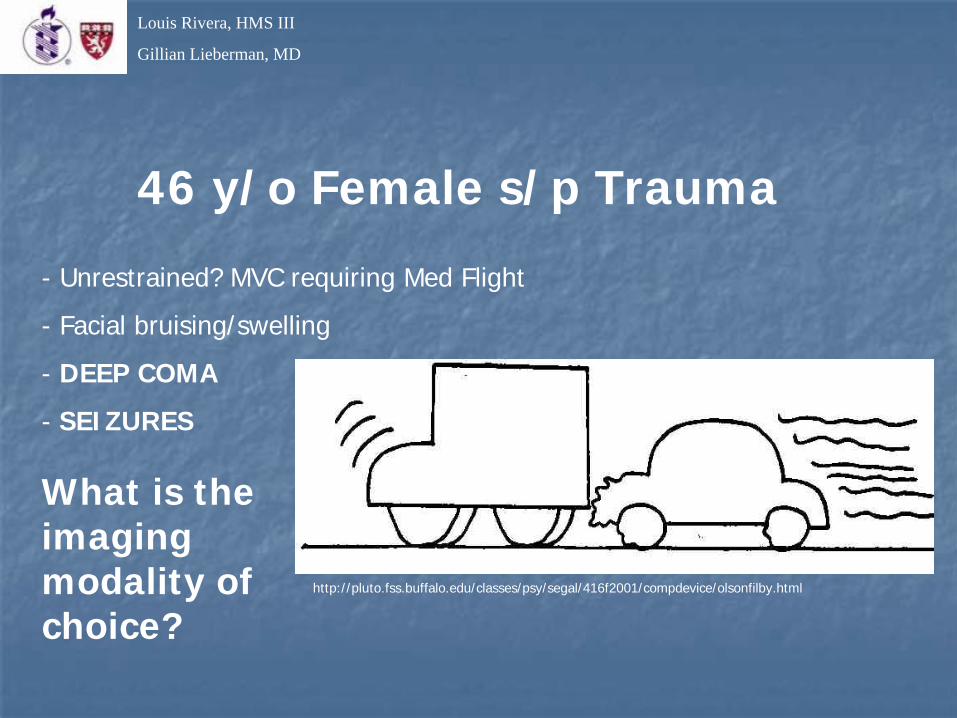
46 y/o Female s/p Trauma
- Unrestrained? MVC requiring Med Flight
- Facial bruising/swelling
- DEEP COMA
- SEIZURES
http://pluto.fss.buffalo.edu/classes/psy/segal/416f2001/compdevice/olsonfilby.html
What is the imaging modality of choice?
Louis Rivera, HMS III
Gillian Lieberman, MD
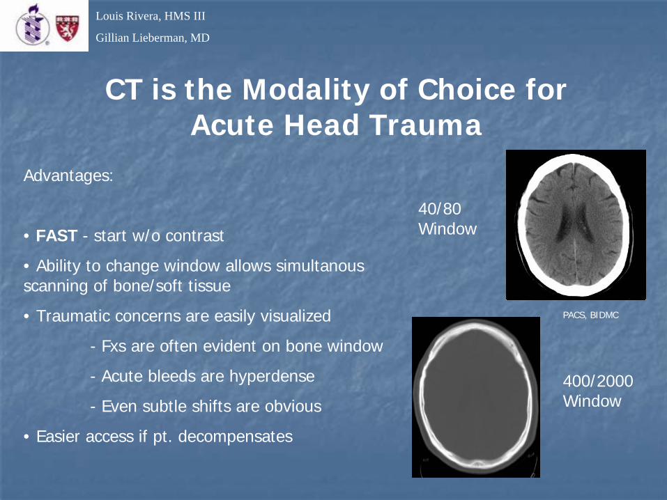
CT is the Modality of Choice for Acute Head Trauma
Advantages:
• FAST - start w/o contrast
• Ability to change window allows simultanous scanning of bone/soft tissue
• Traumatic concerns are easily visualized
- Fxs are often evident on bone window
- Acute bleeds are hyperdense
- Even subtle shifts are obvious
• Easier access if pt. decompensates
PACS, BIDMC
40/80 Window
400/2000 Window
Louis Rivera, HMS III
Gillian Lieberman, MD
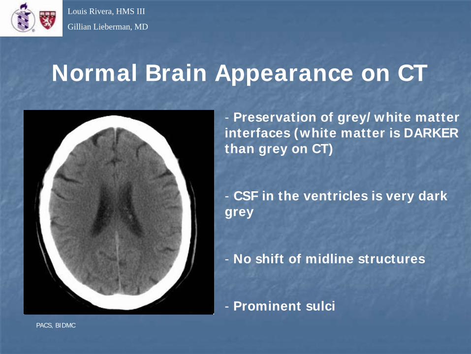
Normal Brain Appearance on CT
- Preservation of grey/white matter interfaces (white matter is DARKER than grey on CT)
- CSF in the ventricles is very dark grey
- No shift of midline structures
- Prominent sulciPACS, BIDMC
Louis Rivera, HMS III
Gillian Lieberman, MD
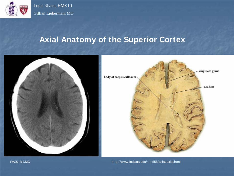
Axial Anatomy of the Superior Cortex
http://www.indiana.edu/~m555/axial/axial.htmlPACS, BIDMC
Louis Rivera, HMS III
Gillian Lieberman, MD
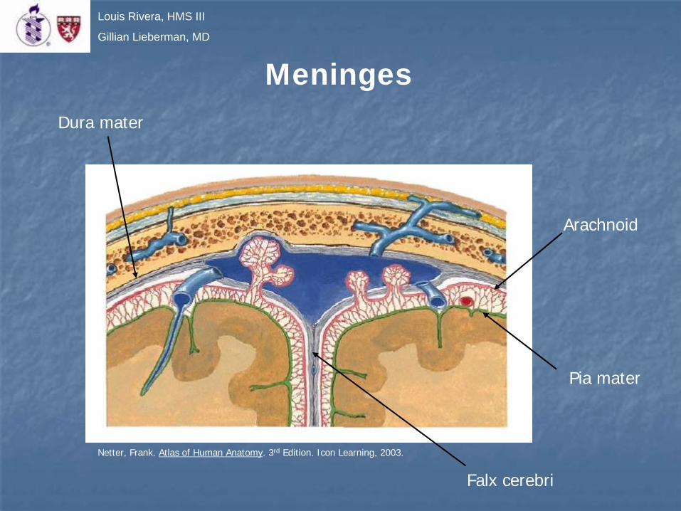
Arachnoid
Pia mater
Falx cerebri
MeningesDura mater
Netter, Frank. Atlas of Human Anatomy. 3rd Edition. Icon Learning, 2003.

Louis Rivera, HMS III
Gillian Lieberman, MD
DDx Traumatic Cranial Bleeds
• Epidural- Well-contained by cranial sutures, can cross midline
- Often “Bi-convex” shape
- Associated with skull Fxs
• Subdural- Contained by falx cerebri and tentorium
- Often “Crescent” shape
- Can have venous OR arterial blood
• Subarachnoid- Extends into CSF space
- Fluid on CT best seen in the cisterns and sulci
Also on the DDx:Tumor AbscessFocal Contusion Hydrocephalus
Netter, Frank. Atlas of Human Anatomy. 3rd Edition. Icon Learning, 2003.

Louis Rivera, HMS III
Gillian Lieberman, MD
Our Patient
• Loss of demarcation of the fissures/sulci
• Loss of grey matter/white matter differentiation
• Blood in the third and lateral ventricles
• Mild shift of the septum pellucidum to the left
What’s the Dx?
PACS, BIDMC
Louis Rivera, HMS III
Gillian Lieberman, MD
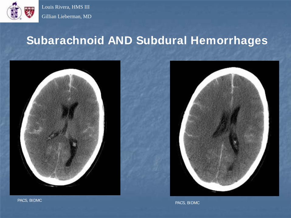
Subarachnoid AND Subdural Hemorrhages
PACS, BIDMC PACS, BIDMC

Louis Rivera, HMS III
Gillian Lieberman, MD
Connecting the Dots
• The amount of subarachnoid blood was out of proportion to the external injuries.
• The presence of both a subarachnoid bleed and a subdural hemorrhage might be due to two separate events.
• EMS reports indicated that patient had struck a parked truck without breaking or swerving.
Follow-up CTs and angiography were ordered…
http://siri.uvm.edu/graphics/Safety_Managment/Detective.gif

Louis Rivera, HMS III
Gillian Lieberman, MD
Day Two – Evolving Infarction
PACS, BIDMC PACS, BIDMC

Louis Rivera, HMS III
Gillian Lieberman, MD
CT Angiography
We now see a small aneurysm in the Circle of Willis…
PACS, BIDMC

Louis Rivera, HMS III
Gillian Lieberman, MD Computerized Reconstruction
Aneurysm is located at the intersection of the R Interior Carotid and the R Posterior Communicating Arteries, directed laterally and inferiorly
PACS, BIDMC

Louis Rivera, HMS III
Gillian Lieberman, MD
Patient Follow-Up
Perhaps an aneurysm ruptured, causing the MVC and subsequent trauma.
Sadly, the injuries were incompatible with life. Patient was pronounced brain dead and removed from life support on day two.

Louis Rivera, HMS III
Gillian Lieberman, MD
Companion Patient 2 – Subdural Hemorrhage
http://www.vh.org/adult/patient/neurosurgery/braininjury/images/http://www.vh.org/adult/patient/neurosurgery/braininjury/images/epihemat.gifepihemat.gif
• 66 Year-old man, unconscious, unknown traumatic history

Louis Rivera, HMS III
Gillian Lieberman, MD
May be classified asMay be classified as
1) 1) hyperacutehyperacute ((HYPOdenseHYPOdense) if ) if less than 12 hoursless than 12 hours
2) acute (2) acute (HYPERdenseHYPERdense) if less ) if less than few daysthan few days
3) 3) subacutesubacute ((ISOdenseISOdense) from a ) from a few days to 2few days to 2--3 weeks3 weeks
4) chronic (4) chronic (HYPOdenseHYPOdense) if more ) if more than 3 weeksthan 3 weeks
Companion Patient 2 – Subdural Hemorrhage
PACS, BIDMC
We need to put this together with clinical observations and Hx to tell for certain if this is hyperacute or chronic.

Louis Rivera, HMS III
Gillian Lieberman, MD
http://www.szote.u-szeged.hu/radio/neuro1/a1neu11b.htm
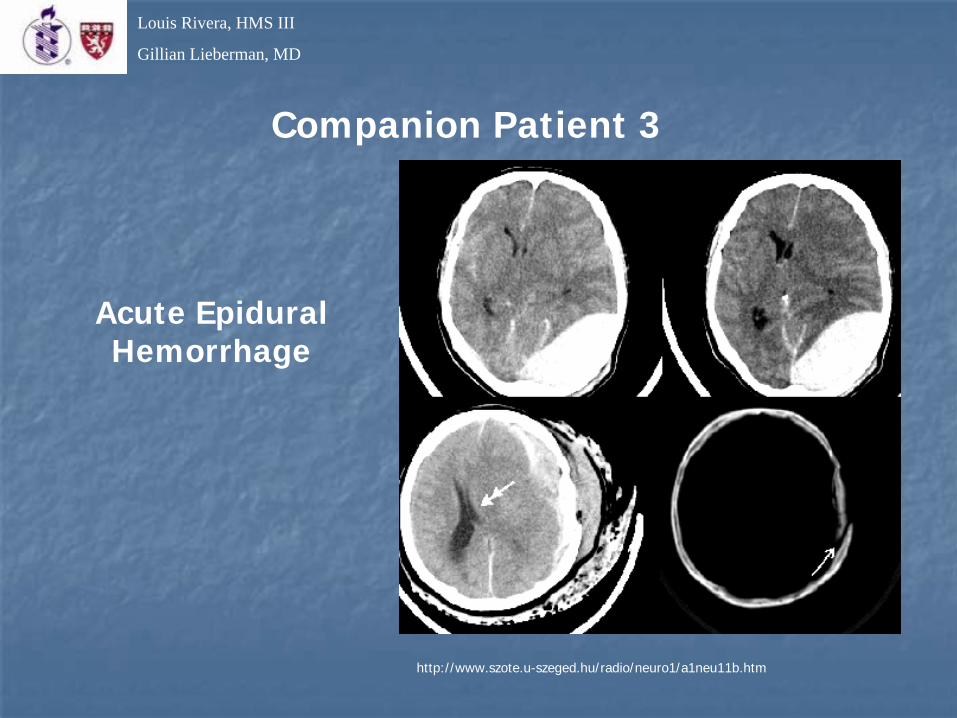
Companion Patient 3
46 y/o man s/p motorcycle crash
• Bi-convex, very large
• Mostly contained within L parieto-occipital region along sutures
• Crosses midline?
• Associated Fx
• Hyperdense
What’s the Dx?
Louis Rivera, HMS III
Gillian Lieberman, MD
http://www.szote.u-szeged.hu/radio/neuro1/a1neu11b.htm
Companion Patient 3
Acute Epidural Hemorrhage
Louis Rivera, HMS III
Gillian Lieberman, MD
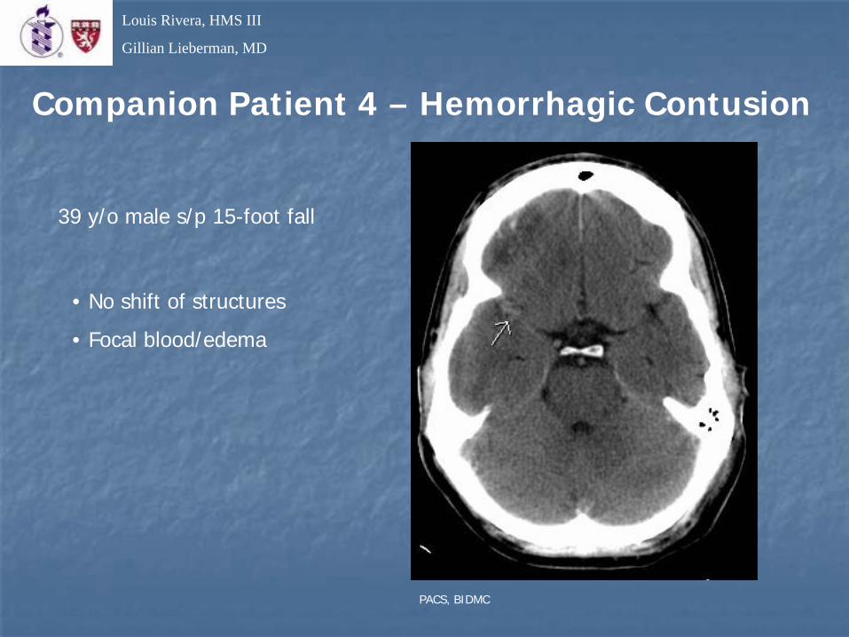
Companion Patient 4 – Hemorrhagic Contusion
39 y/o male s/p 15-foot fall
• No shift of structures
• Focal blood/edema
PACS, BIDMC
Louis Rivera, HMS III
Gillian Lieberman, MD
Traumatic Brain Injury (TBI)
• 1.6 million people/year in the U.S. have a TBI.
• 150,000 of those suffer “severe” (comatose) head injury.
• 50,000 people/year die from TBI.
• CT radiology is the standard for rapid evaluation of the post-trauma brain.
• The most common causes of TBI are MVCs, bicycle/pedestrian accidents, falls, and violence.
• Gunshot wounds are a small percentage of TBIs, but account for half of the fatalities.
• Intracranial pressure monitoring is key in comatose patients.
Louis Rivera, HMS III
Gillian Lieberman, MD
References
• “Acute Epidural Hemorrhage” www.szote.u-szeged.hu/radio/neuro1/a1neu11b.htm
• “Axial Brain Sections” www.indiana.edu/~m555/axial/axial.html
• “BrighamRAD” http://brighamrad.harvard.edu
• Netter, Frank. Atlas of Human Anatomy. 3rd Edition. Icon Learning, 2003.
• Novelline, Robert. Squire’s Fundamentals of Radiology. 6th Ed. Harvard College, 2004.
• “The Brain Trauma Foundation” www.braintrauma.org
• “Traumatic Brain Injury” www.head-trauma-resource.com
• “Virtual Hospital” www.vh.org

Louis Rivera, HMS III
Gillian Lieberman, MD
Acknowledgements
• A.C. Kim, MD for her recommendations of many useful cases
• Pamela Lepkowski
• Gillian Lieberman, MD
• Larry Barbaras, webmaster