Aradhana - IV Gillian Lieberman,...
27
1 The Radiologic Evaluation The Radiologic Evaluation of the Ovary of the Ovary Aradhana Aradhana M. M. Venkatesan Venkatesan , Harvard Medical School Year , Harvard Medical School Year - - IV IV Gillian Lieberman, MD Gillian Lieberman, MD September 2000 Aradhana Venkatesan Gillian Lieberman,
Transcript of Aradhana - IV Gillian Lieberman,...
1
The Radiologic Evaluation The Radiologic Evaluation of the Ovaryof the Ovary
AradhanaAradhana M. M. VenkatesanVenkatesan, Harvard Medical School Year, Harvard Medical School Year-- IVIVGillian Lieberman, MDGillian Lieberman, MD
September 2000Aradhana VenkatesanGillian Lieberman,
2
Menu of Tests
•Trans-abdominal pelvic ultrasound
•Trans-vaginal pelvic ultrasound
•Pelvic MRI- new, promising
Aradhana VenkatesanGillian Lieberman,
3
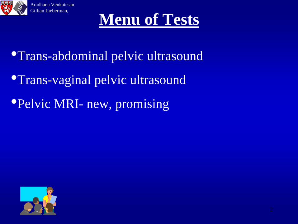
Trans-abdominal Pelvic Ultrasound TECHNIQUE
•A distended bladder is essential - this displaces gas-containing loops of bowel and also places the uterus in a horizontal position relative to the beam from the transducer. This allows optimal resolution of the uterus and adnexa.
•When an ovarian mass is detected, a full abdominal ultrasound is performed to look for hydronephrosis, ascites, or omental “cake”
Midline longitudinal trans-abdominal sonogram of the uterus (u) and vagina (v). S, sacrum
From Callen, P.W. (Ed.) Ultrasonography in Obstetrics and Gynecology. WB Saunders and Co., Philadelphia, 1994, p. 565.
Aradhana VenkatesanGillian Lieberman,
4
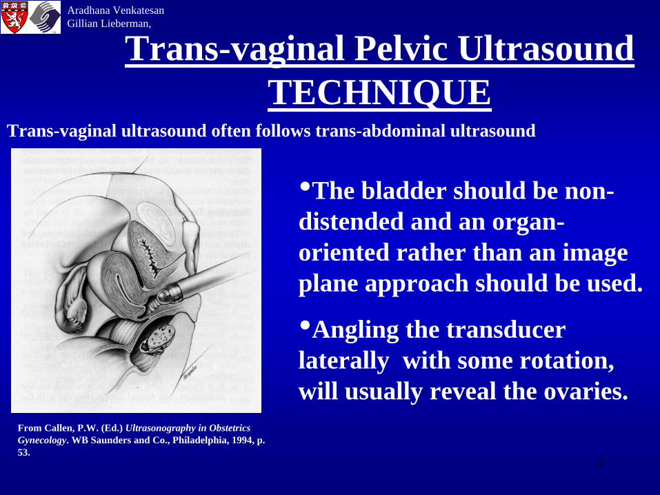
Trans-vaginal Pelvic Ultrasound TECHNIQUE
•The bladder should be non- distended and an organ- oriented rather than an image plane approach should be used.
•Angling the transducer laterally with some rotation, will usually reveal the ovaries.
From Callen, P.W. (Ed.) Ultrasonography in Obstetrics Gynecology. WB Saunders and Co., Philadelphia, 1994, p. 53.
Trans-vaginal ultrasound often follows trans-abdominal ultrasound
Aradhana VenkatesanGillian Lieberman,
5
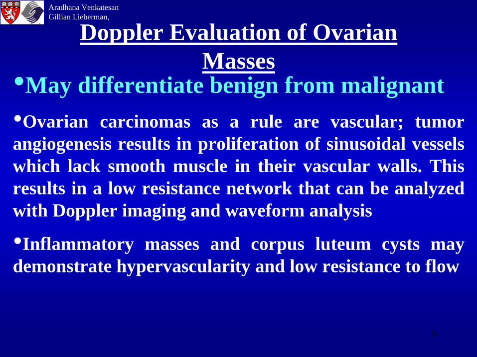
Doppler Evaluation of Ovarian Masses
•May differentiate benign from malignant•Ovarian carcinomas as a rule are vascular; tumor angiogenesis results in proliferation of sinusoidal vessels which lack smooth muscle in their vascular walls. This results in a low resistance network that can be analyzed with Doppler imaging and waveform analysis
•Inflammatory masses and corpus luteum cysts may demonstrate hypervascularity and low resistance to flow
Aradhana VenkatesanGillian Lieberman,
6
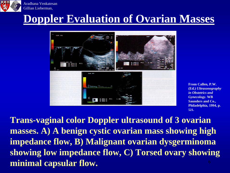
Doppler Evaluation of Ovarian Masses
Trans-vaginal color Doppler ultrasound of 3 ovarian masses. A) A benign cystic ovarian mass showing high impedance flow, B) Malignant ovarian dysgerminoma showing low impedance flow, C) Torsed ovary showing minimal capsular flow.
From Callen, P.W. (Ed.) Ultrasonography in Obstetrics and Gynecology. WB Saunders and Co., Philadelphia, 1994, p. 521.
Aradhana VenkatesanGillian Lieberman,
7
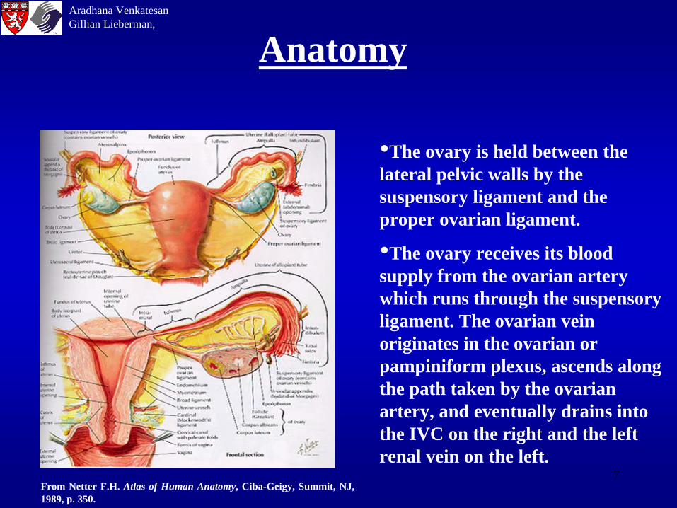
Anatomy
•The ovary is held between the lateral pelvic walls by the suspensory ligament and the proper ovarian ligament.
•The ovary receives its blood supply from the ovarian artery which runs through the suspensory ligament. The ovarian vein originates in the ovarian or pampiniform plexus, ascends along the path taken by the ovarian artery, and eventually drains into the IVC on the right and the left renal vein on the left.
From Netter F.H. Atlas of Human Anatomy, Ciba-Geigy, Summit, NJ, 1989, p. 350.
Aradhana VenkatesanGillian Lieberman,
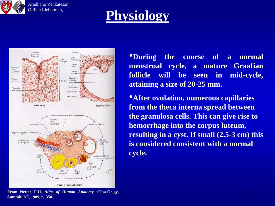
8
Physiology
•During the course of a normal menstrual cycle, a mature Graafian follicle will be seen in mid-cycle, attaining a size of 20-25 mm.
•After ovulation, numerous capillaries from the theca interna spread between the granulosa cells. This can give rise to hemorrhage into the corpus luteum, resulting in a cyst. If small (2.5-3 cm) this is considered consistent with a normal cycle.
From Netter F.H. Atlas of Human Anatomy, Ciba-Geigy, Summit, NJ, 1989, p. 350.
Aradhana VenkatesanGillian Lieberman,
9
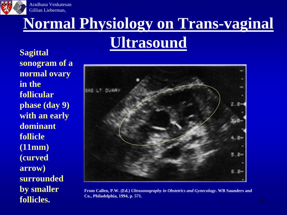
Normal Physiology on Trans-vaginal Ultrasound
Sagittal sonogram of a normal ovary in the follicular phase (day 9) with an early dominant follicle (11mm) (curved arrow) surrounded by smaller follicles.
From Callen, P.W. (Ed.) Ultrasonography in Obstetrics and Gynecology. WB Saunders and Co., Philadelphia, 1994, p. 571.
Aradhana VenkatesanGillian Lieberman,
10
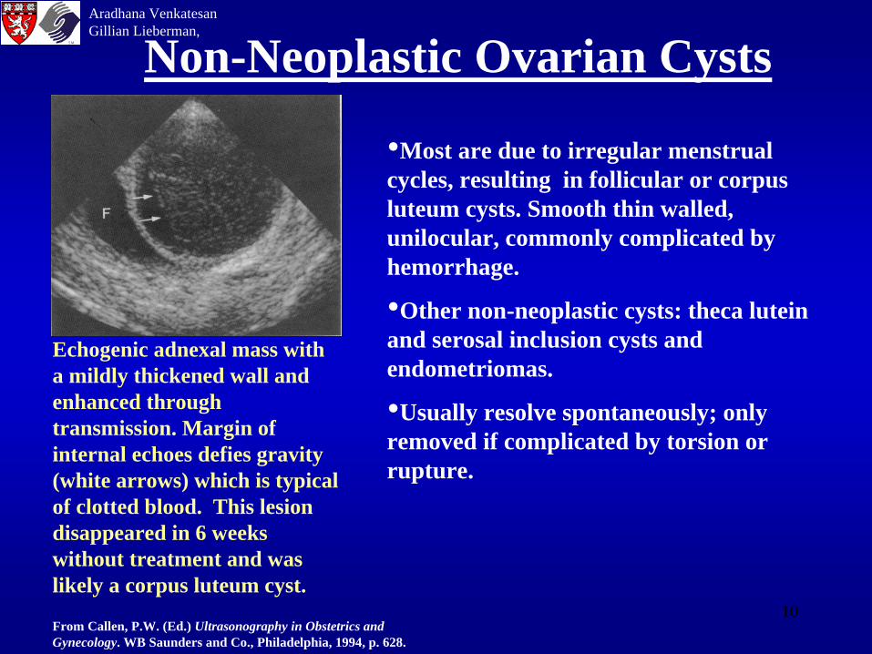
Non-Neoplastic Ovarian Cysts
•Most are due to irregular menstrual cycles, resulting in follicular or corpus luteum cysts. Smooth thin walled, unilocular, commonly complicated by hemorrhage.
•Other non-neoplastic cysts: theca lutein and serosal inclusion cysts and endometriomas.
•Usually resolve spontaneously; only removed if complicated by torsion or rupture.
Echogenic adnexal mass with a mildly thickened wall and enhanced through transmission. Margin of internal echoes defies gravity (white arrows) which is typical of clotted blood. This lesion disappeared in 6 weeks without treatment and was likely a corpus luteum cyst.
From Callen, P.W. (Ed.) Ultrasonography in Obstetrics and Gynecology. WB Saunders and Co., Philadelphia, 1994, p. 628.
Aradhana VenkatesanGillian Lieberman,
11
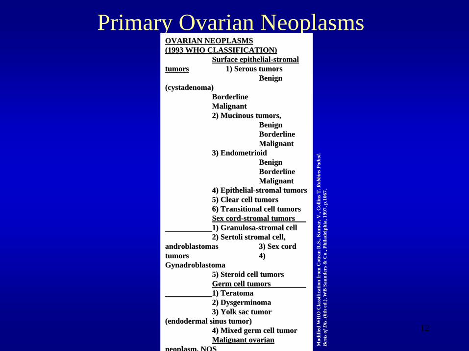
Primary Ovarian Neoplasms
•Primary tumors of the ovary arise from one of the three ovarian components: surface epithelium, germ cells, and the stroma of the ovary (which includes the sex cords).
•Although some ovarian neoplasms are hormonally active, most are non-functional, producing only mild symptoms until they reach a large size. Abdominal pain/distension, urinary or GI tract symptoms due to compressive phenomenon or cancer invasion are the most common presenting symptoms.
Aradhana VenkatesanGillian Lieberman,
12
Primary Ovarian NeoplasmsOVARIAN NEOPLASMS OVARIAN NEOPLASMS (1993 WHO CLASSIFICATION)(1993 WHO CLASSIFICATION)
Surface epithelialSurface epithelial--stromalstromal tumorstumors 1) Serous tumors1) Serous tumors
Benign Benign ((cystadenomacystadenoma))
BorderlineBorderline MalignantMalignant 2) 2) MucinousMucinous tumors, tumors,
BenignBenign BorderlineBorderline MalignantMalignant
3) 3) EndometrioidEndometrioid BenignBenign BorderlineBorderline MalignantMalignant
4) Epithelial4) Epithelial--stromalstromal tumorstumors 5) Clear cell tumors5) Clear cell tumors 6) Transitional cell tumors6) Transitional cell tumors Sex cordSex cord--stromalstromal tumorstumors 1) 1) GranulosaGranulosa--stromalstromal cellcell 2) 2) SertoliSertoli stromalstromal cell, cell,
androblastomasandroblastomas 3) Sex cord 3) Sex cord tumorstumors 4) 4) GynadroblastomaGynadroblastoma
5) Steroid cell tumors5) Steroid cell tumors Germ cell tumorsGerm cell tumors 1) 1) TeratomaTeratoma 2) 2) DysgerminomaDysgerminoma 3) Yolk sac tumor 3) Yolk sac tumor
((endodermalendodermal sinus tumor)sinus tumor) 4) Mixed germ cell tumor4) Mixed germ cell tumor Malignant ovarian Malignant ovarian
neoplasm, NOSneoplasm, NOS
Mod
ified
WH
O C
lass
ifica
tion
from
Cot
ran
R.S
., K
umar
, V.,
Col
lins T
. Rob
bins
Pat
hol.
Bas
is o
f Dis
. (6t
h ed
.), W
B S
aund
ers &
Co.
, Phi
lade
lphi
a, 1
997,
p.1
067.
13
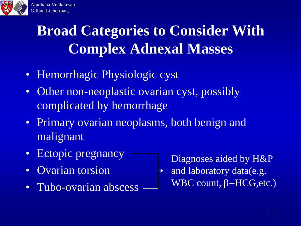
Broad Categories to Consider With Complex Adnexal Masses
• Hemorrhagic Physiologic cyst• Other non-neoplastic ovarian cyst, possibly
complicated by hemorrhage• Primary ovarian neoplasms, both benign and
malignant• Ectopic pregnancy• Ovarian torsion• Tubo-ovarian abscess
Diagnoses aided by H&P and laboratory data(e.g. WBC count, HCG,etc.)
Aradhana VenkatesanGillian Lieberman,
14
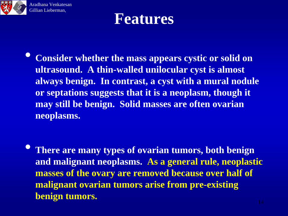
• Consider whether the mass appears cystic or solid on ultrasound. A thin-walled unilocular cyst is almost always benign. In contrast, a cyst with a mural nodule or septations suggests that it is a neoplasm, though it may still be benign. Solid masses are often ovarian neoplasms.
• There are many types of ovarian tumors, both benign and malignant neoplasms. As a general rule, neoplastic masses of the ovary are removed because over half of malignant ovarian tumors arise from pre-existing benign tumors.
FeaturesAradhana VenkatesanGillian Lieberman,
15
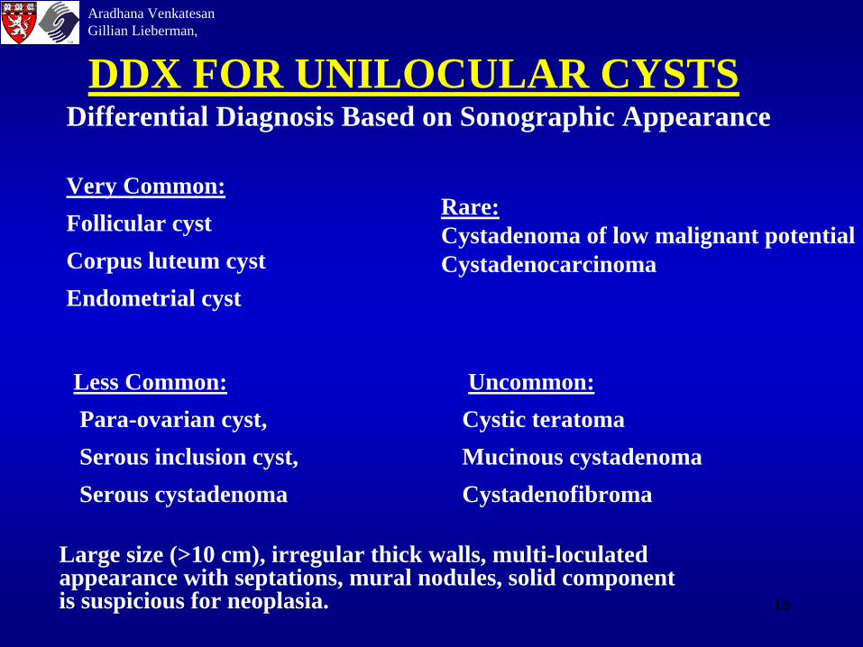
DDX FOR UNILOCULAR CYSTS
Less Common:Para-ovarian cyst,Serous inclusion cyst,Serous cystadenoma
Uncommon: Cystic teratomaMucinous cystadenomaCystadenofibroma
Rare:Cystadenoma of low malignant potentialCystadenocarcinoma
Large size (>10 cm), irregular thick walls, multi-loculated appearance with septations, mural nodules, solid component is suspicious for neoplasia.
Very Common:Follicular cystCorpus luteum cystEndometrial cyst
Aradhana VenkatesanGillian Lieberman,
Differential Diagnosis Based on Sonographic Appearance
16
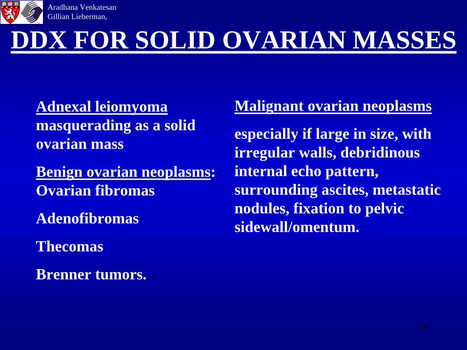
DDX FOR SOLID OVARIAN MASSES
Adnexal leiomyoma masquerading as a solid ovarian mass
Benign ovarian neoplasms: Ovarian fibromas
Adenofibromas
Thecomas
Brenner tumors.
Malignant ovarian neoplasms
especially if large in size, with irregular walls, debridinous internal echo pattern, surrounding ascites, metastatic nodules, fixation to pelvic sidewall/omentum.
Aradhana VenkatesanGillian Lieberman,
17
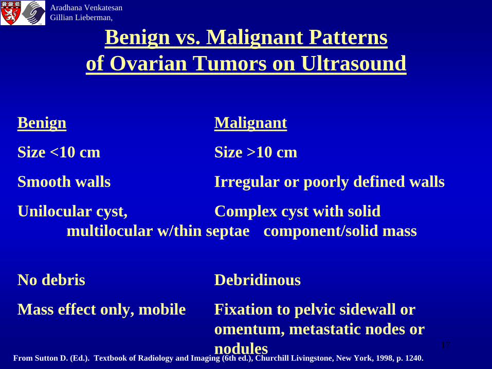
Benign Malignant
Size <10 cm Size >10 cm
Smooth walls Irregular or poorly defined walls
Unilocular cyst, Complex cyst with solid multilocular w/thin septae component/solid mass
No debris Debridinous
Mass effect only, mobile Fixation to pelvic sidewall or omentum, metastatic nodes or nodules
From Sutton D. (Ed.). Textbook of Radiology and Imaging (6th ed.), Churchill Livingstone, New York, 1998, p. 1240.
Benign vs. Malignant Patterns of Ovarian Tumors on Ultrasound
Aradhana VenkatesanGillian Lieberman,
18
PatientsPatient #1: A 32 y.o. F who presented to the BIDMC with LLQ pain of unknown etiology. She had a PMH significant for appendectomy and ovarian cystectomy x 1 (large follicular cyst c/b torsion). A trans-vaginal ultrasound of the left ovary showed the following:
QuickTime™ and aPhoto - JPEG decompressor
are needed to see this picture.
Obtained with permission from the Obstetric and Gynecological Ultrasound Teaching Files, Dept. of Radiology, Beth Israel- Deaconess Medical Center, Boston, MA.
Aradhana VenkatesanGillian Lieberman,
19
Patient #1Impression: A thin-walled unilocular cystic structure in the left ovary measuring 4.6x3.8x3.0 cm, with low-level internal echoes and no evidence of septations, nodules, solid components.
The findings were read as consistent with a hemorrhagic cyst.
F/U: On f/u visit one month later, u/s showed complete resolution of the cyst.
Aradhana VenkatesanGillian Lieberman,
20
PatientsPatient #2: A 34 y.o. F, originally from Ireland, presented to the BIDMC with pelvic pain and increasing abdominal girth. No significant PMH. FH significant for ovarian cancer in paternal grandmother and Hodgkin’s disease in paternal uncle. An abdominal and pelvic CT showed abdominal ascites with omental thickening and enlargement of the R ovary to approx 6.6 cm. F/U trans-vaginal ultrasound was arranged and showed the following in the right ovary:
Aradhana VenkatesanGillian Lieberman,
21
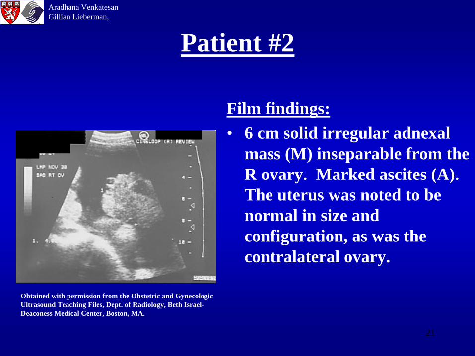
Patient #2
Film findings: • 6 cm solid irregular adnexal
mass (M) inseparable from the R ovary. Marked ascites (A). The uterus was noted to be normal in size and configuration, as was the contralateral ovary.
Obtained with permission from the Obstetric and Gynecologic Ultrasound Teaching Files, Dept. of Radiology, Beth Israel- Deaconess Medical Center, Boston, MA.
Aradhana VenkatesanGillian Lieberman,
M
A
22
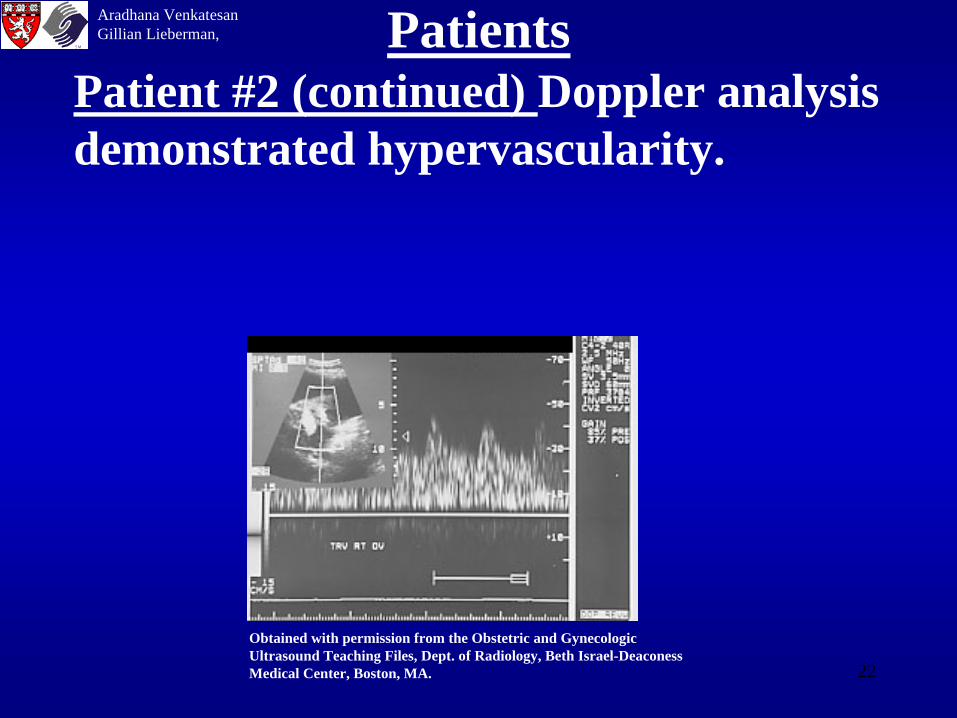
PatientsPatient #2 (continued) Doppler analysis demonstrated hypervascularity.
Obtained with permission from the Obstetric and Gynecologic Ultrasound Teaching Files, Dept. of Radiology, Beth Israel-Deaconess Medical Center, Boston, MA.
Aradhana VenkatesanGillian Lieberman,
23
Patient #2 continued• These findings were read as highly suggestive of malignancy.
• The patient underwent an e-lap and TAH-BSO with omentectomy. The pathology report on the L and R ovaries, fallopian tubes, uterus and omentum were positive for poorly differentiated and invasive ovarian cystadenocarcinoma.
• Surgery was followed by two cycles of taxol/carboplatin.
• Following completion of her second cycle of chemo, the patient elected to move to Ireland to be closer to family. Her prognosis is poor.
Aradhana VenkatesanGillian Lieberman,
24
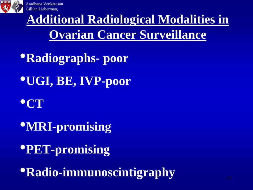
Additional Radiological Modalities in Ovarian Cancer Surveillance
•Radiographs- poor
•UGI, BE, IVP-poor
•CT
•MRI-promising
•PET-promising
•Radio-immunoscintigraphy
Aradhana VenkatesanGillian Lieberman,
25
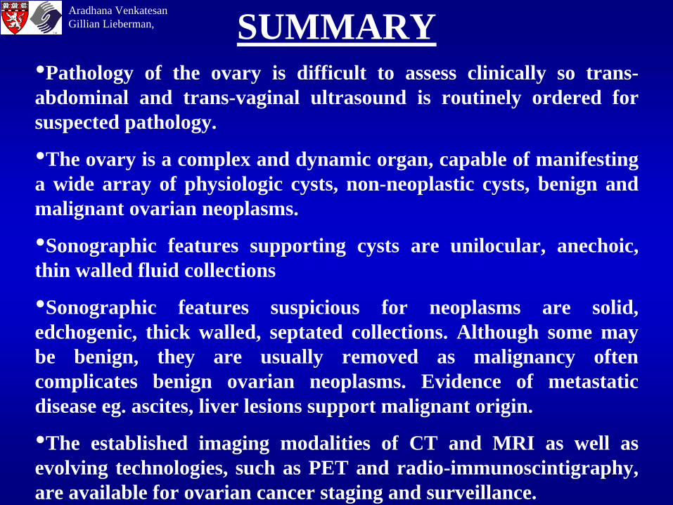
SUMMARY•Pathology of the ovary is difficult to assess clinically so trans- abdominal and trans-vaginal ultrasound is routinely ordered for suspected pathology.
•The ovary is a complex and dynamic organ, capable of manifesting a wide array of physiologic cysts, non-neoplastic cysts, benign and malignant ovarian neoplasms.
•Sonographic features supporting cysts are unilocular, anechoic, thin walled fluid collections
•Sonographic features suspicious for neoplasms are solid, edchogenic, thick walled, septated collections. Although some may be benign, they are usually removed as malignancy often complicates benign ovarian neoplasms. Evidence of metastatic disease eg. ascites, liver lesions support malignant origin.
•The established imaging modalities of CT and MRI as well as evolving technologies, such as PET and radio-immunoscintigraphy, are available for ovarian cancer staging and surveillance.
Aradhana VenkatesanGillian Lieberman,
26
References1) Callen, P.W.(ED.) Ultrasonography in Obstetrics and Gynecology (3rd). W.B. Saunders and Co.,
Philadelphia, 1994, pp. 625-6602) Doubilet P. 1999. Transvaginal sonography vs. transabdominal pelvic sonography. American journal
of Roentgenology, 173(3): 8463) Green V. 1992. Transvaginal ultrasonography. British Medical Journal. 204(6289):7794) Kosmas C., Kalofonoas H.P., Hird V., Epenetos A.A. 1998. Monoclonal antibody targeting of
ovarian cancer. Oncology. 55(5) 435-4465) Kossoff M.B. 1990. Transvaginal Doppler defines pelvic pathology. Diagnostic Imaging (San Fran)
12(9): 96-101.6) Method M.W., Serafini A.N., Averette H.E., Rodriguez M.,Penalver, M.A., Sevin B.U. 1996. The
role of radioimmunoscintigraphy and CT scan prior to reassessment laparotomy of patients with ovarian cancer. A preliminary report. Cancer. 77(11):2286-2293.
7) Netter F.H. Atlas of Human Anatomy. Ciba-Geigy, Summit, N.J. 1989, pp 350-353.8) Putman, C.E., Ravin, C.e. Textbook of Diagnostic Imaging (2nd ed.). W.B. Saunders and Co.,
Philadelphia, 1994, pp. 2088-2096.9) Sivyer P. 2000. Pelvic ultrasound in women. World Journal of Surgery. 24(2): 188-197.10) Sutton D. (Ed.) Textbook of Radiology and Imaging. Churchill Livingston, New York, 1998, 1235-
1271.
Aradhana VenkatesanGillian Lieberman,
27
Acknowledgements• Beverlee Turner for her support and PowerPoint expertise.• Larry Barbaras and Ben Crandall our WebMasters.
Aradhana VenkatesanGillian Lieberman,
The End.