Lithotrophic iron-oxidizing bacteria produce organic stalks to control mineral growth
11
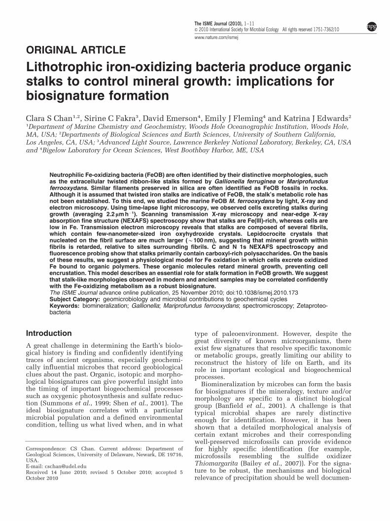
ORIGINAL ARTICLE Lithotrophic iron-oxidizing bacteria produce organic stalks to control mineral growth: implications for biosignature formation Clara S Chan 1,2 , Sirine C Fakra 3 , David Emerson 4 , Emily J Fleming 4 and Katrina J Edwards 2 1 Department of Marine Chemistry and Geochemistry, Woods Hole Oceanographic Institution, Woods Hole, MA, USA; 2 Departments of Biological Sciences and Earth Sciences, University of Southern California, Los Angeles, CA, USA; 3 Advanced Light Source, Lawrence Berkeley National Laboratory, Berkeley, CA, USA and 4 Bigelow Laboratory for Ocean Sciences, West Boothbay Harbor, ME, USA Neutrophilic Fe-oxidizing bacteria (FeOB) are often identified by their distinctive morphologies, such as the extracellular twisted ribbon-like stalks formed by Gallionella ferruginea or Mariprofundus ferrooxydans. Similar filaments preserved in silica are often identified as FeOB fossils in rocks. Although it is assumed that twisted iron stalks are indicative of FeOB, the stalk’s metabolic role has not been established. To this end, we studied the marine FeOB M. ferrooxydans by light, X-ray and electron microscopy. Using time-lapse light microscopy, we observed cells excreting stalks during growth (averaging 2.2 lmh 1 ). Scanning transmission X-ray microscopy and near-edge X-ray absorption fine structure (NEXAFS) spectroscopy show that stalks are Fe(III)-rich, whereas cells are low in Fe. Transmission electron microscopy reveals that stalks are composed of several fibrils, which contain few-nanometer-sized iron oxyhydroxide crystals. Lepidocrocite crystals that nucleated on the fibril surface are much larger (B100 nm), suggesting that mineral growth within fibrils is retarded, relative to sites surrounding fibrils. C and N 1s NEXAFS spectroscopy and fluorescence probing show that stalks primarily contain carboxyl-rich polysaccharides. On the basis of these results, we suggest a physiological model for Fe oxidation in which cells excrete oxidized Fe bound to organic polymers. These organic molecules retard mineral growth, preventing cell encrustation. This model describes an essential role for stalk formation in FeOB growth. We suggest that stalk-like morphologies observed in modern and ancient samples may be correlated confidently with the Fe-oxidizing metabolism as a robust biosignature. The ISME Journal advance online publication, 25 November 2010; doi:10.1038/ismej.2010.173 Subject Category: geomicrobiology and microbial contributions to geochemical cycles Keywords: biomineralization; Gallionella; Mariprofundus ferrooxydans; spectromicroscopy; Zetaproteo- bacteria Introduction A great challenge in determining the Earth’s biolo- gical history is finding and confidently identifying traces of ancient organisms, especially geochemi- cally influential microbes that record geobiological clues about the past. Organic, isotopic and morpho- logical biosignatures can give powerful insight into the timing of important biogeochemical processes such as oxygenic photosynthesis and sulfate reduc- tion (Summons et al., 1999; Shen et al., 2001). The ideal biosignature correlates with a particular microbial population and a defined environmental condition, telling us what lived when, and in what type of paleoenvironment. However, despite the great diversity of known microorganisms, there exist few signatures that resolve specific taxonomic or metabolic groups, greatly limiting our ability to reconstruct the history of life on Earth, and its role in important ecological and biogeochemical processes. Biomineralization by microbes can form the basis for biosignatures if the mineralogy, texture and/or morphology are specific to a distinct biological group (Banfield et al., 2001). A challenge is that typical microbial shapes are rarely distinctive enough for identification. However, it has been shown that a detailed morphological analysis of certain extant microbes and their corresponding well-preserved microfossils can provide evidence for highly specific identification (for example, microfossils resembling the sulfide oxidizer Thiomargarita (Bailey et al., 2007)). For the signa- ture to be robust, the mechanisms and biological relevance of precipitation should be well documen- Received 14 June 2010; revised 5 October 2010; accepted 5 October 2010 Correspondence: CS Chan. Current address: Department of Geological Sciences, University of Delaware, Newark, DE 19716, USA. E-mail: [email protected] The ISME Journal (2010), 1–11 & 2010 International Society for Microbial Ecology All rights reserved 1751-7362/10 www.nature.com/ismej
Transcript of Lithotrophic iron-oxidizing bacteria produce organic stalks to control mineral growth
ORIGINAL ARTICLE
Lithotrophic iron-oxidizing bacteria produce organicstalks to control mineral growth: implications forbiosignature formation
Clara S Chan1,2, Sirine C Fakra3, David Emerson4, Emily J Fleming4 and Katrina J Edwards2
1Department of Marine Chemistry and Geochemistry, Woods Hole Oceanographic Institution, Woods Hole,MA, USA; 2Departments of Biological Sciences and Earth Sciences, University of Southern California,Los Angeles, CA, USA; 3Advanced Light Source, Lawrence Berkeley National Laboratory, Berkeley, CA, USAand 4Bigelow Laboratory for Ocean Sciences, West Boothbay Harbor, ME, USA
Neutrophilic Fe-oxidizing bacteria (FeOB) are often identified by their distinctive morphologies, suchas the extracellular twisted ribbon-like stalks formed by Gallionella ferruginea or Mariprofundusferrooxydans. Similar filaments preserved in silica are often identified as FeOB fossils in rocks.Although it is assumed that twisted iron stalks are indicative of FeOB, the stalk’s metabolic role hasnot been established. To this end, we studied the marine FeOB M. ferrooxydans by light, X-ray andelectron microscopy. Using time-lapse light microscopy, we observed cells excreting stalks duringgrowth (averaging 2.2 lm h�1). Scanning transmission X-ray microscopy and near-edge X-rayabsorption fine structure (NEXAFS) spectroscopy show that stalks are Fe(III)-rich, whereas cells arelow in Fe. Transmission electron microscopy reveals that stalks are composed of several fibrils,which contain few-nanometer-sized iron oxyhydroxide crystals. Lepidocrocite crystals thatnucleated on the fibril surface are much larger (B100 nm), suggesting that mineral growth withinfibrils is retarded, relative to sites surrounding fibrils. C and N 1s NEXAFS spectroscopy andfluorescence probing show that stalks primarily contain carboxyl-rich polysaccharides. On the basisof these results, we suggest a physiological model for Fe oxidation in which cells excrete oxidizedFe bound to organic polymers. These organic molecules retard mineral growth, preventing cellencrustation. This model describes an essential role for stalk formation in FeOB growth. We suggestthat stalk-like morphologies observed in modern and ancient samples may be correlated confidentlywith the Fe-oxidizing metabolism as a robust biosignature.The ISME Journal advance online publication, 25 November 2010; doi:10.1038/ismej.2010.173Subject Category: geomicrobiology and microbial contributions to geochemical cyclesKeywords: biomineralization; Gallionella; Mariprofundus ferrooxydans; spectromicroscopy; Zetaproteo-bacteria
Introduction
A great challenge in determining the Earth’s biolo-gical history is finding and confidently identifyingtraces of ancient organisms, especially geochemi-cally influential microbes that record geobiologicalclues about the past. Organic, isotopic and morpho-logical biosignatures can give powerful insight intothe timing of important biogeochemical processessuch as oxygenic photosynthesis and sulfate reduc-tion (Summons et al., 1999; Shen et al., 2001). Theideal biosignature correlates with a particularmicrobial population and a defined environmentalcondition, telling us what lived when, and in what
type of paleoenvironment. However, despite thegreat diversity of known microorganisms, thereexist few signatures that resolve specific taxonomicor metabolic groups, greatly limiting our ability toreconstruct the history of life on Earth, and itsrole in important ecological and biogeochemicalprocesses.
Biomineralization by microbes can form the basisfor biosignatures if the mineralogy, texture and/ormorphology are specific to a distinct biologicalgroup (Banfield et al., 2001). A challenge is thattypical microbial shapes are rarely distinctiveenough for identification. However, it has beenshown that a detailed morphological analysis ofcertain extant microbes and their correspondingwell-preserved microfossils can provide evidencefor highly specific identification (for example,microfossils resembling the sulfide oxidizerThiomargarita (Bailey et al., 2007)). For the signa-ture to be robust, the mechanisms and biologicalrelevance of precipitation should be well documen-
Received 14 June 2010; revised 5 October 2010; accepted 5October 2010
Correspondence: CS Chan. Current address: Department ofGeological Sciences, University of Delaware, Newark, DE 19716,USA.E-mail: [email protected]
The ISME Journal (2010), 1–11& 2010 International Society for Microbial Ecology All rights reserved 1751-7362/10
www.nature.com/ismej
ted. This is relatively straightforward for intracel-lular mineralization, which is highly controlled(Bazylinski and Frankel, 2003), but challenging incases of extracellular precipitation, which can bestrongly influenced by environmental chemistry.
One potential candidate for a group-specificbiosignature can be found in the distinctive miner-alized filamentous extracellular structures producedby neutrophilic Fe-oxidizing bacteria (FeOB). Thesemicrobes are common in freshwater and marineenvironments in which redox gradients of O2 andFe(II) exist. They biomineralize extensively becauseof their metabolic dependence on Fe(II) oxida-tion combined with the extremely low solubility ofFe(III) at neutral pH. The freshwater Betaproteobac-terium Gallionella ferruginea has been recognizedfor nearly two centuries (first described by Ehren-berg in 1836). Recently, the novel (candidate class)Zetaproteobacterium Mariprofundus ferrooxydansPV-1 was isolated from Fe(III)-rich microbial matsnear Loihi Seamount hydrothermal vents in Hawaii(Emerson and Moyer, 2002; Emerson et al., 2007).These distinct obligate lithoautotrophic FeOB(Hallbeck and Pedersen, 1991; Emerson et al., 2007)produce strikingly similar Fe-rich extracellulartwisted stalks, strongly suggesting that stalk formationhas a core metabolic role in these Fe oxidizers.
These stalks have been suggestively linked toFe(III)-rich filaments observed in geological depositsranging from recent to 1.7 Ga (Alt, 1988; Little et al.,2004; Slack et al., 2007; Hofmann et al., 2008). Fefilaments and stalks could be biosignatures for thepresence and activity of FeOB if (1) they areexclusively produced by FeOB; (2) mineralogicaland/or morphological criteria are established for Femicrofossil identification; and (3) these structuresare linked to Fe-oxidizing metabolism. The firstcriterion is generally supported, and is based onobservations of modern FeOB and on the absenceof known biological and chemical processes tocreate similar Fe-based morphologies (Ghiorse,1984; Garcia-Ruiz et al., 2003; Hofmann et al.,2008). The second criterion has only been partiallymet because of limited detailed study of thesestructures (Hanert, 1999). The third criterion hasnot yet been proven because the role of thesestructures in FeOB metabolism is not established.Here, we address the latter two criteria by combin-ing microscopy and spectroscopy techniques toobserve M. ferrooxydans stalk formation in activecultures and characterize the morphology, physio-logy, mineralogy, and elemental and organic compo-sition of the stalk. We show that the stalk performsan essential role in eliminating the ferric wasteproduct of Fe-oxidation metabolism by encasingFe(III) and transporting it away from the cell.Furthermore, we demonstrate that morphologicaldetails are rooted in physiology and behavior, andrecord relevant information pertaining to environ-mental conditions, making these structures idealsignatures for microaerophilic FeOB.
Materials and methods
CulturingM. ferrooxydans PV-1 was grown from frozen stockcultures in artificial seawater (ASW) media, bufferedby bicarbonate or 2-(N-morpholino)ethanesulfonicacid to pH of 6–6.5 and FeS as an Fe(II) source(Emerson and Floyd, 2005). Samples for transmissionelectron microscopy (TEM) and scanning transmissionX-ray microscopy (STXM) were prepared in gradienttubes (12� 75 mm test tube containing 0.5 ml FeSoverlain by 3.5 ml ASW, sealed with a silicone stopperand incubated horizontally at B101 angle). Sampleholders (TEM grids and Si3N4 membranes for STXM)were placed on an ACLAR (Ted Pella, Redding, CA,USA) strip in the tube and retrieved after 1 day.Cultures for microtomy were grown as follows: 8 ml ofFeS/ASW/1% agarose was overlain by 15 ml ASW in aPetri dish and incubated in a jar with a Campy Pakpouch (BD, Franklin Lakes, NJ, USA). Cultures fortime-lapse microscopy were prepared in rectangularmicrocapillaries (0.2� 2 mm inner diameter, Vitro-com, Mountain Lakes, NJ, USA). These were filledby capillary action in the following order: ASWmedia, inoculum and FeS, leaving a small headspaceof air; both ends were sealed with Vaseline. Theinoculum was PV-1 grown in agarose gradient tubes(ASW with 0.15% low-melt agarose over an FeS/ASW/1% agarose plug). The agarose confined inocu-lum to the end of the microcapillary, keeping the restclear. For bulk culture experiments, log-phase culturesof cells were inoculated into Petri dishes containing20 ml of ASW (10 mM 2-(N-morpholino)ethanesulfo-nic acid) and Fe(0) (200 mesh; Alfa Aesar, Ward Hill,MA, USA) and grown microaerobically (Emerson andFloyd, 2005). After 21.5 h, cultures were homogenizedby repeated pipetting, and analyzed for Fe content,cell number and stalk length.
Fe measurementsBulk culture Fe concentrations were determined bythe ferrozine method (Stookey, 1970). Aliquots(0.1 ml) of cultures were added to 0.5 M HCl (finalconcentration). Fe(II) (78.7±6.7 mM s.e.) was mea-sured in triplicate with ferrozine (100 mM) in anacetate buffer (1.4 M) at 562 nm on a MultiskanMicroplate Reader MCC/340 (Thermo Scientific,Waltham, MA, USA). Total Fe was measured (intriplicate) by reducing Fe(III) with hydroxyl amine(120 mM) before adding ferrozine. Fe(III) was thencalculated as the difference between Fe(II) and totalFe (96.5±4.2 mM s.e.).
Light microscopyMicrocapillary slide cultures were observed usingan Olympus BX 60 light microscope (Center Valley,PA, USA), using brightfield and phase contrastmethods. For time-lapse studies, images werecaptured every 5 min with a QIACAM CCD cameraand QCapture Pro software (Qimaging, Surrey, BC,
Iron-oxidizer stalks control mineral growthCS Chan et al
2
The ISME Journal
Canada). Cultures were also observed by differentialinterference contrast (DIC) using a Zeiss AxioImagerZ1 light microscope, a Zeiss Axiocam Mrm cameraand Axiovision imaging software (Zeiss, Oberkochen,Germany). For cell counting and stalk measurements,bulk culture samples were spread evenly on a 0.5%agarose-coated fluoroslide printed with 10 mmcircles (Thermo Scientific) (Emerson and Moyer,2002). Cells were stained with Syto 13 (Invitrogen,Carlsbad, CA, USA) and counted using fluorescencemicroscopy (1.17� 109±2.92� 107 cells per l s.e.;n¼ 64). Stalk length (31.2±1.4mm s.e.; n¼ 92) wasmeasured between each stalk bifurcation by tracingeach segment with the measure tool in the QCapturePro software. For each 10 mm circle (n¼ 4), at least 10fields were examined.
Stalk length and Fe calculationsThe stalk formation rate was determined by dividingthe stalk length produced by one cell from formationto division (31.2 mm average, n¼ 92) by 12 h, theknown doubling time of M. ferrooxydans (Emersonet al., 2007). The amount of Fe(II) oxidized per cellwas calculated as follows. The total stalk length pervolume (5.93� 1010 mm l�1) was divided by the stalklength produced by one cell (31.2 mm) to determinethe number of individual cells responsiblefor producing the total stalk mass (1.90� 109 cellsper l). Then, the total Fe(III) concentration(1.74� 10�5 mol l�1) was divided by the number ofcells (1.90� 109 cells per l) to determine the amountof Fe oxidized per cell (9.16� 10�15 mol per cell).Note that this assumes that all of the Fe(III) wasbiologically oxidized; the culture time and condi-tions were controlled such that this assumptionwould be valid (or nearly so).
TEMSamples for TEM were prepared in three ways:(1) Formvar-coated copper grids were inserted intocultures to preserve the delicate cell-stalk spatialrelationships. (Normal handling causes cells tobreak free of stalks.) The grids were carefullyremoved, rinsed in distilled water, air-dried andC-coated. (2) Samples were pipetted directly ontogrids, rinsed in distilled water, air-dried andC-coated. (3) Samples were dehydrated in a seriesof solutions of increasing percentages of acetone,embedded in Epon-Araldite resin, ultramicrotomedinto 70–100 nm sections and mounted on C-coated,formvar-coated copper grids. Analyses were per-formed on a Zeiss 10CA TEM (100 kV acceleratingvoltage), Zeiss Libra 120 PLUS (Zeiss) (120 kV) andPhilips (FEI, Hillsboro, OR, USA) CM200 HRTEM(200 kV) with a Link ultrathin-window energy dis-persive X-ray detector.
STXMSTXM analyses were performed on a sample grownon a Si3N4 membrane (Silson Ltd., Northampton,
UK), rinsed in distilled water and air-dried. STXMand near-edge X-ray absorption fine structure (NEX-AFS) spectroscopy measurements were taken onbeamlines 11.0.2 and 5.3.2 of the Advanced LightSource, Lawrence Berkeley National Lab (Kilcoyneet al., 2003). These microscopes use a Fresnel zoneplate lens (25 nm outer zones) to focus a monochro-matic X-ray beam onto a scanned sample to recordtransmission images with a scintillator-photomulti-plier detector assembly. The imaging contrast relieson core electron excitation by X-ray absorption(Stohr, 1992). X-ray images recorded at energies justbelow and at the relevant absorption edges wereconverted into optical density (OD) images and usedto derive elemental maps. OD can be expressed for agiven X-ray energy from the Beer-Lambert law asOD¼�ln(I/Io)¼ mrt, where I, I0, m, r and t aretransmitted intensity through the sample, incidentintensity, mass absorption coefficient, density andsample thickness, respectively. NEXAFS spectrafrom regions of interest were obtained from imagesequences (that is, stacks) collected at energiesspanning the relevant absorption edge. Beamdamage was checked as described by Chan et al.(2009) and was not observed. See SupplementaryMethods for atomic ratio calculations. At least twodifferent sample regions were analyzed for eachelement. The theoretical spectral and spatial resolu-tions were ±100 meV and 30 nm, respectively. Thephoton energy was calibrated at the C 1s edge usingthe Rydberg transitions of gaseous CO2 at 292.74 eV
(C 1s-3s (n¼ 0)) and 294.96 eV (C 1s-3p (n¼ 0)),at the N 1s edge using the N 1s-3s (n¼ 0) transitionat 401.1 eV of gaseous N2, at the O 1s edge using theO 1s-3s transition at 538.9 eV of gaseous CO2 and atFe 2p edge using ferrihydrite Fe 2p3/2 resonance setat 709.5 eV. All data processing was carried out usingIDL aXis2000 software (Hitchcock, an IDL-basedanalytical package, http://unicorn.mcmaster.ca).
Results
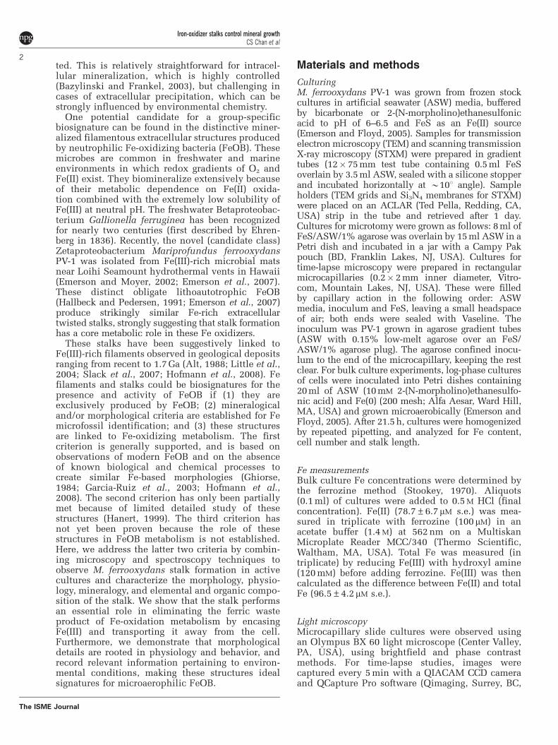
Correlating stalk formation and cell growthMicroslide cultures enabled visualization ofM. ferrooxydans cell and stalk growth in opposinggradients of O2 and Fe(II). Cell and stalk growth isobserved within 1–2 days and continued for a weekor longer, consistent with results of test tubegradient cultures (Emerson et al., 2007). A singlecell is located at the end of a stalk. Stalk formationbegins with attachment (to glass, in this case);the holdfast is characterized by dense mineralprecipitation (Supplementary Figure S1a). Cellgrowth coincides with translocation of the cell,although the stalk remains stationary (Figure 1 andSupplementary Video S1). As cells grow, theyrotate, resulting in twisted or coiled stalks. Celldivision results in bifurcation, sequential cell divi-sion results in multiple branching and cells mayleave stalks (see Supplementary Video S2). The
Iron-oxidizer stalks control mineral growthCS Chan et al
3
The ISME Journal
cell–stalk connection is fragile; typical handlingof samples (for example, pipetting) causes most cellsto break away from stalks. Stalk formation ismeasured between 0.6 and 5.5 mm h�1 (n¼ 15),averaging 2.2 mm h�1. A Petri dish-based liquidculture yielded an average stalk formation rate of2.6 mm cell�1 h�1, which agrees well with the micro-slide-based rate. Each cell oxidized 9.2� 10�15 molFe per cell and produced a stalk averaging 31 mm inlength between divisions.
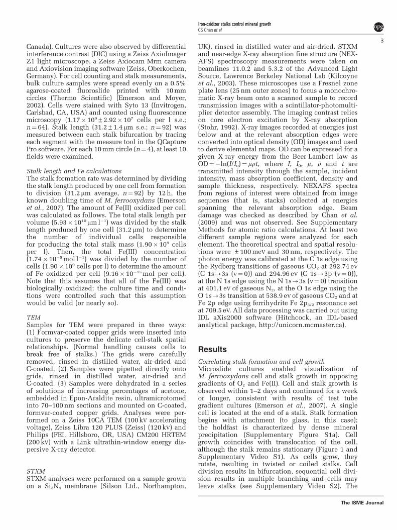
Cell–stalk–mineral spatial relationshipsThe cells are shaped in the form of beans or bent rods;stalk formation occurs within the concavity of the cell.A range of cell lengths and stalk morphologies areobserved (Figures 1, 2 and Supplementary Figure S1).Cell lengths varied from 0.8 to 4.7mm (in TEM),occasionally up to 11mm (in microslides). Stalkwidths ranged from 0.3 to 1.8mm (in TEM), occasion-ally up to 8mm wide (in microslides). Smaller cellsoccur in pairs, attached to the ends of a branched stalk(Supplementary Figure S1c and Video S2). The widthof the stalk near the cell correlates with the cell length.The stalk gradually widens from the holdfast to thecell, as illustrated in Figure 1b, SupplementaryFigures S1d and S2. The stalks are composed of2–30 parallel nanometer-sized fibrils. Stalk fibrils areelectron dense and sometimes have a smooth, less-electron-dense layer at the ends closest to the cell, atthe edges of the fibrils (Figure 2b). At the cell surface,the fibrils are typically thin and widen substantiallyover the first few 100 nm (Figure 2 and SupplementaryFigure S1b). Away from the cell, individual fibrilsexhibit more subtle increases in width with increasingdistance from the cell (Supplementary Figure S3). Thenumber of fibrils increases with increasing stalk width(Figure 1b, Supplementary Figures S1d and S2),
with branching of individual fibrils coinciding withincreased stalk width.
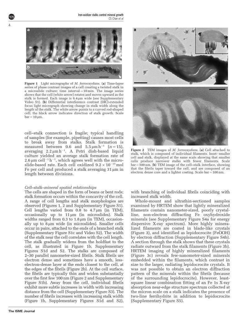
Whole-mount and ultrathin-sectioned samplesexamined by HRTEM show that lightly mineralizedfilaments contain nanometer-sized, poorly crystal-line, non-electron diffracting Fe oxyhydroxideminerals (see Supplementary Figure S4a for energydispersive X-ray spectrum). More highly minera-lized filaments are coated in blade-like crystals(Figure 3), and identified as lepidocrocite (FeOOH)by electron diffraction (Supplementary Figure S4b).A section through the stalk shows that these crystalsradiate outward from the stalk filaments (Figure 3b).HRTEM imaging of highly mineralized filaments(Figure 3c) reveals few-nanometer-sized mineralsembedded within the filaments, which contrast insize to the larger, radiating lepidocrocite crystals. Itwas not possible to obtain an electron diffractionpattern of the minerals within the fibrils (becauseof the surrounding lepidocrocite). However, least-square linear combination fitting of an Fe 1s X-rayabsorption near-edge structure spectrum collected atthe micron scale on a stalk indicates the presence oftwo-line ferrihydrite in addition to lepidocrocite(Supplementary Figure S5).
Figure 2 TEM images of M. ferrooxydans. (a) Cell attached tostalk, which is composed of individual filaments. Inset: smallercell and stalk, displayed at the same scale showing that smallercells produce narrower stalks with fewer filaments. Scalebar¼500 nm. (b) TEM image of the cell–stalk interface, showingthat the fibrils taper toward the cell, and are composed of anelectron dense core and a lighter coating. Scale bar¼ 100 nm.
Figure 1 Light micrographs of M. ferrooxydans. (a) Time-lapseseries of phase contrast images of a cell creating a twisted stalk ina microslide culture; time interval¼10 min. The image seriesshows that the cell (white arrow) rotates and moves upward as thestalk is formed. Each image is 6.4mm wide (see SupplementaryVideo S1). (b) Differential interference contrast (DIC)-extendedfocus light micrograph showing change in stalk width along thelength of the stalk. The white arrow points to a curved rod-shapedcell; the black arrow indicates direction of stalk growth. Scalebar¼10mm.
Iron-oxidizer stalks control mineral growthCS Chan et al
4
The ISME Journal
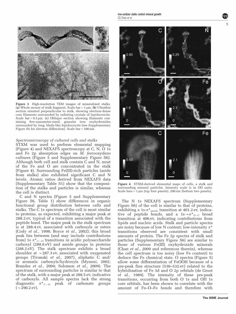
Spectromicroscopy of cultured cells and stalksSTXM was used to perform elemental mapping(Figure 4) and NEXAFS spectroscopy at C, N, O 1sand Fe 2p absorption edges on M. ferrooxydanscultures (Figure 5 and Supplementary Figure S6).Although both cell and stalk contain C and N, mostof the Fe and O are concentrated in the stalk(Figure 4). Surrounding Fe(III)-rich particles (asidefrom stalks) also exhibited significant C and Nlevels. Atomic ratios derived from NEXAFS data(Supplementary Table S1) show that the composi-tion of the stalks and particles is similar, whereasthe cell is distinct.
C and N spectra (Figure 5 and SupplementaryFigure S6, Table 1) show differences in organicfunctional group distribution between cells andstalks. The C 1s spectrum of the cell is most similarto proteins, as expected, exhibiting a major peak at288.2 eV, typical of a transition associated with thepeptide bond. The major peak in the stalk spectrumis at 288.4 eV, associated with carboxyls or esters(Cody et al., 1998; Boyce et al., 2002); this broadpeak lies between (and may include contributionsfrom) 1s p*C¼O transitions in acidic polysaccharidecarboxyl (288.6 eV) and amide groups in proteins(288.2 eV). The stalk spectrum exhibits a broadshoulder at B287.3 eV, associated with oxygenatedgroups (Tivanski et al., 2007), aliphatic C and/or aromatic carbonyls/hydroxyls (Myneni, 2002;Brandes et al., 2004; Solomon et al., 2009). Thespectrum of surrounding particles is similar to thatof the stalk, with a major peak at 288.5 eV, indicativeof carboxyls. All sample spectra lack the strongdiagnostic p*C¼O peak of carbonate groups(B290.2 eV).
The N 1s NEXAFS spectrum (SupplementaryFigure S6) of the cell is similar to that of proteins,exhibiting a 1s-p*amide transition at 401.2 eV, indica-tive of peptide bonds, and a 1s-s*N�C broadtransition at 406 eV, indicating contributions fromlipids and nucleic acids. Stalk and particle spectraare noisy because of low N content; low-intensity p*transitions observed are consistent with smallamounts of protein. The Fe 2p spectra of stalk andparticles (Supplementary Figure S6) are similar tothose of various Fe(III) oxyhydroxide minerals(Chan et al., 2009 and references therein), whereasthe cell spectrum is too noisy (low Fe content) todeduce the Fe chemical state. O spectra (Figure 5)allow some differentiation of FeOOH because of apre-peak fine structure (530–532 eV) related to thehybridization of Fe 3d and O 2p orbitals (de Grootet al., 1989). The intensity of these pre-peaktransitions, occurring from both O 1s and OH 1score orbitals, has been shown to correlate with theamount of Fe–O–Fe bonds and therefore with
Figure 4 STXM-derived elemental maps of cells, a stalk andsurrounding mineral particles. Intensity scale is in OD units.Scale bars¼ 1 mm (top four panels), 200 nm (bottom two panels).
Figure 3 High-resolution TEM images of mineralized stalks.(a) Whole mount of stalk fragment. Scale bar¼ 1 mm. (b) Ultrathinsection oriented perpendicular to stalk, showing electron-densecore filaments surrounded by radiating crystals of lepidocrocite.Scale bar¼0.5 mm. (c) Oblique section showing filaments con-taining few-nanometer-sized, granular iron oxyhydroxidessurrounded by long, blade-like lepidocrocite (see SupplementaryFigure S4 for electron diffraction). Scale bar¼ 100 nm.
Iron-oxidizer stalks control mineral growthCS Chan et al
5
The ISME Journal
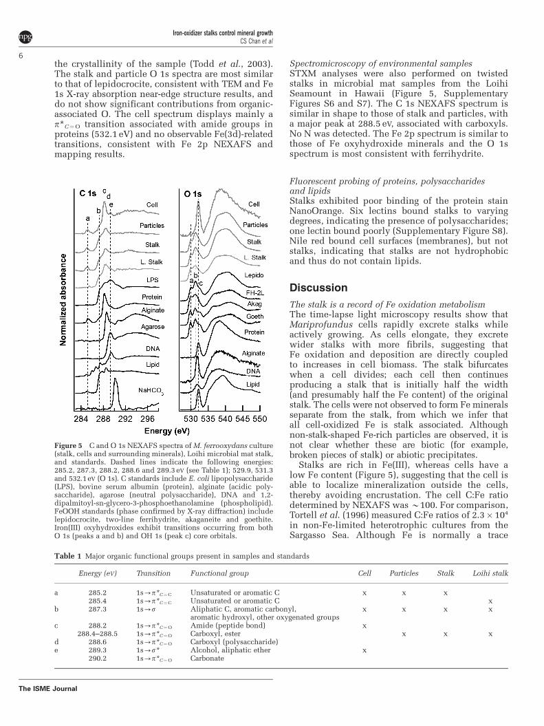
the crystallinity of the sample (Todd et al., 2003).The stalk and particle O 1s spectra are most similarto that of lepidocrocite, consistent with TEM and Fe1s X-ray absorption near-edge structure results, anddo not show significant contributions from organic-associated O. The cell spectrum displays mainly ap*C¼O transition associated with amide groups inproteins (532.1 eV) and no observable Fe(3d)-relatedtransitions, consistent with Fe 2p NEXAFS andmapping results.
Spectromicroscopy of environmental samplesSTXM analyses were also performed on twistedstalks in microbial mat samples from the LoihiSeamount in Hawaii (Figure 5, SupplementaryFigures S6 and S7). The C 1s NEXAFS spectrum issimilar in shape to those of stalk and particles, witha major peak at 288.5 eV, associated with carboxyls.No N was detected. The Fe 2p spectrum is similar tothose of Fe oxyhydroxide minerals and the O 1sspectrum is most consistent with ferrihydrite.
Fluorescent probing of proteins, polysaccharidesand lipidsStalks exhibited poor binding of the protein stainNanoOrange. Six lectins bound stalks to varyingdegrees, indicating the presence of polysaccharides;one lectin bound poorly (Supplementary Figure S8).Nile red bound cell surfaces (membranes), but notstalks, indicating that stalks are not hydrophobicand thus do not contain lipids.
Discussion
The stalk is a record of Fe oxidation metabolismThe time-lapse light microscopy results show thatMariprofundus cells rapidly excrete stalks whileactively growing. As cells elongate, they excretewider stalks with more fibrils, suggesting thatFe oxidation and deposition are directly coupledto increases in cell biomass. The stalk bifurcateswhen a cell divides; each cell then continuesproducing a stalk that is initially half the width(and presumably half the Fe content) of the originalstalk. The cells were not observed to form Fe mineralsseparate from the stalk, from which we infer thatall cell-oxidized Fe is stalk associated. Althoughnon-stalk-shaped Fe-rich particles are observed, it isnot clear whether these are biotic (for example,broken pieces of stalk) or abiotic precipitates.
Stalks are rich in Fe(III), whereas cells have alow Fe content (Figure 5), suggesting that the cell isable to localize mineralization outside the cells,thereby avoiding encrustation. The cell C:Fe ratiodetermined by NEXAFS was B100. For comparison,Tortell et al. (1996) measured C:Fe ratios of 2.3� 104
in non-Fe-limited heterotrophic cultures from theSargasso Sea. Although Fe is normally a trace
Table 1 Major organic functional groups present in samples and standards
Energy (eV) Transition Functional group Cell Particles Stalk Loihi stalk
a 285.2 1s-p*C¼C Unsaturated or aromatic C x x x285.4 1s-p*C¼C Unsaturated or aromatic C x
b 287.3 1s-s Aliphatic C, aromatic carbonyl,aromatic hydroxyl, other oxygenated groups
x x x x
c 288.2 1s-p*C¼O Amide (peptide bond) x288.4–288.5 1s-p*C¼O Carboxyl, ester x x x
d 288.6 1s-p*C¼O Carboxyl (polysaccharide)e 289.3 1s-s* Alcohol, aliphatic ether x
290.2 1s-p*C¼O Carbonate
Figure 5 C and O 1s NEXAFS spectra of M. ferrooxydans culture(stalk, cells and surrounding minerals), Loihi microbial mat stalk,and standards. Dashed lines indicate the following energies:285.2, 287.3, 288.2, 288.6 and 289.3 eV (see Table 1); 529.9, 531.3and 532.1eV (O 1s). C standards include E. coli lipopolysaccharide(LPS), bovine serum albumin (protein), alginate (acidic poly-saccharide), agarose (neutral polysaccharide), DNA and 1,2-dipalmitoyl-sn-glycero-3-phosphoethanolamine (phospholipid).FeOOH standards (phase confirmed by X-ray diffraction) includelepidocrocite, two-line ferrihydrite, akaganeite and goethite.Iron(III) oxyhydroxides exhibit transitions occurring from bothO 1s (peaks a and b) and OH 1s (peak c) core orbitals.
Iron-oxidizer stalks control mineral growthCS Chan et al
6
The ISME Journal

metal in a cell, the Fe content of Mariprofundus isrelatively high, possibly because of adsorption to thecell surface. On the basis of Fe elemental map andspectra, we conclude that most of the Fe(III) frommetabolic Fe oxidation (and abiotic mineralization)is located on the stalk.
Fe-binding organics retard mineral growth in stalkfibrilsThe radiating habit of the lepidocrocite crystals(Figure 3) indicates that they nucleated on the fibrilsurface and grew outward. Newly formed stalks donot have lepidocrocite, although stalks gain thickerencrustations with time, suggesting that this miner-alization ‘overprint’ begins after stalk formation. Feoxyhydroxide minerals within cultured stalk fibrilsare much smaller than the lepidocrocite on thesurface, suggesting the presence of a stalk compo-nent responsible for retarding crystal growth(including growth associated with transformation).The light smooth coating at the fibril tips possiblyrepresents organic polymers (Figure 2b). The Celemental maps show that stalks indeed containC throughout the fibrils (Figure 4 and Supplemen-tary Figure S7). The stalk spectrum exhibits peakssimilar to biomolecule standards, but lack carbo-nate, suggesting that C is organic. The dominant288.4 eV peak (p*C¼O) is close in energy to the proteinamide peak (288.2 eV) and the acidic polysaccharidecarboxyl peak (288.6 eV) (Urquhart and Ade, 2002).However, little N is detected in cultured stalks (nonein Loihi stalks) and the stalks bind NanoOrangeprotein stain weakly, from which we conclude thatprotein could at most account for a small portion ofthe stalk organic matter. On the basis of C:N ratios,Hallbeck and Pedersen (1995) also concluded thatprotein is a minor component of the G. ferrugineastalk, although there are no studies specificallyexamining the protein content of stalks. There is alsoa prominent shoulder at B287.3 eV, similar to the onein the lipid spectrum; however, the stalks do not bindNile Red, ruling out lipids as a significant component.The stalks do bind several lectins; thus, in light of theabove evidence, we conclude that the stalk organicmatter is dominated by acidic polysaccharides rich incarboxyls. Previous work on freshwater FeOB systemsalso found the predominance of acidic polysac-charides associated with Fe-mineralized extracellularstructures (Chan et al., 2009). At the circumneutralpH values found in neutrophilic FeOB habitats and inour cultures, these carboxyls would be deprotonatedand would have a high affinity for Fe. Furtheranalysis will be required to determine a more specificcomposition of the organic polymers, as they likelycontain variations of our biomolecule standards ormay even contain novel components that are not wellrepresented by our reference materials.
Fe(III) occurs throughout the stalk fibrils, notas a coating (Figure 3b); thus, it is an integral partof the stalk. In experiments in which Fe isdissolved, stalks disappear (Emerson et al., 2007).
As a multivalent cation, Fe3þ can crosslinkcarboxyl-rich polymers; thus, we conclude that stalkfibrils are a matrix of Fe and organics binding oneanother. Ligands retard mineral growth and it hasbeen suggested by Kennedy et al. (2004) and Toneret al. (2009) that organics are responsible for thepoor crystallinity of microbe-associated Fe minerals.Although we did not quantify the amount of C in thestalk, it appears (in OD units) to be significant,suggesting that these autotrophic organisms expenda significant amount of energy to create the stalk.The necessity of removing waste products andpreventing mineral growth near the cell presents agood justification for such an expenditure.
A biomineralization modelThis work demonstrates that the stalk ofM. ferrooxydans is a well-coordinated biomineralstructure controlled by the cell. We combine ourresults into a model of stalk formation and biomi-neralization, presented in the following sequence(Figure 6):
(1) Cells attach to a surface, which may be an existingstalk produced by another cell. Fe is oxidized andexcreted from discrete locations on the cell sur-face, bound to carboxyl groups of organic polymers(for example, acidic polysaccharides). Fe(III) cross-links polymers, aiding in fibril formation. Cellsgrow, elongate and oxidize more Fe(II), resulting inwider stalks containing more fibrils.
(2) As cells continue to form new stalk materialand move away from older portions of stalks,
Figure 6 Model of stalk formation and mineralization process.Iron oxidation is coupled to O2 reduction (exact location of ironoxidation is not known). (1) Fe(III)-polysaccharide (EPS) isexcreted from the cell as fibrils. (2) Over time, Fe(III) precipitatesas Fe oxyhydroxides. (3) As stalks age, lepidocrocite nucleates onfibril surfaces.
Iron-oxidizer stalks control mineral growthCS Chan et al
7
The ISME Journal
the Fe(III) in the fibrils precipitates as Feoxyhydroxide minerals. The timing of precipita-tion and extent of mineral growth within fibrilshave not yet been determined, but our observa-tions and previous studies on environmentalsamples suggest that they remain nanocrystallineover the timescale of months or longer (Toneret al., 2009).
(3) As stalks age, Fe oxyhydroxide minerals (forexample, lepidocrocite) nucleate on the surfacesof fibrils and grow into larger crystals, whereasthe minerals within the fibrils remain small.Abiotic Fe oxidation is typical of FeOB cultures,partly because of autocatalysis of Fe oxidationby the biotic, stalk-bound oxides (Rentz et al.,2007; Druschel et al., 2008). Although mineralsinside fibrils could in theory nucleate furthermineralization, the limited space within thepolymer network would preclude formation oflarger crystals.
Physiological implications of stalk formationConsidering this model, what are the physiologicalroles of the stalk? The stalk includes a holdfast,which anchors the cell in favorable environmentalgradients of O2 and Fe (II). As the cell excretes thesemirigid stalk, the cell translocates away from theholdfast, whereas the stalk remains stationary. In asense, this provides a form of motility to the cell. Acomparable set of functions can be found in sulfuroxidizers Thiovulum and Thioturbo, which colonizesulfide/O2 gradient environments with thin, thread-like ‘mucous’ stalks, forming ‘veils’ in which condi-tions are optimal (Fenchel, 1994; Thar and Kuhl, 2002;Muyzer et al., 2005). Similar to Mariprofundus, thecells rotate about the axis parallel to the stalk; thisrotation has been associated with aerotaxis and fluidmixing. Thar and Kuhl (2003) suggested that cells cansense gradients through a combination of helicalmotion and sensors located at cell poles. The stalkitself is also used in motility, as the length iscontrolled to track the position of the oxic/anoxicinterface (Thar and Kuhl, 2002).
Perhaps, more importantly, the mineralized stalkserves as an organized structure for depositing solidmetabolic products. A similar phenomenon isobserved for the sulfur oxidizer Arcobacter sulfidi-cus, which excretes S(0)-rich mineral filaments thatform microbial mats (Sievert et al., 2007). Sievertet al. suggested that the rapid sulfur filamentformation is related to the fact that A. sulfidicuslives in extremely high sulfide environments. Thus,the formation of mineralized filaments may be ageneral adaptation of microbes that rapidly producelarge quantities of solid metabolic waste. In the caseof Mariprofundus, polymers encase and carry Fe(III)away from the cell; in this way, the cell escapesencrustation by its waste products. This model maybe generalized to other stalk-forming FeOB, namely,
G. ferruginea, and more universally to any FeOBthat excrete Fe-mineralized polymers.
Cells that do not normally produce minerals seemto lack such adaptations to prevent cell encrustation(for example, Williams et al., 2005). Althoughsurface and periplasmic precipitation may notcompletely preclude survival (Miot et al., 2009), itdoes present a severe challenge, as encrustationwould obstruct solute uptake and excretion, andpotentially damage the cell wall. Non-structure-forming FeOB, such as microaerophiles Sideroxy-dans sp., Ferritrophicum radicicola and Gallionellacapsiferriformans (Emerson and Moyer, 1997; Weisset al., 2007) and the phototroph Rhodobacterferrooxidans (Schadler et al., 2009), likely haveless-obvious mechanisms for escaping total encrus-tation. Minerals may be nucleated on less-organizedpolymers and periodically shed, or, as Schadleret al. (2009) suggest, there may be a combination oflocal acidification and ligand binding (to keep Fe insolution), as well as cell surface charge adaptations.Mariprofundus may use some of these other adapta-tions in addition to stalk formation.
Supporting the case for an FeOB biosignatureDespite the abundance of Fe and the significance ofits redox state in Earth’s history, there is a paucityof biosignatures established for Fe-oxidizing andreducing microorganisms. This limits our ability topiece together the history of Fe biogeochemicalcycling, although there is some geochemical andbiological evidence that Fe-based metabolismscould have evolved early in Earth’s history (Vargaset al., 1998; Johnson et al., 2008; Croal et al., 2009).Fe reducers and anaerobic FeOB (including photo-trophs) are not known to form easily distinguishableFe mineral morphologies, and there have not beenany unique organic biomarkers or isotopic signa-tures established (Kappler and Newman, 2004).Filamentous Fe microfossils corresponding to aero-bic stalk-forming FeOB represent the best potentialsignatures of Fe-cycling microbes thus far. Further-more, because only aerobic FeOB produce stalks, Fefilaments are indicators of locally oxygenatedenvironments and thus could be important markersof the Earth’s oxygen history.
Biosignatures based on morphology are oftendebated because of the possibility that chemicalprocesses may form similar shapes. This is particu-larly a problem for microbial signatures because ofthe small size and simplicity of microbial forms.An often-cited example is a twisted morphologycomposed of silica and witherite, a barium carbo-nate (Garcia-Ruiz et al., 2003, 2009). When imagedstrategically, this precipitate appears very similar tothe twisted stalks of Gallionella and Mariprofundus,although closer inspection reveals key morphologi-cal differences that allow distinction of the chemicalprecipitates from biological ones. In contrast tothe fibrillar, ribbon-like stalks, these chemical
Iron-oxidizer stalks control mineral growthCS Chan et al
8
The ISME Journal
precipitates are rope-like, and emanate fromlarge, clearly abiological aggregates (Figure 1 ofGarcia-Ruiz et al., 2003). Furthermore, chemicalsynthesis of these biomorphs requires environ-mentally unrealistic conditions and concentrations,inconsistent with what is known about theconditions of Fe oxide deposition. Nevertheless,this work reminds us that abiotic precipitation canresult in very biological-looking forms.
In this study of the marine FeOB M. ferrooxydansstalk, we have compiled multiple criteria forbiosignature distinction that, when consideredcollectively, delineate them clearly from purelychemical precipitates. We have strengthened itsutility as a biosignature by showing that it has vitalroles in FeOB metabolism and by linking multiplestalk features (for example, width, branching,twisting and composition) to biological functions.The ultrastructural description can form the basis ofmorphological criteria for filamentous Fe microfo-ssils. A well-preserved stalk would appear as atwisted or coiled ribbon with a fibrillar substructure;stalks that have undergone recrystallization mightlose these details, but width and branching informa-tion could still be preserved. As it is a consequenceof reproduction and growth—fundamental charac-teristics of life—multiple branching, especially atregular intervals, would present strong evidence fora biological origin.
The oldest known Fe oxide filaments that may beplausibly linked to FeOB are 1.7 Ga, in the JeromeJasper (Slack et al., 2007). A careful study of theseand other putative Fe microfossils will likely revealcharacteristics similar to those observed in Maripro-fundus stalks. Earth’s oceans were once ferruginous,and, as oxygen was produced, it was initiallyconsumed by Fe oxidation (chemical and/or biolo-gical) before global oxygenation of the atmosphereand surface oceans, B2.3–2.4 Ga (Farquhar et al.,2000; Bekker et al., 2004). Such a scenario would beextremely conducive to microaerophilic Fe oxidi-zers; therefore, the question is whether olderfilaments exist or, if not, is this an indication thatO2 levels were not high enough to support microbialFe filament formation before this time? Could theseFeOB be the original aerobic microbes, present inlocalized microaerobic environments before theatmosphere was fully oxygenated? Although we donot have the answers yet, reliable FeOB biosigna-tures, such as FeOB stalks, will provide a tool toaddress these fundamental questions.
Acknowledgements
TEM analyses were performed at the Berkeley NationalCenter for Electron Microscopy, at the Marine BiologicalLaboratory and at the University of Delaware BioimagingFacility. We thank Reena Zalpuri (Berkeley) for ultra-microtoming samples, Natalie Villa (University of Dela-ware) for performing lectin studies and Shawn French
(University of Guelph) for providing LPS standard. Wethank T Tyliszczak and ALD Kilcoyne for support at theAdvanced Light Source BL11.0.2 and 5.3.2, respectively,and Jill Banfield for helpful discussions. Funding wasprovided by a NSF Ridge 2000 postdoctoral fellowship(CSC, KJE), by the NSF Microbial Observatories Program(KJE, DE) and by the NASA Astrobiology Institute (KJE,DE). ALS is supported by the Office of Science, BasicEnergy Sciences, Division of Materials Science ofthe United States Department of Energy (DE-AC02-05CH11231).
References
Alt JC. (1988). Hydrothermal oxide and nontro-nite deposits on seamounts in the eastern Pacific.Mar Geol 81: 227–239.
Bailey JV, Joye SB, Kalanetra KM, Flood BE, Corsetti FA.(2007). Evidence of giant sulphur bacteria in Neopro-terozoic phosphorites. Nature 445: 198–201.
Banfield JF, Moreau JW, Chan CS, Welch SA. (2001).Mineralogical biosignatures and the search for life onMars. Astrobiology 1: 447–465.
Bazylinski DA, Frankel RB. (2003). Biologically controlledmineralization in prokaryotes. Rev Mineral Geochem54: 217–247.
Bekker A, Holland HD, Wang PL, Rumble D, Stein HJ,Hannah JL et al. (2004). Dating the rise of atmosphericoxygen. Nature 427: 117–120.
Boyce CK, Cody GD, Feser M, Jacobsen C, Knoll AH,Wirick S. (2002). Organic chemical differentiationwithin fossil plant cell walls detected with x-rayspectromicroscopy. Geology 30: 1039–1042.
Brandes J, Lee C, Wakeham S, Peterson M, Jacobsen C,Wirick S et al. (2004). Examining marine particulateorganic matter at sub-micron scales using scanningtransmission x-ray microscopy and carbon x-rayabsorption near edge structure spectroscopy. MarChem 92: 107–121.
Chan CS, Fakra SC, Edwards DC, Emerson D, Banfield JF.(2009). Iron oxyhydroxide mineralization on microbialextracellular polysaccharides. Geochim CosmochimActa 73: 3807–3818.
Cody G, Ade H, Wirick S, Mitchell GD, Davis A. (1998).Determination of chemical-structural changes invitrinite accompanying luminescence alteration usingC-NEXAFS analysis. Org Geochem 28: 441–455.
Croal LR, Jiao Y, Kappler A, Newman DK. (2009).Phototrophic Fe(II) oxidation in an atmosphere of H2:implications for archean banded iron formations.Geobiology 7: 21–24.
de Groot F, Grioni M, Fuggle J, Ghijsen J, Sawatzky G,Petersen H. (1989). Oxygen 1s x-ray-absorption edgesof transition-metal oxides. Physical Review B 40:5715–5723.
Druschel G, Emerson D, Sutka R, Suchecki P, Luther GW.(2008). Low-oxygen and chemical kinetic constraintson the geochemical niche of neutrophilic iron(II)oxidizing microorganisms. Geochim Cosmochim Acta72: 3358–3370.
Emerson D, Floyd MM. (2005). Enrichment and isolationof iron-oxidizing bacteria at neutral pH. MethodsEnzymol 397: 112–123.
Emerson D, Moyer C. (1997). Isolation and characteriza-tion of novel iron-oxidizing bacteria that grow at
Iron-oxidizer stalks control mineral growthCS Chan et al
9
The ISME Journal
circumneutral pH. Appl Environ Microbiol 63:4784–4792.
Emerson D, Moyer CL. (2002). Neutrophilic Fe-oxidizingbacteria are abundant at the Loihi Seamount hydro-thermal vents and play a major role in Fe oxidedeposition. Appl Environ Microbiol 68: 3085–3093.
Emerson D, Rentz JA, Lilburn T, Davis R, Aldrich H, ChanC et al. (2007). A novel lineage of Proteobacteriainvolved in formation of marine Fe-oxidizing micro-bial mat communities. PLoS ONE 2: e667.
Farquhar J, Bao H, Thiemens M. (2000). Atmosphericinfluence of Earth0s earliest sulfur cycle. Science 289:756–758.
Fenchel T. (1994). Motility and chemosensory behaviourof the sulphur bacterium Thiovulum majus. Micro-biology 140: 3109–3116.
Garcia-Ruiz JM, Hyde ST, Carnerup AM, Christy AG, VanKranendonk MJ, Welham NJ. (2003). Self-assembledsilica-carbonate structures and detection of ancientmicrofossils. Science 302: 1194–1197.
Garcia-Ruiz JM, Melero-Garcia E, Hyde ST. (2009).Morphogenesis of self-assembled nanocrystallinematerials of barium carbonate and silica. Science323: 362–365.
Ghiorse WC. (1984). Biology of iron-depositing andmanganese-depositing bacteria. Annu Rev Microbiol38: 515–550.
Hallbeck L, Pedersen K. (1991). Autotrophic and mixo-trophic growth of Gallionella ferruginea. J Gen Micro-biol 137: 2657–2661.
Hallbeck L, Pedersen K. (1995). Benefits associated with thestalk of Gallionella ferruginea, evaluated by comparisonof a stalk-forming and a non-stalk- forming strain andbiofilm studies in-situ. Microb Ecol 30: 257–268.
Hanert HH. (1999). The genus Gallionella. In: Dworkin M,Falkow S, Rosenberg E, Schleifer K-H, Stackebrandt E(eds). The Prokaryotes: An Evolving ElectronicResource for the Microbiological Community. Springer-Verlag: New York.
Hofmann BA, Farmer JD, Von Blanckenburg F, Fallick AE.(2008). Subsurface filamentous fabrics: an evaluationof origins based on morphological and geochemicalcriteria, with implications for exopaleontology. Astro-biology 8: 87–117.
Johnson CM, Beard BL, Roden EE. (2008). The iron isotopefingerprints of redox and biogeochemical cycling inmodern and ancient Earth. Annu Rev Earth Planet Sci36: 457–493.
Kappler A, Newman DK. (2004). Formation of Fe(III)-minerals by Fe(II)-oxidizing photoautotrophic bacteria.Geochim Cosmochim Acta 68: 1217–1226.
Kennedy CB, Scott SD, Ferris FG. (2004). Hydrothermalphase stabilization of 2-line ferrihydrite by bacteria.Chem Geol 212: 269–277.
Kilcoyne ALD, Tyliszczak T, Steele WF, Fakra S, Hitch-cock P, Franck K et al. (2003). Interferometer-controlled scanning transmission x-ray microscopesat the advanced light source. J Synchrotron Radiat 10:125–136.
Little CTS, Glynn SEJ, Mills RA. (2004). Four-hundred-and-ninety-million-year record of bacteriogenic ironoxide precipitation at sea-floor hydrothermal vents.Geomicrobiol J 21: 415–429.
Miot J, Benzerara K, Obst M, Kappler A, Hegler F,Schadler S et al. (2009). Extracellular iron biominer-alization by photoautotrophic iron-oxidizing bacteria.Appl Environ Microbiol 75: 5586–5591.
Muyzer G, Yildirim E, van Dongen U, Kuhl M, Thar R.(2005). Identification of ‘Candidatus Thioturbo danicus,’a microaerophilic bacterium that builds conspicuousveils on sulfidic sediments. Appl Environ Microbiol71: 8929–8933.
Myneni SCB. (2002). Soft x-ray spectroscopy and spectro-microscopy studies of organic molecules in theenvironment. Rev Mineral Geochem 49: 485–579.
Rentz JA, Kraiya C, Luther GW, Emerson D. (2007). Controlof ferrous iron oxidation within circumneutral micro-bial iron mats by cellular activity and autocatalysis.Environ Sci Technol 41: 6084–6089.
Schadler S, Burkhardt C, Hegler F, Straub KL, Miot J,Benzerara K et al. (2009). Formation of cell-iron-mineral aggregates by phototrophic and nitrate-reducinganaerobic Fe(II)-oxidizing bacteria. Geomicrobiol J 26:93–103.
Shen Y, Buick R, Canfield DE. (2001). Isotopic evidencefor microbial sulphate reduction in the early Archaeanera. Nature 410: 77–81.
Sievert SM, Wieringa E, Wirsen CO, Taylor CD. (2007).Growth and mechanism of filamentous-sulfur forma-tion by Candidatus Arcobacter sulfidicus in oppo-sing oxygen-sulfide gradients. Environ Microbiol 9:271–276.
Slack JF, Grenne T, Bekker A, Rouxel OJ, Lindberg PA.(2007). Suboxic deep seawater in the late Paleoproter-ozoic: evidence from hematitic chert and ironformation related to seafloor-hydrothermal sulfidedeposits, central Arizona, USA. Earth Planet Sci Lett255: 243–256.
Solomon D, Lehmann J, Kinyangi J, Liang B, Heymann K,Dathe L et al. (2009). Carbon (1 s) NEXAFS spectro-scopy of biogeochemically relevant reference organiccompounds. Soil Sci Soc Am J 73: 1817–1830.
Stohr J. (1992). NEXAFS Spectroscopy. Springer-Verlag:Berlin, pp 402.
Stookey LL. (1970). Ferrozine–a new spectrophotometricreagent for iron. Anal Chem 42: 779–781.
Summons RE, Jahnke LL, Hope JM, Logan GA. (1999).2-Methylhopanoids as biomarkers for cyanobacterialoxygenic photosynthesis. Nature 400: 554–557.
Thar R, Kuhl M. (2002). Conspicuous veils formed byvibrioid bacteria on sulfidic marine sediment.Appl Environ Microbiol 68: 6310–6320.
Thar R, Kuhl M. (2003). Bacteria are not too small forspatial sensing of chemical gradients: an experimentalevidence. Proc Natl Acad Sci 100: 5748–5753.
Tivanski AV, Hopkins RJ, Tyliszczak T, Gilles MK. (2007).Oxygenated interface on biomass burn tar ballsdetermined by single particle scanning transmissionx-ray microscopy. J Phys Chem A 111: 5448–5458.
Todd E, Sherman DM, Purton JA. (2003). Surface oxida-tion of pyrite under ambient atmospheric and aqueous(pH ¼ 2 to 10) conditions: electronic structure andmineralogy from x-ray absorption spectroscopy.Geochim Cosmochim Acta 67: 881–893.
Toner BM, Santelli CM, Marcus MA, Wirth R, Chan CS,McCollom T et al. (2009). Biogenic iron oxyhydroxideformation at mid-ocean ridge hydrothermal vents:Juan de Fuca Ridge. Geochim Cosmochim Acta 73:388–403.
Tortell PD, Maldonado MT, Price NM. (1996). The role ofheterotrophic bacteria in iron-limited ocean ecosys-tems. Nature 383: 330–332.
Urquhart SG, Ade H. (2002). Trends in the carbonyl core(C 1S, O 1S) p*C¼O transition in the near-edge x-ray
Iron-oxidizer stalks control mineral growthCS Chan et al
10
The ISME Journal
absorption fine structure spectra of organic molecules.The J Phys Chem B 106: 8531–8538.
Vargas M, Kashefi K, Blunt-Harris EL, Lovley DR. (1998).Microbiological evidence for Fe (III) reduction onearly Earth. Nature 395: 65–67.
Weiss JV, Rentz JA, Plaia T, Neubauer SC, Merrill-Floyd M,Lilburn T et al. (2007). Characterization of neutro-philic Fe(II)-oxidizing bacteria isolated from the
rhizosphere of wetland plants and description ofFerritrophicum radicicola gen. nov. sp. nov., andSideroxydans paludicola sp. nov. Geomicrobiol J 24:559–570.
Williams KH, Ntarlagiannis D, Slater LD, Dohnalkova A,Hubbard SS, Banfield JF. (2005). Geophysical imagingof stimulated microbial biomineralization. Environ SciTechnol 39: 7592–7600.
Supplementary Information accompanies the paper on The ISME Journal website (http://www.nature.com/ismej)
Iron-oxidizer stalks control mineral growthCS Chan et al
11
The ISME Journal