InsightsintoStructureandFunctionoftheActiveSiteof … · · 2011-11-10phototrophic...
11
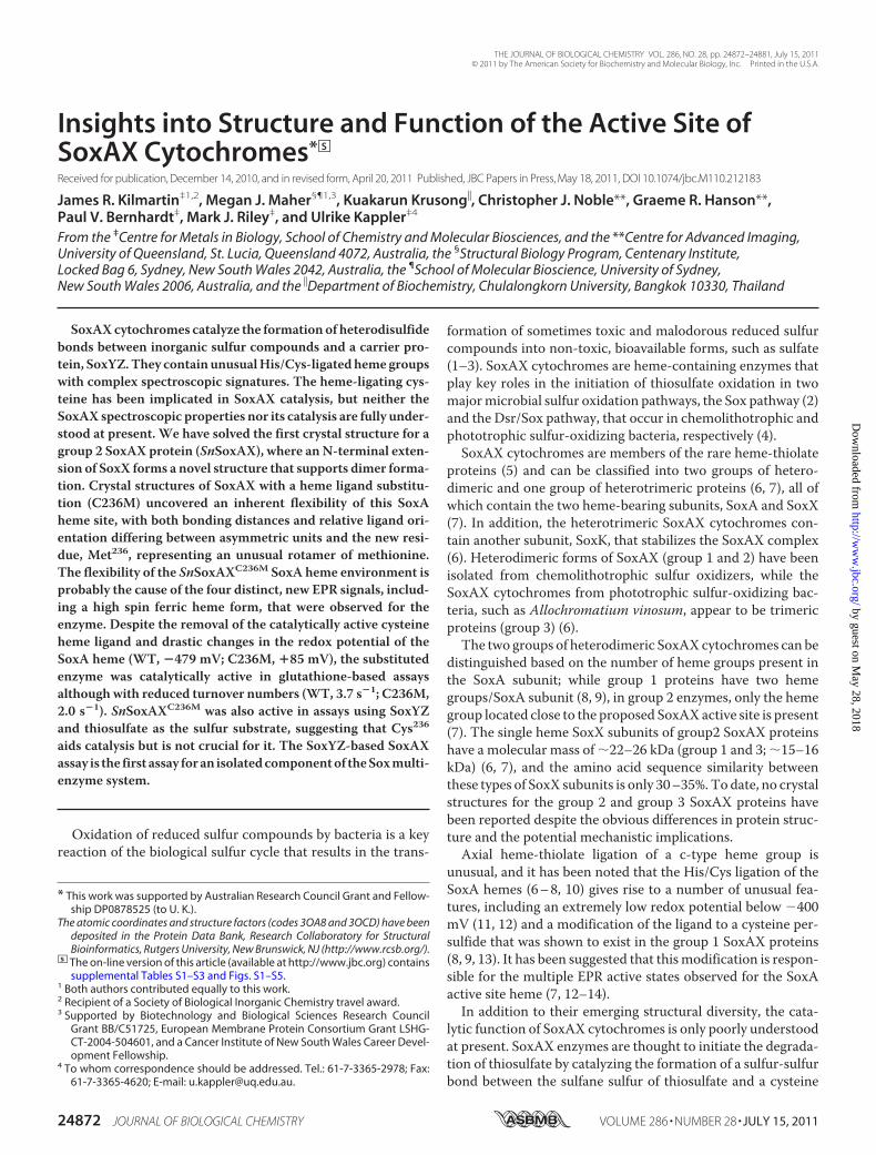
Insights into Structure and Function of the Active Site of SoxAX Cytochromes * □ S Received for publication, December 14, 2010, and in revised form, April 20, 2011 Published, JBC Papers in Press, May 18, 2011, DOI 10.1074/jbc.M110.212183 James R. Kilmartin ‡1,2 , Megan J. Maher §¶1,3 , Kuakarun Krusong , Christopher J. Noble**, Graeme R. Hanson**, Paul V. Bernhardt ‡ , Mark J. Riley ‡ , and Ulrike Kappler ‡4 From the ‡ Centre for Metals in Biology, School of Chemistry and Molecular Biosciences, and the **Centre for Advanced Imaging, University of Queensland, St. Lucia, Queensland 4072, Australia, the § Structural Biology Program, Centenary Institute, Locked Bag 6, Sydney, New South Wales 2042, Australia, the ¶ School of Molecular Bioscience, University of Sydney, New South Wales 2006, Australia, and the Department of Biochemistry, Chulalongkorn University, Bangkok 10330, Thailand SoxAX cytochromes catalyze the formation of heterodisulfide bonds between inorganic sulfur compounds and a carrier pro- tein, SoxYZ. They contain unusual His/Cys-ligated heme groups with complex spectroscopic signatures. The heme-ligating cys- teine has been implicated in SoxAX catalysis, but neither the SoxAX spectroscopic properties nor its catalysis are fully under- stood at present. We have solved the first crystal structure for a group 2 SoxAX protein (SnSoxAX), where an N-terminal exten- sion of SoxX forms a novel structure that supports dimer forma- tion. Crystal structures of SoxAX with a heme ligand substitu- tion (C236M) uncovered an inherent flexibility of this SoxA heme site, with both bonding distances and relative ligand ori- entation differing between asymmetric units and the new resi- due, Met 236 , representing an unusual rotamer of methionine. The flexibility of the SnSoxAX C236M SoxA heme environment is probably the cause of the four distinct, new EPR signals, includ- ing a high spin ferric heme form, that were observed for the enzyme. Despite the removal of the catalytically active cysteine heme ligand and drastic changes in the redox potential of the SoxA heme (WT, 479 mV; C236M, 85 mV), the substituted enzyme was catalytically active in glutathione-based assays although with reduced turnover numbers (WT, 3.7 s 1 ; C236M, 2.0 s 1 ). SnSoxAX C236M was also active in assays using SoxYZ and thiosulfate as the sulfur substrate, suggesting that Cys 236 aids catalysis but is not crucial for it. The SoxYZ-based SoxAX assay is the first assay for an isolated component of the Sox multi- enzyme system. Oxidation of reduced sulfur compounds by bacteria is a key reaction of the biological sulfur cycle that results in the trans- formation of sometimes toxic and malodorous reduced sulfur compounds into non-toxic, bioavailable forms, such as sulfate (1–3). SoxAX cytochromes are heme-containing enzymes that play key roles in the initiation of thiosulfate oxidation in two major microbial sulfur oxidation pathways, the Sox pathway (2) and the Dsr/Sox pathway, that occur in chemolithotrophic and phototrophic sulfur-oxidizing bacteria, respectively (4). SoxAX cytochromes are members of the rare heme-thiolate proteins (5) and can be classified into two groups of hetero- dimeric and one group of heterotrimeric proteins (6, 7), all of which contain the two heme-bearing subunits, SoxA and SoxX (7). In addition, the heterotrimeric SoxAX cytochromes con- tain another subunit, SoxK, that stabilizes the SoxAX complex (6). Heterodimeric forms of SoxAX (group 1 and 2) have been isolated from chemolithotrophic sulfur oxidizers, while the SoxAX cytochromes from phototrophic sulfur-oxidizing bac- teria, such as Allochromatium vinosum, appear to be trimeric proteins (group 3) (6). The two groups of heterodimeric SoxAX cytochromes can be distinguished based on the number of heme groups present in the SoxA subunit; while group 1 proteins have two heme groups/SoxA subunit (8, 9), in group 2 enzymes, only the heme group located close to the proposed SoxAX active site is present (7). The single heme SoxX subunits of group2 SoxAX proteins have a molecular mass of 22–26 kDa (group 1 and 3; 15–16 kDa) (6, 7), and the amino acid sequence similarity between these types of SoxX subunits is only 30 –35%. To date, no crystal structures for the group 2 and group 3 SoxAX proteins have been reported despite the obvious differences in protein struc- ture and the potential mechanistic implications. Axial heme-thiolate ligation of a c-type heme group is unusual, and it has been noted that the His/Cys ligation of the SoxA hemes (6 – 8, 10) gives rise to a number of unusual fea- tures, including an extremely low redox potential below 400 mV (11, 12) and a modification of the ligand to a cysteine per- sulfide that was shown to exist in the group 1 SoxAX proteins (8, 9, 13). It has been suggested that this modification is respon- sible for the multiple EPR active states observed for the SoxA active site heme (7, 12–14). In addition to their emerging structural diversity, the cata- lytic function of SoxAX cytochromes is only poorly understood at present. SoxAX enzymes are thought to initiate the degrada- tion of thiosulfate by catalyzing the formation of a sulfur-sulfur bond between the sulfane sulfur of thiosulfate and a cysteine * This work was supported by Australian Research Council Grant and Fellow- ship DP0878525 (to U. K.). The atomic coordinates and structure factors (codes 3OA8 and 3OCD) have been deposited in the Protein Data Bank, Research Collaboratory for Structural Bioinformatics, Rutgers University, New Brunswick, NJ (http://www.rcsb.org/). □ S The on-line version of this article (available at http://www.jbc.org) contains supplemental Tables S1–S3 and Figs. S1–S5. 1 Both authors contributed equally to this work. 2 Recipient of a Society of Biological Inorganic Chemistry travel award. 3 Supported by Biotechnology and Biological Sciences Research Council Grant BB/C51725, European Membrane Protein Consortium Grant LSHG- CT-2004-504601, and a Cancer Institute of New South Wales Career Devel- opment Fellowship. 4 To whom correspondence should be addressed. Tel.: 61-7-3365-2978; Fax: 61-7-3365-4620; E-mail: [email protected]. THE JOURNAL OF BIOLOGICAL CHEMISTRY VOL. 286, NO. 28, pp. 24872–24881, July 15, 2011 © 2011 by The American Society for Biochemistry and Molecular Biology, Inc. Printed in the U.S.A. 24872 JOURNAL OF BIOLOGICAL CHEMISTRY VOLUME 286 • NUMBER 28 • JULY 15, 2011 by guest on May 28, 2018 http://www.jbc.org/ Downloaded from
Transcript of InsightsintoStructureandFunctionoftheActiveSiteof … · · 2011-11-10phototrophic...
Insights into Structure and Function of the Active Site ofSoxAX Cytochromes*□S
Received for publication, December 14, 2010, and in revised form, April 20, 2011 Published, JBC Papers in Press, May 18, 2011, DOI 10.1074/jbc.M110.212183
James R. Kilmartin‡1,2, Megan J. Maher§¶1,3, Kuakarun Krusong�, Christopher J. Noble**, Graeme R. Hanson**,Paul V. Bernhardt‡, Mark J. Riley‡, and Ulrike Kappler‡4
From the ‡Centre for Metals in Biology, School of Chemistry and Molecular Biosciences, and the **Centre for Advanced Imaging,University of Queensland, St. Lucia, Queensland 4072, Australia, the §Structural Biology Program, Centenary Institute,Locked Bag 6, Sydney, New South Wales 2042, Australia, the ¶School of Molecular Bioscience, University of Sydney,New South Wales 2006, Australia, and the �Department of Biochemistry, Chulalongkorn University, Bangkok 10330, Thailand
SoxAXcytochromes catalyze the formation of heterodisulfidebonds between inorganic sulfur compounds and a carrier pro-tein, SoxYZ.They containunusualHis/Cys-ligatedhemegroupswith complex spectroscopic signatures. The heme-ligating cys-teine has been implicated in SoxAX catalysis, but neither theSoxAX spectroscopic properties nor its catalysis are fully under-stood at present. We have solved the first crystal structure for agroup 2 SoxAX protein (SnSoxAX), where anN-terminal exten-sion of SoxX forms a novel structure that supports dimer forma-tion. Crystal structures of SoxAX with a heme ligand substitu-tion (C236M) uncovered an inherent flexibility of this SoxAheme site, with both bonding distances and relative ligand ori-entation differing between asymmetric units and the new resi-due, Met236, representing an unusual rotamer of methionine.The flexibility of the SnSoxAXC236M SoxA heme environment isprobably the cause of the four distinct, new EPR signals, includ-ing a high spin ferric heme form, that were observed for theenzyme. Despite the removal of the catalytically active cysteineheme ligand and drastic changes in the redox potential of theSoxA heme (WT, �479 mV; C236M, �85 mV), the substitutedenzyme was catalytically active in glutathione-based assaysalthough with reduced turnover numbers (WT, 3.7 s�1; C236M,2.0 s�1). SnSoxAXC236M was also active in assays using SoxYZand thiosulfate as the sulfur substrate, suggesting that Cys236
aids catalysis but is not crucial for it. The SoxYZ-based SoxAXassay is the first assay foran isolatedcomponentof theSoxmulti-enzyme system.
Oxidation of reduced sulfur compounds by bacteria is a keyreaction of the biological sulfur cycle that results in the trans-
formation of sometimes toxic and malodorous reduced sulfurcompounds into non-toxic, bioavailable forms, such as sulfate(1–3). SoxAX cytochromes are heme-containing enzymes thatplay key roles in the initiation of thiosulfate oxidation in twomajormicrobial sulfur oxidation pathways, the Sox pathway (2)and the Dsr/Sox pathway, that occur in chemolithotrophic andphototrophic sulfur-oxidizing bacteria, respectively (4).SoxAX cytochromes are members of the rare heme-thiolate
proteins (5) and can be classified into two groups of hetero-dimeric and one group of heterotrimeric proteins (6, 7), all ofwhich contain the two heme-bearing subunits, SoxA and SoxX(7). In addition, the heterotrimeric SoxAX cytochromes con-tain another subunit, SoxK, that stabilizes the SoxAX complex(6). Heterodimeric forms of SoxAX (group 1 and 2) have beenisolated from chemolithotrophic sulfur oxidizers, while theSoxAX cytochromes from phototrophic sulfur-oxidizing bac-teria, such as Allochromatium vinosum, appear to be trimericproteins (group 3) (6).The two groups of heterodimeric SoxAXcytochromes can be
distinguished based on the number of heme groups present inthe SoxA subunit; while group 1 proteins have two hemegroups/SoxA subunit (8, 9), in group 2 enzymes, only the hemegroup located close to the proposed SoxAXactive site is present(7). The single heme SoxX subunits of group2 SoxAX proteinshave a molecular mass of �22–26 kDa (group 1 and 3; �15–16kDa) (6, 7), and the amino acid sequence similarity betweenthese types of SoxX subunits is only 30–35%.To date, no crystalstructures for the group 2 and group 3 SoxAX proteins havebeen reported despite the obvious differences in protein struc-ture and the potential mechanistic implications.Axial heme-thiolate ligation of a c-type heme group is
unusual, and it has been noted that the His/Cys ligation of theSoxA hemes (6–8, 10) gives rise to a number of unusual fea-tures, including an extremely low redox potential below �400mV (11, 12) and a modification of the ligand to a cysteine per-sulfide that was shown to exist in the group 1 SoxAX proteins(8, 9, 13). It has been suggested that thismodification is respon-sible for the multiple EPR active states observed for the SoxAactive site heme (7, 12–14).In addition to their emerging structural diversity, the cata-
lytic function of SoxAX cytochromes is only poorly understoodat present. SoxAX enzymes are thought to initiate the degrada-tion of thiosulfate by catalyzing the formation of a sulfur-sulfurbond between the sulfane sulfur of thiosulfate and a cysteine
* This work was supported by Australian Research Council Grant and Fellow-ship DP0878525 (to U. K.).
The atomic coordinates and structure factors (codes 3OA8 and 3OCD) have beendeposited in the Protein Data Bank, Research Collaboratory for StructuralBioinformatics, Rutgers University, New Brunswick, NJ (http://www.rcsb.org/).
□S The on-line version of this article (available at http://www.jbc.org) containssupplemental Tables S1–S3 and Figs. S1–S5.
1 Both authors contributed equally to this work.2 Recipient of a Society of Biological Inorganic Chemistry travel award.3 Supported by Biotechnology and Biological Sciences Research Council
Grant BB/C51725, European Membrane Protein Consortium Grant LSHG-CT-2004-504601, and a Cancer Institute of New South Wales Career Devel-opment Fellowship.
4 To whom correspondence should be addressed. Tel.: 61-7-3365-2978; Fax:61-7-3365-4620; E-mail: [email protected].
THE JOURNAL OF BIOLOGICAL CHEMISTRY VOL. 286, NO. 28, pp. 24872–24881, July 15, 2011© 2011 by The American Society for Biochemistry and Molecular Biology, Inc. Printed in the U.S.A.
24872 JOURNAL OF BIOLOGICAL CHEMISTRY VOLUME 286 • NUMBER 28 • JULY 15, 2011
by guest on May 28, 2018
http://ww
w.jbc.org/
Dow
nloaded from
residue present on the SoxYZ carrier protein (Reaction 1) (2, 8,12).
SoxZY-SH � S-SO32� � 2 ferricytochrome c
3 SoxZY-S-S-SO3� � 2 ferrocytochrome c
REACTION 1
A sulfur transferase-like mechanism has been proposed forSoxAX, where thiosulfate becomes bound to the heme-ligatingcysteine before being transferred to SoxYZ (8). However, thismechanismdoes not explain how two electrons liberated by theformation of a disulfide bond can be even temporarily stored inthe SoxAX protein, given that the SoxA heme is unlikely toaccept electrons under physiological conditions due to its lowredox potential and there is no evidence of radical formation.Recently, the group 2 SoxAX protein from Starkeya novella(SnSoxAX)5 has been shown to bind 1 eq of copper/enzymemolecule (12), and line broadening observed for the SoxA EPRsignature suggests that the copper is located close to the SoxAheme in the active site (12). Copper-loaded SoxAX showedenhanced activity in an in vitro assay that uses glutathione asthe sulfur substrate and cytochrome c as an electron acceptor,leading to the formation of oxidized glutathione (12). The pres-ence of an additional, non-heme redox center can explain howSoxAX can store the two electrons that are released during itsreaction (12); however, it does not explain the role of the SoxAheme cysteine ligand in catalysis or how it could become mod-ified to a persulfide form.In this paper, we have explored this issue by investigating the
structural, physical, and kinetic changes in SoxAX propertiesassociatedwith a substitution of the heme-ligating cysteine res-idue. As part of this work, the first crystal structure for both thewild type and substituted group2 SoxAX protein were solvedand provide unique insights into the structural changes under-lying the catalytic and spectroscopic properties of SoxAXcytochromes.
EXPERIMENTAL PROCEDURES
Bacterial Strains and Growth Conditions—Escherichia colistrains S17-1 (15) andDH5� (Invitrogen) were routinely grownon liquid or solid LB medium at 37 °C (16); for Rhodobactercapsulatus strains, TYS (17), or RCVmedium (18) and incuba-tion at 30 °C were used. R. capsulatus was grown either anaer-obically under phototrophic conditions or aerobically in thedark using shake flasks.Where appropriate,mediawere supple-mented with antibiotics (given in �g/ml; numbers in parenthe-ses refer to R. capsulatus): ampicillin 100 (�), tetracycline 10(1), and gentamicin � (4).Expression and Purification of Recombinant Proteins—SoxAX
proteins from S. novella were expressed in R. capsulatus andpurified as described (14); SoxYZ from S. novellawas expressedin E. coli and purified as described (12).
Generation of a Site-directed Mutation in SoxAX—A muta-tion in the heme-ligating cysteine, Cys236, was created usingprimers SOXAC236MF (CAC CGC ATG TGG GAC ATGTAC CGC CAG ATG CGC) and SOXAC236MR (GCG CATCTG GCG GTA CAT GTC CCA CAT GCG GTG). Mutagen-esis was performed using the pDorex-SoxAX plasmid (14) asthe template essentially as described (19). Inserts carrying themutationwere identified by sequencing followed by subcloninginto pRK415 and transfer into R. capsulatus as in Ref. 14.Protein Characterization Methods—SDS-PAGE was per-
formed according to the method of Ref. 20; in-gel heme stainsused the method of Ref. 21. Protein concentrations were deter-mined using either the BCA-1 kit (Sigma-Aldrich) or the2D-Quant kit (GE Healthcare). Mass fingerprints of proteinsseparated on SDS-polyacrylamide gels were prepared as in Ref.7 and analyzed using a VoyagerSTR MALDI-TOF mass spec-trometer (Applied Biosystems). Electrospray mass spectrome-try was performed on a Q-Star mass spectrometer (AppliedBiosystems) essentially as in Ref. 7. Molecular mass determina-tion of native, purified proteins by multiangle laser light scat-tering was carried out as in Refs. 22 and 23. A sample volume of50 �l of an �200 �M protein solution was used per injection,and experimental errors are reported as S.D. of the molecularmass estimate. Copper loading of purified SoxAX was carriedout as in Ref. 12; metal content analysis of purified proteins wascarried out using ICP-MS at the ENTOX Centre (University ofQueensland); the heme content of recombinant SoxAX pro-teins was determined using alkaline hemochrome spectra as inRef. 24.Mass Spectra of whole proteins and tryptic digests werecollected at the School of Chemistry and Molecular BiologyProteomics facility. Modification of SoxYZ by sulfur com-pounds was tested using 30 �M protein samples and a 5 mM
concentration of each sulfur compound, and the experimentswere conducted at room temperature and 37 °C for 1 h beforeanalysis using mass spectrometry. Thiol content of treatedpreparations was estimated using the method of Ref. 25.Heme redox potentials were determined by optical redox
potentiometry essentially as in Ref. 12. All potentials have beencorrected relative to NHE. The experiments employed 5 �M
enzyme solutions in 20 mM Tris buffer (pH 8.0), and the solu-tion potentials were stabilized using high potential iron com-plexes as mediators (26). Changes in the electronic absorptionspectrum were monitored continuously with an Ocean OpticsUSB4000 fiber optic spectrometer. A global analysis of thespectra was performed with the program SPECFIT/32 (Dr.R. A. Binstead, Spectrum Software Associates, Marlborough,MA) for a three-component model (Fe(III)/Fe(III), Fe(III)/Fe(II), and Fe(II)/Fe(II)) comprising two consecutive singleelectron redox reactions.Assays determining the reduction of SoxAX by SoxYZ (12)
used 10 �M SoxAX in 20 mM Tris-Cl, pH 8.0, and a 6:1 ratio ofpurified SoxYZ to SoxAX.All SoxYZ sampleswere stored in thepresence of reductant (DTT; 5 mM), which was removed fromthe sample immediately prior to use by passing it through aPD-10 column (GE Healthcare). Electronic absorption spectra(300–700 nm) of the reaction mixture were recorded every 2min using a Cary50 spectrophotometer (Varian). Kinetic assayscontained 20�M horse heart cytochrome c (Sigma-Aldrich cat-
5 The abbreviations used are: SnSoxAX, S. novella SoxAX protein; SnSoxA,S. novella SoxA protein; SnSoxX, S. novella SoxX protein; RsSoxAX, R. sulfi-dophilum SoxAX protein; PpSoxAX, P. pantotrophus SoxAX protein; CT,charge transfer; mT, millitesla(s); MCD, magnetic circular dichroism.
Structure and Function of SoxAX Cytochromes
JULY 15, 2011 • VOLUME 286 • NUMBER 28 JOURNAL OF BIOLOGICAL CHEMISTRY 24873
by guest on May 28, 2018
http://ww
w.jbc.org/
Dow
nloaded from
alog no. C7752), 20 mMMES, pH 6.0, or 20 mM Tris-Cl, pH 7.0,and reduced glutathione (final concentration 0.15–2 mM;standard assays used 1mM), from a stock solution titrated to pH7.0 as described (12). Assays using SoxYZ contained 20 mM
MES, pH 6.0, 300 �M thiosulfate, 38.5 �M cytochrome c, and4.85 �M reduced SoxYZ. Reduced SoxYZ refers to SoxYZ pro-teinwhere the cysteine is detectable by 5,5�-dithiobis(nitroben-zoic acid)-based thiol assays. In both cases, reduction of cyto-chrome c was monitored at 550 nm, and data were fitted to theMichaelis-Menten equation using SigmaPlot (Sysstat Inc.) orIGOR Pro (WaveMetrics Inc.).MCD Spectroscopy—MCD spectroscopy was carried out as
described (27). Samples contained 20 mM Tris-Cl, pH 8 (sam-ples for the IRwere prepared in deuterium oxide) and 60% (v/v)glycerol as a glassing agent. Protein concentrations of 25 and100 �M were used for the visible and infrared regions,respectively.EPR Spectroscopy—Continuous wave X-band EPR spectra
were recorded as in Ref. 12.Crystallization and Data Collection—Conditions for the
crystallization of SnSoxAX (32 mg/ml) were screened at 293 Kaccording to the sparsematrixmethod (28) using commerciallyavailable screens. Screens were set up in 96-well plates (F-bot-tom Microplates, Greiner) using a Mosquito nanoliter liquid-handling robot (TTP LabTech). Equal volumes of protein (100nl) and reservoir were mixed. Initial crystals were observed incondition 77 of the index screen (Hampton Research, 25% PEG3350, 0.2 M lithium sulfate, 0.1 M Tris-Cl, pH 8.5). Crystal opti-mization trialswere thenperformed in 24-well plates by varyingthe concentration of precipitant (PEG 3350, 24–29%) and thepH (0.1 M Tris-Cl, pH 8.3–8.9). In addition, an additive screen(Hampton Research) was carried out to improve the diffractionproperties of the SnSoxAX crystals. The best crystals weregrown in PEG3350 (25%), lithium sulfate (0.2 M), Tris-Cl (0.1 M,pH 8.5), and spermidine (0.01 M) at 25 °C (298 K). The crystalsreached maximum size in 3 weeks. Prior to x-ray data collec-tion, crystals were quickly transferred into mother liquor con-taining 30% glycerol and frozen in a cold nitrogen stream (100K). Data were collected using aMar 345 image plate detector ateither BM14 or ID29 at the European Synchrotron RadiationFacility or with copper K� X-rays from a Rigaku RU-200 rotat-ing anode generator focused using Osmic mirror optics. Alldatawere processed and scaledwithDENZOandSCALEPACK(HKL program suite) or with HKL2000. As prepared, SoxAXcontains �10–20% copper. Crystals of copper-loaded SoxAXproteinwere obtained but failed to diffract. Similarly, soaking ofSoxAX crystals in CuSO4 solution resulted in crystals withinsufficient diffraction properties.Structure Solution and Refinement—The structure of SoxAX
was solved bymultiple wavelength anomalous dispersion usingdata collected at the ironK-edge. Twodata setswere used in thestructure solution: a peak data set and a remote data set (col-lected at 1.739 and 0.954 Å, respectively). The peak data set wasused to determine the positions of 8 iron atoms/asymmetricunit using SHELXD (29). Phaseswere calculatedwith both peakand remote data sets using SHARP (30) to the resolution limitof the remote data (2.20Å). The automatic densitymodificationprotocol within autoSHARP (using DM and SOLOMON (31))
yielded an interpretable electron density map. Model buildingwas carried out automatically withARP/wARP (31, 32). Furthermodel building was carried out manually in COOT (33). Thisstructure was used to determine the positions of three SoxAXheterodimers in the asymmetric unit of a high resolution, nativedata set (1.77 Å, SnSoxAX in supplemental Table S1) bymolec-ular replacement with PHASER (34). All three copies of SoxAXin the P1 asymmetric unit are identical within experimentalerror (r.m.s. deviation of 0.3–0.6 Å for the superposition ofSoxA residues 46–274 and SoxX residues 40–208).Refinement of the native structure was carried out with
REFMAC5 (35), with manual adjustments incorporated inCOOT. Tight NCS restraints were maintained throughout therefinement process until the final cycle, where they wereremoved completely. The structure of the C236Mmutant pro-tein (SnSoxAXC236M; supplemental Table S1) was solved bymolecular replacement using the coordinates of the nativeSnSoxAX structure as a search model. Refinement and modelbuilding were carried out as for the native SnSoxAX structure,with the addition of twin refinement in REFMAC5. For allstructures, water molecules were incorporated into the modelsautomatically with ARP/wARP and later confirmed manuallyby inspection of electron density maps in COOT, with consid-eration of conservative hydrogen-bonding criteria. Residuenumbering for all structures is based on the sequence of theunprocessed protein (accession number Q7BQR6).
RESULTS
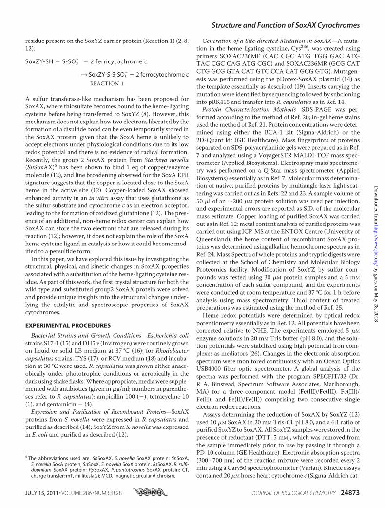
Crystal Structure of the S. novella SoxAX Protein—Three dif-ferent groups of SoxAX proteins are known to exist, and whilethe structure of two representatives of the group 1, trihemeSoxAX proteins from Rhodovulum sulfidophilum (RsSoxAX,Protein Data Bank code 2OZ1) and Paracoccus pantotrophus(PpSoxXA, Protein Data Bank code 2C1D) have been solvedpreviously (8, 9), we have solved the first structure for a group 2,diheme SoxAX protein. SoxAX from S. novella (SnSoxAX)crystallized in space group P1 (1.77 Å resolution) with threeheterodimers per asymmetric unit (supplemental Table S1).Similar to what has been described for the group 1 SoxAXstructures, the SnSoxA subunit (A46–A274) can be dividedinto two structurally similar domains, A46–A152 and A153–A274, (r.m.s. deviation of 1.86Å, superposition of 84 alignedC�atoms)which introduces a pseudo-2-fold symmetry to this sub-unit (Fig. 1 and supplemental Fig. S1). Although RsSoxA andPpSoxA are diheme proteins, the single heme prosthetic groupfound in SnSoxA is coordinated by HisA187 and residue A236(discussed below), while a disulfide bridge between CysA74 andCysA110 replaces the second heme group, thus confirming ear-lier observations using mass spectrometry (6, 7). Surface loopswith different relative conformations (residues A187–A207and A65–A86, respectively) border the SnSoxA heme groupand the disulfide bond. The heme loop has an open conforma-tion that “covers” the heme cofactor, while the disulfide loopreaches in toward the core of the molecule, filling the spacecreated by the absence of the heme group (Fig. 1B). In contrast,both the RsSoxA and PpSoxA proteins show open loop struc-tures about both heme-binding sites.
Structure and Function of SoxAX Cytochromes
24874 JOURNAL OF BIOLOGICAL CHEMISTRY VOLUME 286 • NUMBER 28 • JULY 15, 2011
by guest on May 28, 2018
http://ww
w.jbc.org/
Dow
nloaded from
The SnSoxAheme ligand, residueA236, has beenmodeled asan equal mixture of Cys and the post-translationally modifiedcysteine persulfide (supplemental Fig. S2) residues as a result ofcareful analysis of difference Fourier electron density maps fora variety of models and occupancy refinement with PHENIX(36). Using this refinement, the average coordination distancesfor the three SnSoxA molecules in the asymmetric unit are asfollows: HisA187 NE2-Fe � 2.1 Å; cysteine persulfide A236SD-Fe� 2.3Å;CysA236 SG-Fe� 2.5, with a bond length error of�0.15 Å.The SnSoxX Subunit—The SnSoxX structure (residues B29–
B208) can be partitioned into a tightly folded heme-bindingdomain (residues B96–B208) and an N-terminal extension(residues B29–B95) that is unique to group 2 SoxAX proteins.SnSoxX contains a single heme cofactor that is bound by aCXXCHmotif (residues B125–129) and is axially ligated by res-idues HisB129 and MetB187 (HisB129 NE2-Fe � 2.09 Å; MetB178SD-Fe � 2.36 Å).The core of the SnSoxX model (residues B96–B208) can be
superposed on the corresponding subunit structures of RsSoxX(Protein Data Bank code 1H33, 92 aligned residues, r.m.s. devi-ation of 1.6 Å) and PpSoxX (Protein Data Bank code 2C1D, 90
aligned residues, r.m.s. deviation of 1.5 Å) (Fig. 1 and supple-mental Fig. S1). However, the mature SnSoxX protein is 70residues longer than its homologues, and most of that differ-ence lies in the SnSoxX N-terminal extension (B29–B95) aswell as in some loop structures (e.g. RsSoxX B97–B119 andSnSoxXB109–B121) that are unique to each protein (Fig. 1 andsupplemental Fig. S1).The SnSoxX N-terminal extension (B29–B95) is tethered to
the heme-binding domain through a disulfide bridge (CysB64–CysB175) and three hydrogen-bonding interactions between theN-terminal extension and the main body of SnSoxX (ArgB54–GlyB111; AspB58–ArgB180; ThrB60–GluB83). The N-terminalextension shows no significant structural homology to domainsof proteins deposited within the PDB, and it is unlikely that itrepresents an independently folded domain. Because theN-ter-minal extension mediates a significant portion of the interac-tions between SnSoxX and SnSoxA, its structure may dependon these interactions.The SnSoxAX Dimer—The SnSoxAX heterodimer has
approximate dimensions of 70 � 35 � 35 Å (Fig. 1 and supple-mental Fig. S1), with a buried surface area of �2100 Å2 permonomer (19%) and a shape complementarity statistic of 0.75,in keeping with values from other known permanent heterodi-meric complexes (37). The interactions aremediatedmainly byan association of theN-terminal extension structure of SnSoxX(residues B29–B95) with a helix and two loops from theSnSoxA subunit (residues A158–A174, A174–A183, andA241–A250) as well as some loop structures (supplementalTable S2).In contrast, the RsSoxAX and PpSoxXA structures only have
buried surface areas of �1500 Å2 per monomer (�16% persubunit). The larger interface for SnSoxAX presumably trans-lates to greater stability of the heterodimer as a whole.The active site of SoxAXproteins is located at the interface of
the two subunits, where the two heme cofactors of SnSoxAXare located. The edge-to-edge separation of the heme cofactors(distance between thioether groups) is 5.9 Å (Fe-Fe distance, 19Å), and they have solvent-accessible areas of 86 and 113 Å2,respectively, which include the heme propionate groups (Fig.1). The thioether groups of the heme cofactors point towardeach other across the dimer interface; in fact, association ofSnSoxAX in the dimer reduces the solvent-accessible area ofthe SnSoxX heme from 144Å2 (SnSoxX subunit only) to 113Å2
(SnSoxAX dimer).The SnSoxAXC236M Cysteine Ligand Mutant—The cysteine
ligand to the SnSoxA heme has been proposed to play a crucialrole in catalysis by providing an intermediate binding site forsubstrate molecules, such as thiosulfate (8). Breakdown of thethiosulfate-SoxA complex without turnover has been postu-lated to lead to the persulfide modification of this cysteineobserved in all available crystal structures of SoxAXproteins (8,9). In order to investigate the importance of this residue forcatalysis, we have created a substitution of this cysteine,C236M, in the SnSoxAX protein. If this cysteine residue is a keycomponentintheSoxAXreaction, in theSnSoxAXC236M-substi-tuted protein, catalysis should be impaired, and the spectro-scopic signature of the protein should be less complex because
FIGURE 1. Structure of the SnSoxAX. A, overall structure of SnSoxAX. Green,SnSoxA; gray, SnSoxX; orange, heme groups. B, domain structure of SnSoxA.Turquoise, domain 1; gray, domain 2; orange, heme group; yellow, disulfidebond. C, structure of SnSoxX. Gray, heme-binding core; blue, N-terminalextension. D and E, electrostatic surface of the SnSoxAX dimer. Both the SoxA(D) and SoxX (E) hemes are solvent-exposed. E shows the SoxAX dimer rotatedby 180° around the horizontal (x) axis.
Structure and Function of SoxAX Cytochromes
JULY 15, 2011 • VOLUME 286 • NUMBER 28 JOURNAL OF BIOLOGICAL CHEMISTRY 24875
by guest on May 28, 2018
http://ww
w.jbc.org/
Dow
nloaded from
the substituted enzyme contains only two His/Met-ligatedheme groups.The substituted protein, SnSoxAXC236M, contained a full
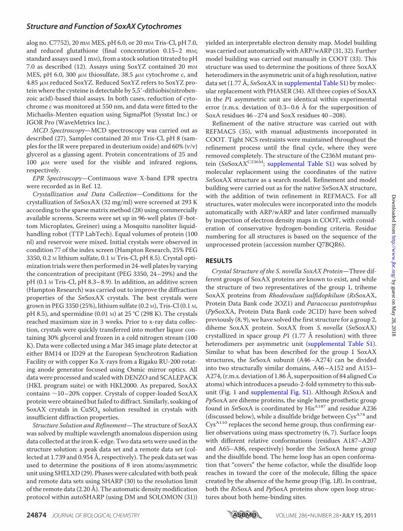
complement of redox cofactors (1.9 heme groups/molecule),and both subunits could be stained for heme-linked peroxidaseactivity (data not shown). SnSoxAXC236M was also capable ofbinding �1.2 eq of copper/molecule, which is comparable withwhat was observed for SnSoxAXWT (12).By a global analysis of the spectra obtained during optical
redox titrations of SnSoxAXC236M (supplemental Fig. S3), theredox potentials of the two hemes were determined to be E1 �162 � 12 mV and E2 � 85 � 15 mV, which is within the rangeexpected for His/Met-ligated heme groups (38). Because E1 israther close to the value previously determined for the SnSoxXheme group (12), we have tentatively assigned this potential tothe SnSoxX heme. The spectra of the diferric, ferric/ferrous,and diferrous SnSoxAXC236M were also obtained as a result ofthe global analysis (Fig. 2), with the intermediate ferric/ferrousSnSoxAXC236M displaying a Soret band (417 nm) with approx-imately half the intensity of the diferrous form. Lowering thesolution potentials of SnSoxAXC236M below �400 mV did notresult in any further changes to the electronic absorption spec-trum (data not shown).
Structural Implications of the SnSoxAXC236M Substitution—SnSoxAXC236M crystallized in space group P1, with two het-erodimers per asymmetric unit, and the refinement convergedwith residuals r � 0.251 and Rfree � 0.314 for all data to 2.25 Åresolution (supplemental Table S1). In the SnSoxAXC236M
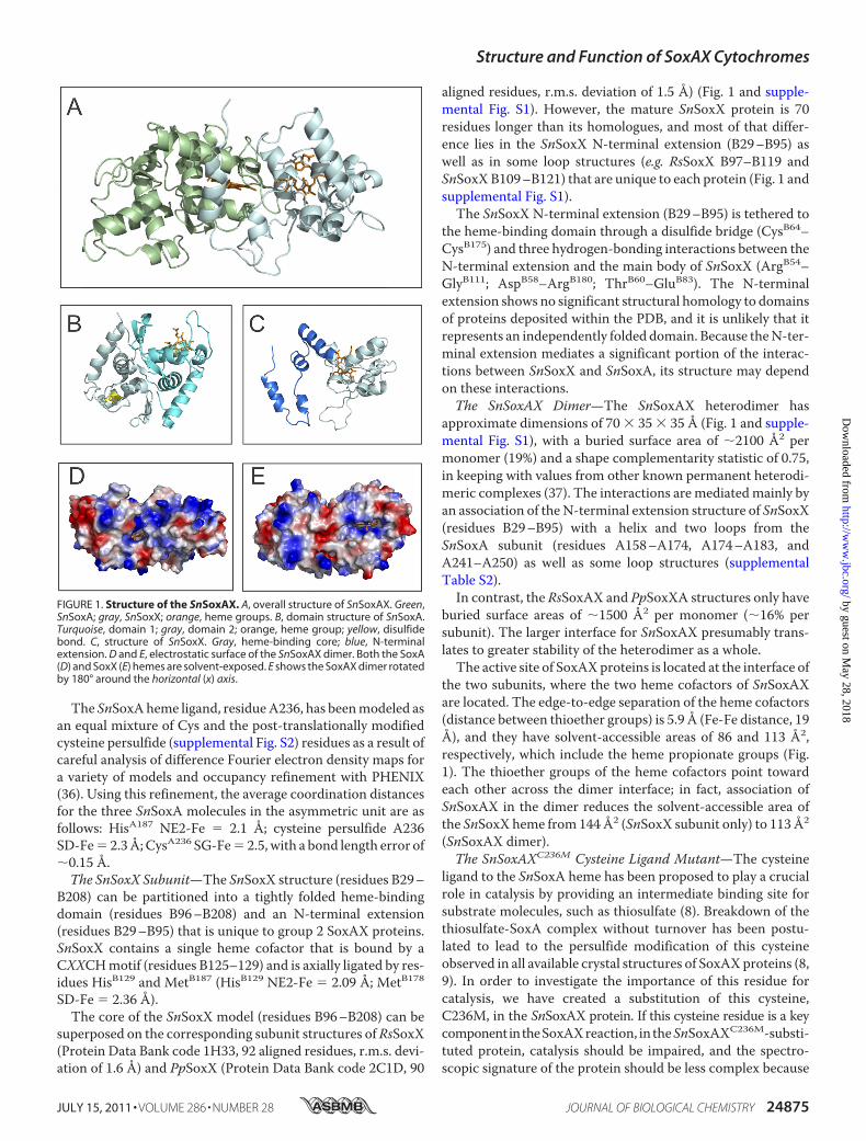
SoxA subunit (A46–A274), the heme cofactor is coordinatedby residues HisA187 and MetA236, while heme coordination forthe SoxX subunit (B30–B208) is unchanged.Superposition of the SnSoxAXC236M and SnSoxAXWT struc-
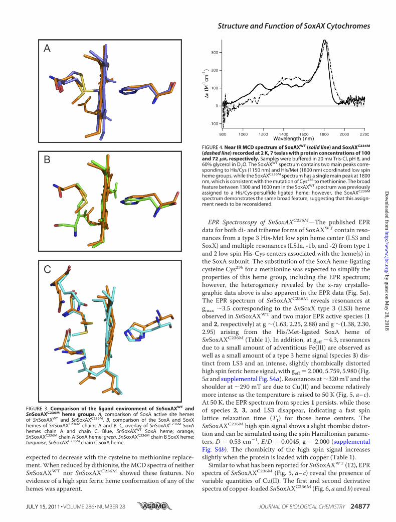
tures (chains A and B only, r.m.s. deviation of 0.75 Å for 414 C�positions) shows that structural differences are confined to theN andC termini of the respectivemolecules, and only negligibleadjustment of the surrounding polypeptide structure hasaccompanied the substitution of residue A236. In fact, thestructure of Met236 is clearly constrained by the surroundingpolypeptide, and analysis of the conformation of the Met236ligand (33) revealed it to be a relatively uncommon rotamer,occurring in only 3% of protein crystal structures. Further com-parisons revealed that although theHis ligands andheme cofac-tors of the two SnSoxAXC236M heme groups overlay wellbetween the two subunits (Fig. 3), the polypeptide structuresharboring theMet ligands are completely different. This is alsoreflected in the coordination distances of the heme ligands. Forthe SnSoxAXC236M SoxA subunit, these are HisA187 NE2-Fe �2.4 Å, MetA236 SD-Fe � 2.56 Å, HisC187 NE2-Fe � 2.1 Å, andMetC236 SD-Fe � 2.9 Å, while SoxX subunit distances ofHisB129 NE2-Fe � 2.1 Å, MetB178 SD-Fe � 2.4 Å, HisD129 NE2-Fe� 2.1Å, andMetD178 SD-Fe� 2.6Åwith a bond length errorof �0.2 Å were observed. These data clearly show that differ-ences between the general structures of the heme environmentsin the two heterodimers in the SnSoxAXC236M asymmetric unitexist.In addition to revealing the differing Met236 to iron bond
lengths, the superposition of the SoxAC236M heme groups(chains A and C) shows that the relative orientations of the HisandMet ligands differ (Fig. 3). In chain A, the planes of the twoligands are close to perpendicular, while in chain C they areparallel. Orientations of the side chains of Arg232 and Gln239and the heme propionate groups also differ in chains A and C,which further suggests that the heme environment in the SoxAsubunit of SnSoxAXC236M has some inherent flexibility.MCD Spectroscopy—The UV-visible MCD spectra of
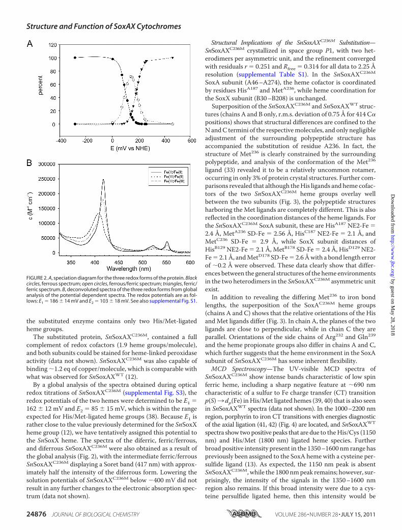
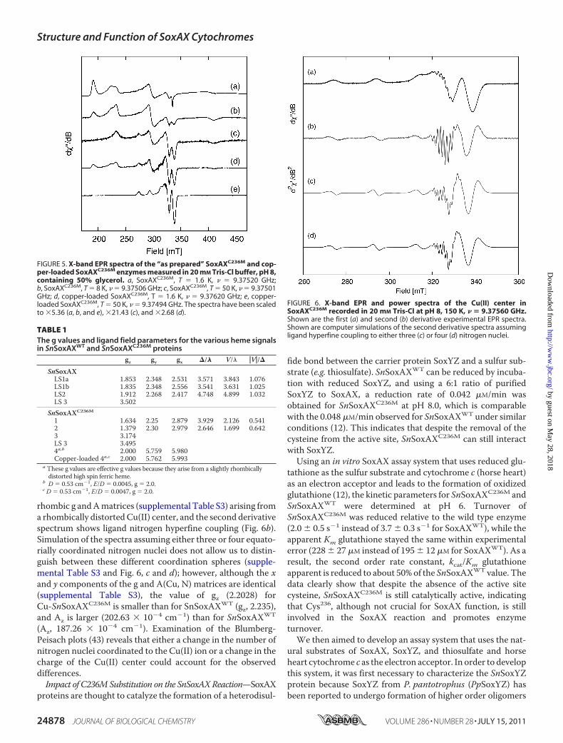
SnSoxAXC236M show intense bands characteristic of low spinferric heme, including a sharp negative feature at �690 nmcharacteristic of a sulfur to Fe charge transfer (CT) transitionp(S)3 dp(Fe) inHis/Met ligated hemes (39, 40) that is also seenin SnSoxAXWT spectra (data not shown). In the 1000–2200 nmregion, porphyrin to iron CT transitions with energies diagnosticof the axial ligation (41, 42) (Fig. 4) are located, and SnSoxAXWT
spectra show twopositive peaks that are due to theHis/Cys (1150nm) and His/Met (1800 nm) ligated heme species. Furtherbroad positive intensity present in the 1350–1600 nm range haspreviously been assigned to the SoxA hemewith a cysteine per-sulfide ligand (13). As expected, the 1150 nm peak is absentSnSoxAXC236M,while the 1800 nmpeak remains; however, sur-prisingly, the intensity of the signals in the 1350–1600 nmregion also remains. If this broad intensity were due to a cys-teine persulfide ligated heme, then this intensity would be
FIGURE 2. A, speciation diagram for the three redox forms of the protein. Blackcircles, ferrous spectrum; open circles, ferrous/ferric spectrum; triangles, ferric/ferric spectrum. B, deconvoluted spectra of the three redox forms from globalanalysis of the potential dependent spectra. The redox potentials are as fol-lows: E1 � 186 � 14 mV and E2 � 103 � 18 mV. See also supplemental Fig. S1.
Structure and Function of SoxAX Cytochromes
24876 JOURNAL OF BIOLOGICAL CHEMISTRY VOLUME 286 • NUMBER 28 • JULY 15, 2011
by guest on May 28, 2018
http://ww
w.jbc.org/
Dow
nloaded from
expected to decrease with the cysteine to methionine replace-ment.When reduced by dithionite, theMCD spectra of neitherSnSoxAXWT nor SnSoxAXC236M showed these features. Noevidence of a high spin ferric heme conformation of any of thehemes was apparent.
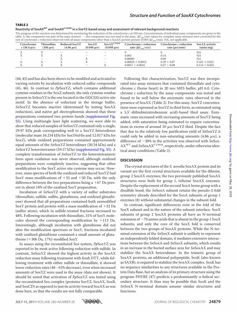
EPR Spectroscopy of SnSoxAXC236M—The published EPRdata for both di- and triheme forms of SoxAXWT contain reso-nances from a type 3 His-Met low spin heme center (LS3 andSoxX) and multiple resonances (LS1a, -1b, and -2) from type 1and 2 low spin His-Cys centers associated with the heme(s) inthe SoxA subunit. The substitution of the SoxA heme-ligatingcysteine Cys236 for a methionine was expected to simplify theproperties of this heme group, including the EPR spectrum;however, the heterogeneity revealed by the x-ray crystallo-graphic data above is also apparent in the EPR data (Fig. 5a).The EPR spectrum of SnSoxAXC236M reveals resonances atgmax �3.5 corresponding to the SnSoxX type 3 (LS3) hemeobserved in SnSoxAXWT and two major EPR active species (1and 2, respectively) at g �(1.63, 2.25, 2.88) and g �(1.38, 2.30,2.95) arising from the His/Met-ligated SoxA heme ofSnSoxAXC236M (Table 1). In addition, at geff �4.3, resonancesdue to a small amount of adventitious Fe(III) are observed aswell as a small amount of a type 3 heme signal (species 3) dis-tinct from LS3 and an intense, slightly rhombically distortedhigh spin ferric heme signal, with geff � 2.000, 5.759, 5.980 (Fig.5a and supplemental Fig. S4a). Resonances at�320mTand theshoulder at �290 mT are due to Cu(II) and become relativelymore intense as the temperature is raised to 50 K (Fig. 5, a–c).At 50 K, the EPR spectrum from species 1 persists, while thoseof species 2, 3, and LS3 disappear, indicating a fast spinlattice relaxation time (T1) for those heme centers. TheSnSoxAXC236M high spin signal shows a slight rhombic distor-tion and can be simulated using the spin Hamiltonian parame-ters, D � 0.53 cm�1, E/D � 0.0045, g � 2.000 (supplementalFig. S4b). The rhombicity of the high spin signal increasesslightly when the protein is loaded with copper (Table 1).Similar to what has been reported for SnSoxAXWT (12), EPR
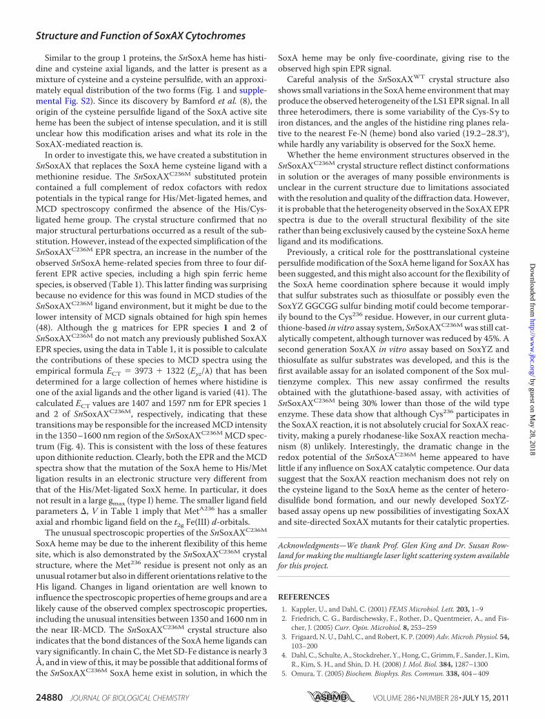
spectra of SnSoxAXC236M (Fig. 5, a–c) reveal the presence ofvariable quantities of Cu(II). The first and second derivativespectra of copper-loaded SnSoxAXC236M (Fig. 6, a and b) reveal
FIGURE 3. Comparison of the ligand environment of SnSoxAXWT andSnSoxAXC236M heme groups. A, comparison of SoxA active site hemesof SnSoxAXWT and SnSoxAXC236M. B, comparison of the SoxA and SoxXhemes of SnSoxAXC236M chains A and B. C, overlay of SnSoxAXC236M SoxAhemes chain A and chain C. Blue, SnSoxAXWT SoxA heme; orange,SnSoxAXC236M chain A SoxA heme; green, SnSoxAXC236M chain B SoxX heme;turquoise, SnSoxAXC236M chain C SoxA heme.
FIGURE 4. Near IR MCD spectrum of SoxAXWT (solid line) and SoxAXC236M
(dashed line) recorded at 2 K, 7 teslas with protein concentrations of 100and 72 �M, respectively. Samples were buffered in 20 mM Tris-Cl, pH 8, and60% glycerol in D2O. The SoxAXWT spectrum contains two main peaks corre-sponding to His/Cys (1150 nm) and His/Met (1800 nm) coordinated low spinheme groups, while the SoxAXC236M spectrum has a single main peak at 1800nm, which is consistent with the mutation of Cys236 to methionine. The broadfeature between 1300 and 1600 nm in the SoxAXWT spectrum was previouslyassigned to a His/Cys-persulfide ligated heme; however, the SoxAXC236M
spectrum demonstrates the same broad feature, suggesting that this assign-ment needs to be reconsidered.
Structure and Function of SoxAX Cytochromes
JULY 15, 2011 • VOLUME 286 • NUMBER 28 JOURNAL OF BIOLOGICAL CHEMISTRY 24877
by guest on May 28, 2018
http://ww
w.jbc.org/
Dow
nloaded from
rhombic g andAmatrices (supplemental Table S3) arising froma rhombically distortedCu(II) center, and the second derivativespectrum shows ligand nitrogen hyperfine coupling (Fig. 6b).Simulation of the spectra assuming either three or four equato-rially coordinated nitrogen nuclei does not allow us to distin-guish between these different coordination spheres (supple-mental Table S3 and Fig. 6, c and d); however, although the xand y components of the g and A(Cu, N) matrices are identical(supplemental Table S3), the value of gz (2.2028) forCu-SnSoxAXC236M is smaller than for SnSoxAXWT (gz, 2.235),and Az is larger (202.63 � 10�4 cm�1) than for SnSoxAXWT
(Az, 187.26 � 10�4 cm�1). Examination of the Blumberg-Peisach plots (43) reveals that either a change in the number ofnitrogen nuclei coordinated to the Cu(II) ion or a change in thecharge of the Cu(II) center could account for the observeddifferences.Impact of C236MSubstitution on the SnSoxAXReaction—SoxAX
proteins are thought to catalyze the formation of a heterodisul-
fide bond between the carrier protein SoxYZ and a sulfur sub-strate (e.g. thiosulfate). SnSoxAXWT can be reduced by incuba-tion with reduced SoxYZ, and using a 6:1 ratio of purifiedSoxYZ to SoxAX, a reduction rate of 0.042 �M/min wasobtained for SnSoxAXC236M at pH 8.0, which is comparablewith the 0.048�M/min observed for SnSoxAXWT under similarconditions (12). This indicates that despite the removal of thecysteine from the active site, SnSoxAXC236M can still interactwith SoxYZ.Using an in vitro SoxAX assay system that uses reduced glu-
tathione as the sulfur substrate and cytochrome c (horse heart)as an electron acceptor and leads to the formation of oxidizedglutathione (12), the kinetic parameters for SnSoxAXC236M andSnSoxAXWT were determined at pH 6. Turnover ofSnSoxAXC236M was reduced relative to the wild type enzyme(2.0 � 0.5 s�1 instead of 3.7 � 0.3 s�1 for SoxAXWT), while theapparent Km glutathione stayed the same within experimentalerror (228� 27 �M instead of 195� 12 �M for SoxAXWT). As aresult, the second order rate constant, kcat/Km glutathioneapparent is reduced to about 50%of the SnSoxAXWT value. Thedata clearly show that despite the absence of the active sitecysteine, SnSoxAXC236M is still catalytically active, indicatingthat Cys236, although not crucial for SoxAX function, is stillinvolved in the SoxAX reaction and promotes enzymeturnover.We then aimed to develop an assay system that uses the nat-
ural substrates of SoxAX, SoxYZ, and thiosulfate and horseheart cytochrome c as the electron acceptor. In order to developthis system, it was first necessary to characterize the SnSoxYZprotein because SoxYZ from P. pantotrophus (PpSoxYZ) hasbeen reported to undergo formation of higher order oligomers
FIGURE 5. X-band EPR spectra of the “as prepared” SoxAXC236M and cop-per-loaded SoxAXC236M enzymes measured in 20 mM Tris-Cl buffer, pH 8,containing 50% glycerol. a, SoxAXC236M, T � 1.6 K, � � 9.37520 GHz;b, SoxAXC236M, T � 8 K, � � 9.37506 GHz; c, SoxAXC236M, T � 50 K, � � 9.37501GHz; d, copper-loaded SoxAXC236M, T � 1.6 K, � � 9.37620 GHz; e, copper-loaded SoxAXC236M, T � 50 K, � � 9.37494 GHz. The spectra have been scaledto �5.36 (a, b, and e), �21.43 (c), and �2.68 (d).
TABLE 1The g values and ligand field parameters for the various heme signalsin SnSoxAXWT and SnSoxAXC236M proteins
gz gy gx �/� V/� �V�/�
SnSoxAXLS1a 1.853 2.348 2.531 3.571 3.843 1.076LS1b 1.835 2.348 2.556 3.541 3.631 1.025LS2 1.912 2.268 2.417 4.748 4.899 1.032LS 3 3.502
SnSoxAXC236M
1 1.634 2.25 2.879 3.929 2.126 0.5412 1.379 2.30 2.979 2.646 1.699 0.6423 3.174LS 3 3.4954a,b 2.000 5.759 5.980Copper-loaded 4a,c 2.000 5.762 5.993
a These g values are effective g values because they arise from a slightly rhombicallydistorted high spin ferric heme.
b D � 0.53 cm�1, E/D � 0.0045, g � 2.0.c D � 0.53 cm�1, E/D � 0.0047, g � 2.0.
FIGURE 6. X-band EPR and power spectra of the Cu(II) center inSoxAXC236M recorded in 20 mM Tris-Cl at pH 8, 150 K, � � 9.37560 GHz.Shown are the first (a) and second (b) derivative experimental EPR spectra.Shown are computer simulations of the second derivative spectra assumingligand hyperfine coupling to either three (c) or four (d) nitrogen nuclei.
Structure and Function of SoxAX Cytochromes
24878 JOURNAL OF BIOLOGICAL CHEMISTRY VOLUME 286 • NUMBER 28 • JULY 15, 2011
by guest on May 28, 2018
http://ww
w.jbc.org/
Dow
nloaded from
(44, 45) and has also been shown to bemodified and activated tovarying extents by incubation with reduced sulfur compounds(45, 46). In contrast to PpSoxYZ, which contains additionalcysteine residues in the SoxZ subunit, the only cysteine residuepresent in SnSoxYZ is the one located in theGGCGGactive sitemotif. In the absence of reductant in the storage buffer,SnSoxYZ becomes inactive (determined by testing SoxAXreduction), and native gel electrophoresis showed that thesepreparations contained two protein bands (supplemental Fig.S5). Using multiangle laser light scattering, we were able toshow that reduced samples of SnSoxYZ contained only a single,29.97 kDa peak corresponding well to a SoxYZ heterodimer(molecular mass 16.254 kDa for SoxY6xHis and 12.017 kDa forSoxZ), while oxidized preparations contained approximatelyequal amounts of the SnSoxYZ heterodimer (30.34 kDa) and aSnSoxYZ heterotetramer (59.57 kDa) (supplemental Fig. S5). Acomplete transformation of SnSoxYZ to the heterotetramericform upon oxidation was never observed, although oxidizedpreparations were completely inactive, suggesting that othermodification to the SoxY active site cysteine may occur. How-ever, mass spectra of both the oxidized and reduced SoxYZ hadSoxY mass modifications of �33 and �50 Da, with the onlydifference between the two preparations being a �67 Da pres-ent in about 18% of the oxidized SoxY preparation.Incubation of SnSoxYZ with a variety of sulfur substrates
(thiosulfate, sulfide, sulfite, glutathione, and oxidized glutathi-one) showed that all preparations contained both unmodifiedSoxY protein and protein with a mass modification of �32 Da(sulfur atom), which in sulfide-treated fractions increased to48%. Following incubation with thiosulfate, 31% of SoxY mole-cules showed the corresponding modification by �113 Da.Interestingly, although incubation with glutathione did notalter the modification spectrum or SoxY, fractions incubatedwith oxidized glutathione contained a small amount of gluta-thione (�306 Da, 17%)-modified SoxY.In assays using the reconstituted Sox system, PpSoxYZ was
reported to be most active following reduction with sulfide. Incontrast, SnSoxYZ showed the highest activity in the SoxAXreduction assay following treatment with fresh DTT, while fol-lowing treatment with either sulfide or thiosulfate, it showedlower reduction rates (48–63% decrease), even when increasedamounts of SoxYZ were used in the assay (data not shown). Itshould be noted that activation of PpSoxYZ was tested usingthe reconstituted Sox complex (proteins SoxYZ, SoxAX, SoxB,and SoxCD) as opposed to just its activity toward SoxAX as wasdone here, so that the results are not fully comparable.
Following this characterization, SoxYZ was then incorpo-rated into assay mixtures that contained thiosulfate and cyto-chrome c (horse heart) in 20 mM MES buffer, pH 6.0. Cyto-chrome c reduction by the assay components was tested andfound to be well below the enzymatic rates observed in thepresence of SoxAX (Table 2). For this assay, SoxYZ concentra-tions were expressed as SoxYZ in thiol form, as estimated usinga 5,5�-dithiobis(nitrobenzoic acid)-based thiol assay. Enzy-matic rates increased with increasing amounts of SoxYZ beingadded, with saturation being estimated to require concentra-tions in excess of around 10 �M SoxYZ thiol. Despite the factthat due to the relatively low purification yield of SnSoxYZ itcould only be added in non-saturating amounts (4.86 �M), adifference of �30% in the activities was observed with SnSox-AXWT and SnSoxAXC236M, respectively, under otherwise iden-tical assay conditions (Table 2).
DISCUSSION
The crystal structures of the S. novella SoxAX protein and itsvariant are the first crystal structures available for the diheme,group 2 SoxAX enzymes; the two previously published SoxAXstructures were both for group 1, triheme SoxAX enzymes.Despite the replacement of the second SoxA heme groupwith adisulfide bond, the SnSoxA subunit retains the pseudo-2-foldsymmetry already described for the SoxA subunits of group 1enzymes (8) without substantial changes in the subunit fold.In contrast, significant differences exist in the fold of the
SoxX subunit and in the nature of the subunit interface. SoxXsubunits of group 2 SoxAX proteins all have an N-terminalextension of�70 amino acids that is absent in the group 1 SoxXsubunits, and only the core of the SoxX fold is conservedbetween the two groups of SoxAX proteins. While the N-ter-minal extension of the SnSoxX subunit is unlikely to representan independently folded domain, it mediates extensive interac-tions between the SnSoxA and SnSoxX subunits, which resultsin an increase in the buried surface area for SnSoxAX and maystabilize the SoxAX heterodimer. In the trimeric group ofSoxAX proteins, an additional polypeptide, SoxK (also knownas SAXB), is required to stabilize the SoxAX complex. SoxK hasno sequence similarities to any structures available in the Pro-tein Data Base, but an analysis of its primary structure using theprogram PHYRE (47) predicts a predominantly �-helical sec-ondary structure. It thus may be possible that SoxK and theSnSoxX N-terminal domain assume similar structures andfunctions.
TABLE 2Reactivity of SoxAXWT and SoxAXC236M in a SoxYZ-based assay and assessment of relevant background reactionsThe progress of the reaction was determined by monitoring the reduction of the cytochrome c at 550 nm. Concentrations of individual assay components are given in thetable. X, the component was part of the assay mixture. �, the component was not used in the assay. �E550/min values for complete assay mixtures were corrected for therate of cytchrome c reduction observed with all assay components other than a SoxAX protein present (0.003 �E550/min). NA, not applicable.
Cytochromec (38.4 �M)
Thiosulfate(300 �M)
Reduced SoxYZ(4.86 �M)
SoxAX(0.048 �M)
SoxAXC236M
(0.048 �M)Cytochrome c reduction
rate (�E550/min)Cytochrome c reduction
rate (�M/min)SoxAX activity(units/mg)
X X � � � 0 0 NAX X X � � 0.003 0.14 NA� � X X � 0.00085 0.04 NAX X X X � 0.00824 � 0.0015 0.39 � 0.07 0.165 � 0.021X X X � X 0.0057 � 0.0016 0.27 � 0.08 0.114 � 0.022
Structure and Function of SoxAX Cytochromes
JULY 15, 2011 • VOLUME 286 • NUMBER 28 JOURNAL OF BIOLOGICAL CHEMISTRY 24879
by guest on May 28, 2018
http://ww
w.jbc.org/
Dow
nloaded from
Similar to the group 1 proteins, the SnSoxA heme has histi-dine and cysteine axial ligands, and the latter is present as amixture of cysteine and a cysteine persulfide, with an approxi-mately equal distribution of the two forms (Fig. 1 and supple-mental Fig. S2). Since its discovery by Bamford et al. (8), theorigin of the cysteine persulfide ligand of the SoxA active siteheme has been the subject of intense speculation, and it is stillunclear how this modification arises and what its role in theSoxAX-mediated reaction is.In order to investigate this, we have created a substitution in
SnSoxAX that replaces the SoxA heme cysteine ligand with amethionine residue. The SnSoxAXC236M substituted proteincontained a full complement of redox cofactors with redoxpotentials in the typical range for His/Met-ligated hemes, andMCD spectroscopy confirmed the absence of the His/Cys-ligated heme group. The crystal structure confirmed that nomajor structural perturbations occurred as a result of the sub-stitution.However, instead of the expected simplification of theSnSoxAXC236M EPR spectra, an increase in the number of theobserved SnSoxA heme-related species from three to four dif-ferent EPR active species, including a high spin ferric hemespecies, is observed (Table 1). This latter finding was surprisingbecause no evidence for this was found in MCD studies of theSnSoxAXC236M ligand environment, but it might be due to thelower intensity of MCD signals obtained for high spin hemes(48). Although the g matrices for EPR species 1 and 2 ofSnSoxAXC236M do not match any previously published SoxAXEPR species, using the data in Table 1, it is possible to calculatethe contributions of these species to MCD spectra using theempirical formula ECT � 3973 � 1322 (Eyz/�) that has beendetermined for a large collection of hemes where histidine isone of the axial ligands and the other ligand is varied (41). Thecalculated ECT values are 1407 and 1597 nm for EPR species 1and 2 of SnSoxAXC236M, respectively, indicating that thesetransitionsmay be responsible for the increasedMCD intensityin the 1350–1600 nm region of the SnSoxAXC236MMCD spec-trum (Fig. 4). This is consistent with the loss of these featuresupon dithionite reduction. Clearly, both the EPR and theMCDspectra show that the mutation of the SoxA heme to His/Metligation results in an electronic structure very different fromthat of the His/Met-ligated SoxX heme. In particular, it doesnot result in a large gmax (type I) heme. The smaller ligand fieldparameters �, V in Table 1 imply that MetA236 has a smalleraxial and rhombic ligand field on the t2g Fe(III) d-orbitals.The unusual spectroscopic properties of the SnSoxAXC236M
SoxA heme may be due to the inherent flexibility of this hemesite, which is also demonstrated by the SnSoxAXC236M crystalstructure, where the Met236 residue is present not only as anunusual rotamer but also in different orientations relative to theHis ligand. Changes in ligand orientation are well known toinfluence the spectroscopic properties of heme groups and are alikely cause of the observed complex spectroscopic properties,including the unusual intensities between 1350 and 1600 nm inthe near IR-MCD. The SnSoxAXC236M crystal structure alsoindicates that the bond distances of the SoxA heme ligands canvary significantly. In chainC, theMet SD-Fe distance is nearly 3Å, and in view of this, itmay be possible that additional forms ofthe SnSoxAXC236M SoxA heme exist in solution, in which the
SoxA heme may be only five-coordinate, giving rise to theobserved high spin EPR signal.Careful analysis of the SnSoxAXWT crystal structure also
shows small variations in the SoxAheme environment thatmayproduce the observed heterogeneity of the LS1 EPR signal. In allthree heterodimers, there is some variability of the Cys-S� toiron distances, and the angles of the histidine ring planes rela-tive to the nearest Fe-N (heme) bond also varied (19.2–28.3°),while hardly any variability is observed for the SoxX heme.Whether the heme environment structures observed in the
SnSoxAXC236M crystal structure reflect distinct conformationsin solution or the averages of many possible environments isunclear in the current structure due to limitations associatedwith the resolution and quality of the diffraction data.However,it is probable that the heterogeneity observed in the SoxAXEPRspectra is due to the overall structural flexibility of the siterather than being exclusively caused by the cysteine SoxA hemeligand and its modifications.Previously, a critical role for the posttranslational cysteine
persulfidemodification of the SoxA heme ligand for SoxAXhasbeen suggested, and thismight also account for the flexibility ofthe SoxA heme coordination sphere because it would implythat sulfur substrates such as thiosulfate or possibly even theSoxYZ GGCGG sulfur binding motif could become temporar-ily bound to the Cys236 residue. However, in our current gluta-thione-based in vitro assay system, SnSoxAXC236Mwas still cat-alytically competent, although turnover was reduced by 45%. Asecond generation SoxAX in vitro assay based on SoxYZ andthiosulfate as sulfur substrates was developed, and this is thefirst available assay for an isolated component of the Sox mul-tienzyme complex. This new assay confirmed the resultsobtained with the glutathione-based assay, with activities ofSnSoxAXC236M being 30% lower than those of the wild typeenzyme. These data show that although Cys236 participates inthe SoxAX reaction, it is not absolutely crucial for SoxAX reac-tivity, making a purely rhodanese-like SoxAX reaction mecha-nism (8) unlikely. Interestingly, the dramatic change in theredox potential of the SnSoxAC236M heme appeared to havelittle if any influence on SoxAX catalytic competence. Our datasuggest that the SoxAX reaction mechanism does not rely onthe cysteine ligand to the SoxA heme as the center of hetero-disulfide bond formation, and our newly developed SoxYZ-based assay opens up new possibilities of investigating SoxAXand site-directed SoxAXmutants for their catalytic properties.
Acknowledgments—We thank Prof. Glen King and Dr. Susan Row-land for making the multiangle laser light scattering system availablefor this project.
REFERENCES1. Kappler, U., and Dahl, C. (2001) FEMS Microbiol. Lett. 203, 1–92. Friedrich, C. G., Bardischewsky, F., Rother, D., Quentmeier, A., and Fis-
cher, J. (2005) Curr. Opin. Microbiol. 8, 253–2593. Frigaard, N. U., Dahl, C., and Robert, K. P. (2009)Adv.Microb. Physiol. 54,
103–2004. Dahl, C., Schulte, A., Stockdreher, Y., Hong, C., Grimm, F., Sander, J., Kim,
R., Kim, S. H., and Shin, D. H. (2008) J. Mol. Biol. 384, 1287–13005. Omura, T. (2005) Biochem. Biophys. Res. Commun. 338, 404–409
Structure and Function of SoxAX Cytochromes
24880 JOURNAL OF BIOLOGICAL CHEMISTRY VOLUME 286 • NUMBER 28 • JULY 15, 2011
by guest on May 28, 2018
http://ww
w.jbc.org/
Dow
nloaded from
6. Ogawa, T., Furusawa, T., Nomura, R., Seo, D., Hosoya-Matsuda, N., Saku-rai, H., and Inoue, K. (2008) J. Bacteriol. 190, 6097–6110
7. Kappler, U., Aguey-Zinsou, K. F., Hanson, G. R., Bernhardt, P. V., andMcEwan, A. G. (2004) J. Biol. Chem. 279, 6252–6260
8. Bamford, V. A., Bruno, S., Rasmussen, T., Appia-Ayme, C., Cheesman,M. R., Berks, B. C., and Hemmings, A. M. (2002) EMBO J. 21, 5599–5610
9. Dambe, T., Quentmeier, A., Rother, D., Friedrich, C., and Scheidig, A. J.(2005) J. Struct. Biol. 152, 229–234
10. Hensen, D., Sperling, D., Truper, H. G., Brune, D. C., and Dahl, C. (2006)Mol. Microbiol. 62, 794–810
11. Reijerse, E. J., Sommerhalter, M., Hellwig, P., Quentmeier, A., Rother, D.,Laurich, C., Bothe, E., Lubitz,W., and Friedrich, C. G. (2007) Biochemistry46, 7804–7810
12. Kappler, U., Bernhardt, P. V., Kilmartin, J., Riley, M. J., Teschner, J., McK-enzie, K. J., and Hanson, G. R. (2008) J. Biol. Chem. 283, 22206–22214
13. Cheesman, M. R., Little, P. J., and Berks, B. C. (2001) Biochemistry 40,10562–10569
14. Kappler, U., Hanson, G. R., Jones, A., andMcEwan, A. G. (2005) FEBS Lett.579, 2491–2498
15. Simon, R., Priefer, U., and Puhler, A. (1983) Bio/Technology 1, 784–79116. Ausubel, F. M., Brent, R., Kingston, R. E., Moore, D. D., Seidman, J. G.,
Smith, J. A., and Struhl, K. (2005) inCurrent Protocols inMolecular Biology(Janssen, K., ed) John Wiley & Sons, Inc., Hoboken, NJ
17. Beringer, J. E. (1974) J. Gen. Microbiol. 84, 188–19818. Weaver, P. F., Wall, J. D., and Gest, H. (1975) Arch. Microbiol. 105,
207–21619. Kappler, U., Bailey, S., Feng, C., Honeychurch, M. J., Hanson, G. R., Bern-
hardt, P. V., Tollin, G., and Enemark, J. H. (2006) Biochemistry 45,9696–9705
20. Laemmli, U. K. (1970) Nature 227, 680–68521. Thomas, P. E., Ryan, D., and Levin,W. (1976)Anal. Biochem. 75, 168–17622. Wilson, J. J., and Kappler, U. (2009) Biochim. Biophys. Acta 1787,
1516–152523. Rowland, S. L., Burkholder, W. F., Cunningham, K. A., Maciejewski,
M. W., Grossman, A. D., and King, G. F. (2004)Mol. Cell 13, 689–70124. Berry, E. A., and Trumpower, B. L. (1987) Anal. Biochem. 161, 1–1525. Riddles, P.W., Blakeley, R. L., and Zerner, B. (1983)Methods Enzymol. 91,
49–6026. Bernhardt, P. V., Chen, K. I., and Sharpe, P. C. (2006) J. Biol. Inorg. Chem.
11, 930–93627. Kappler, U. (2008) inMicrobial SulfurMetabolism, pp. 151–169, Springer,
Berlin28. Jancarik, J., and Kim, S. H. (1991) J. Appl. Crystallogr. 24, 409–41129. Sheldrick, G. M. (2008) Acta Crystallogr. A 64, 112–12230. delaFortelle, E., and Bricogne, G. (1997) Macromol. Crystallogr. A 276,
472–49431. CCP4 (1994) Acta Crystallogr. D 50, 760–76332. Langer, G., Cohen, S. X., Lamzin, V. S., and Perrakis, A. (2008)Nat. Protoc.
3, 1171–117933. Emsley, P., and Cowtan, K. (2004)Acta Crystallogr. D Biol. Crystallogr. 60,
2126–213234. McCoy, A. J., Grosse-Kunstleve, R. W., Adams, P. D., Winn, M. D., Sto-
roni, L. C., and Read, R. J. (2007) J. Appl. Crystallogr. 40, 658–67435. Murshudov, G. N., Vagin, A. A., and Dodson, E. J. (1997)Acta Crystallogr.
D 53, 240–25536. Adams, P.D., Afonine, P. V., Bunkoczi, G., Chen,V. B., Davis, I.W., Echols,
N., Headd, J. J., Hung, L. W., Kapral, G. J., Grosse-Kunstleve, R. W., Mc-Coy, A. J., Moriarty, N. W., Oeffner, R., Read, R. J., Richardson, D. C.,Richardson, J. S., Terwilliger, T. C., and Zwart, P. H. (2010) Acta Crystal-logr. D Biol. Crystallogr. 66, 213–221
37. Jones, S., andThornton, J.M. (1996)Proc.Natl. Acad. Sci. U.S.A.93, 13–2038. Frausto da Silva, J. J. R., andWilliams, R. J. P. (2001) The Biological Chem-
istry of the Elements: The Inorganic Chemistry of Life, pp. 370–399, OxfordUniversity Press, Oxford
39. McKnight, J., Cheesman, M. R., Thomson, A. J., Miles, J. S., and Munro,A. W. (1993) Eur. J. Biochem. 213, 683–687
40. Spilotros, A., Levantino, M., and Cupane, A. (2010) Biophys. Chem. 147,8–12
41. Gadsby, P. M. A., and Thomson, A. J. (1990) J. Am. Chem. Soc. 112,5003–5011
42. Thomson, A. J., and Gadsby, P. M. A. (1990) J. Chem. Soc. Dalton Trans.,1921–1928
43. Peisach, J., and Blumberg, W. E. (1974) Arch. Biochem. Biophys. 165,691–708
44. Quentmeier, A., Li, L., and Friedrich, C. G. (2008) FEBS Lett. 582,3701–3704
45. Quentmeier, A., Janning, P., Hellwig, P., and Friedrich, C. G. (2007) Bio-chemistry 46, 10990–10998
46. Quentmeier, A., and Friedrich, C. G. (2001) FEBS Lett. 503, 168–17247. Kelley, L. A., and Sternberg, M. J. E. (2009) Nat. Protoc. 4, 363–37148. Cheesman, M. R., Greenwood, C., and Thomson, A. J. (1991) Adv. Inorg.
Chem. 36, 201–205
Structure and Function of SoxAX Cytochromes
JULY 15, 2011 • VOLUME 286 • NUMBER 28 JOURNAL OF BIOLOGICAL CHEMISTRY 24881
by guest on May 28, 2018
http://ww
w.jbc.org/
Dow
nloaded from
R. Hanson, Paul V. Bernhardt, Mark J. Riley and Ulrike KapplerJames R. Kilmartin, Megan J. Maher, Kuakarun Krusong, Christopher J. Noble, Graeme
Insights into Structure and Function of the Active Site of SoxAX Cytochromes
doi: 10.1074/jbc.M110.212183 originally published online May 18, 20112011, 286:24872-24881.J. Biol. Chem.
10.1074/jbc.M110.212183Access the most updated version of this article at doi:
Alerts:
When a correction for this article is posted•
When this article is cited•
to choose from all of JBC's e-mail alertsClick here
Supplemental material:
http://www.jbc.org/content/suppl/2011/05/19/M110.212183.DC1
http://www.jbc.org/content/286/28/24872.full.html#ref-list-1
This article cites 44 references, 5 of which can be accessed free at
by guest on May 28, 2018
http://ww
w.jbc.org/
Dow
nloaded from